Product: Phospho-IRS1 (Ser307) Antibody
Catalog: AF3272
Description: Rabbit polyclonal antibody to Phospho-IRS1 (Ser307)
Application: WB IHC IF/ICC
Reactivity: Human, Mouse, Rat, Monkey
Prediction: Pig, Zebrafish, Bovine, Horse, Sheep, Rabbit, Dog
Mol.Wt.: 180kDa; 132kD(Calculated).
Uniprot: P35568
RRID: AB_2834329

View similar products>>

   Size Price Inventory
 100ul $280 In stock
 200ul $350 In stock

Lead Time: Same day delivery

For pricing and ordering contact:
Local distributors

Product Info

Source:
Rabbit
Application:
WB 1:500-1:2000, IHC 1:50-1:200, IF/ICC 1:100-1:500
*The optimal dilutions should be determined by the end user.
*Tips:

WB: For western blot detection of denatured protein samples. IHC: For immunohistochemical detection of paraffin sections (IHC-p) or frozen sections (IHC-f) of tissue samples. IF/ICC: For immunofluorescence detection of cell samples. ELISA(peptide): For ELISA detection of antigenic peptide.

Reactivity:
Human,Mouse,Rat,Monkey
Prediction:
Pig(100%), Zebrafish(82%), Bovine(100%), Horse(100%), Sheep(100%), Rabbit(100%), Dog(100%)
Clonality:
Polyclonal
Specificity:
Phospho-IRS1 (Ser307) Antibody detects endogenous levels of IRS1 only when phosphorylated at Serine 307.
RRID:
AB_2834329
Cite Format: Affinity Biosciences Cat# AF3272, RRID:AB_2834329.
Conjugate:
Unconjugated.
Purification:
The antibody is from purified rabbit serum by affinity purification via sequential chromatography on phospho-peptide and non-phospho-peptide affinity columns.
Storage:
Rabbit IgG in phosphate buffered saline , pH 7.4, 150mM NaCl, 0.02% sodium azide and 50% glycerol. Store at -20 °C. Stable for 12 months from date of receipt.
Alias:

Fold/Unfold

HIRS 1; HIRS1; Insulin receptor substrate 1; IRS 1; IRS-1; IRS1; IRS1_HUMAN; OTTHUMP00000164234;

Immunogens

Immunogen:
Uniprot:
Gene(ID):
Description:
IRS-1 is an adaptor protein that is one of the major substrates of the insulin receptor kinase. Contains multiple tyrosine phosphorylation motifs that serve as docking sites for SH2-domain-containing proteins including phosphatidylinositol 3-kinase p85 subunit and GRB-2.
Sequence:
MASPPESDGFSDVRKVGYLRKPKSMHKRFFVLRAASEAGGPARLEYYENEKKWRHKSSAPKRSIPLESCFNINKRADSKNKHLVALYTRDEHFAIAADSEAEQDSWYQALLQLHNRAKGHHDGAAALGAGGGGGSCSGSSGLGEAGEDLSYGDVPPGPAFKEVWQVILKPKGLGQTKNLIGIYRLCLTSKTISFVKLNSEAAAVVLQLMNIRRCGHSENFFFIEVGRSAVTGPGEFWMQVDDSVVAQNMHETILEAMRAMSDEFRPRSKSQSSSNCSNPISVPLRRHHLNNPPPSQVGLTRRSRTESITATSPASMVGGKPGSFRVRASSDGEGTMSRPASVDGSPVSPSTNRTHAHRHRGSARLHPPLNHSRSIPMPASRCSPSATSPVSLSSSSTSGHGSTSDCLFPRRSSASVSGSPSDGGFISSDEYGSSPCDFRSSFRSVTPDSLGHTPPARGEEELSNYICMGGKGPSTLTAPNGHYILSRGGNGHRCTPGTGLGTSPALAGDEAASAADLDNRFRKRTHSAGTSPTITHQKTPSQSSVASIEEYTEMMPAYPPGGGSGGRLPGHRHSAFVPTRSYPEEGLEMHPLERRGGHHRPDSSTLHTDDGYMPMSPGVAPVPSGRKGSGDYMPMSPKSVSAPQQIINPIRRHPQRVDPNGYMMMSPSGGCSPDIGGGPSSSSSSSNAVPSGTSYGKLWTNGVGGHHSHVLPHPKPPVESSGGKLLPCTGDYMNMSPVGDSNTSSPSDCYYGPEDPQHKPVLSYYSLPRSFKHTQRPGEPEEGARHQHLRLSTSSGRLLYAATADDSSSSTSSDSLGGGYCGARLEPSLPHPHHQVLQPHLPRKVDTAAQTNSRLARPTRLSLGDPKASTLPRAREQQQQQQPLLHPPEPKSPGEYVNIEFGSDQSGYLSGPVAFHSSPSVRCPSQLQPAPREEETGTEEYMKMDLGPGRRAAWQESTGVEMGRLGPAPPGAASICRPTRAVPSSRGDYMTMQMSCPRQSYVDTSPAAPVSYADMRTGIAAEEVSLPRATMAAASSSSAASASPTGPQGAAELAAHSSLLGGPQGPGGMSAFTRVNLSPNRNQSAKVIRADPQGCRRRHSSETFSSTPSATRVGNTVPFGAGAAVGGGGGSSSSSEDVKRHSSASFENVWLRPGELGGAPKEPAKLCGAAGGLENGLNYIDLDLVKDFKQCPQECTPEPQPPPPPPPHQPLGSGESSSTRRSSEDLSAYASISFQKQPEDRQ

Predictions

Predictions:

Score>80(red) has high confidence and is suggested to be used for WB detection. *The prediction model is mainly based on the alignment of immunogen sequences, the results are for reference only, not as the basis of quality assurance.

Species
Results
Score
Pig
100
Horse
100
Bovine
100
Sheep
100
Dog
100
Rabbit
100
Zebrafish
82
Xenopus
0
Chicken
0
Model Confidence:
High(score>80) Medium(80>score>50) Low(score<50) No confidence

PTMs - P35568 As Substrate

Site PTM Type Enzyme
Phosphorylation
S3 Phosphorylation
S24 Phosphorylation P17252 (PRKCA) , P53350 (PLK1) , P67870 (CSNK2B)
Y46 Phosphorylation
Y47 Phosphorylation
K52 Acetylation
K61 Acetylation
Y87 Phosphorylation
T88 Phosphorylation P67870 (CSNK2B)
S99 Phosphorylation P67870 (CSNK2B)
K169 Ubiquitination
Y183 Phosphorylation
S268 Phosphorylation O14920 (IKBKB)
S270 Phosphorylation Q13535 (ATR) , P23443 (RPS6KB1) , O14920 (IKBKB)
S272 Phosphorylation O14920 (IKBKB)
S273 Phosphorylation
S274 Phosphorylation O14920 (IKBKB)
S303 Phosphorylation
T305 Phosphorylation
S307 Phosphorylation Q02750 (MAP2K1) , Q05655 (PRKCD) , P45984 (MAPK9) , P23443 (RPS6KB1) , O14920 (IKBKB) , P45983 (MAPK8) , P42345 (MTOR)
T309 Phosphorylation
T311 Phosphorylation
S312 Phosphorylation P28482 (MAPK1) , O14733 (MAP2K7) , O14920 (IKBKB) , P45983 (MAPK8)
S315 Phosphorylation P45983 (MAPK8) , P45984 (MAPK9)
S323 Phosphorylation Q05655 (PRKCD) , P05771 (PRKCB) , Q05513 (PRKCZ)
S329 Phosphorylation
S330 Phosphorylation P67870 (CSNK2B)
T335 Phosphorylation
S337 Phosphorylation
S341 Phosphorylation O14920 (IKBKB)
S345 Phosphorylation O14920 (IKBKB)
S348 Phosphorylation
S350 Phosphorylation
T351 Phosphorylation
S362 Phosphorylation Q05655 (PRKCD)
S372 Phosphorylation
R373 Methylation
S374 Phosphorylation
S383 Phosphorylation
S385 Phosphorylation
S388 Phosphorylation
S391 Phosphorylation
S417 Phosphorylation
S419 Phosphorylation
Y431 Phosphorylation
S441 Phosphorylation Q05655 (PRKCD) , P05771 (PRKCB) , Q05513 (PRKCZ)
S444 Phosphorylation
T446 Phosphorylation
S449 Phosphorylation
T453 Phosphorylation
S463 Phosphorylation
Y465 Phosphorylation P08069 (IGF1R)
Y483 Phosphorylation
S486 Phosphorylation
T495 Phosphorylation
T498 Phosphorylation
T502 Phosphorylation P68400 (CSNK2A1)
S503 Phosphorylation
T525 Phosphorylation
S527 Phosphorylation P23443 (RPS6KB1) , O14920 (IKBKB)
T530 Phosphorylation
S531 Phosphorylation O14920 (IKBKB)
T533 Phosphorylation
S574 Phosphorylation Q05655 (PRKCD)
S581 Phosphorylation
S603 Phosphorylation
S604 Phosphorylation
T605 Phosphorylation
T608 Phosphorylation
Y612 Phosphorylation P23458 (JAK1) , P08069 (IGF1R) , P45984 (MAPK9)
S616 Phosphorylation P27361 (MAPK3) , P28482 (MAPK1) , P45983 (MAPK8) , Q05655 (PRKCD)
S624 Phosphorylation
S629 Phosphorylation P31749 (AKT1)
Y632 Phosphorylation P45984 (MAPK9) , P23458 (JAK1) , P08069 (IGF1R)
S636 Phosphorylation P23443 (RPS6KB1) , O75116 (ROCK2) , P45983 (MAPK8) , P28482 (MAPK1)
S639 Phosphorylation P28482 (MAPK1)
Y662 Phosphorylation P08069 (IGF1R) , P23458 (JAK1)
S666 Phosphorylation
S668 Phosphorylation
S672 Phosphorylation
S680 Phosphorylation
S694 Phosphorylation
Y695 Phosphorylation
Y732 Phosphorylation P23458 (JAK1) , P08069 (IGF1R)
S736 Phosphorylation
S766 Phosphorylation
K772 Methylation
K772 Ubiquitination
S794 Phosphorylation Q9H0K1 (SIK2)
T811 Phosphorylation P67870 (CSNK2B)
T847 Phosphorylation
T859 Phosphorylation
S862 Phosphorylation
K867 Ubiquitination
S892 Phosphorylation
Y896 Phosphorylation P06241 (FYN) , P08069 (IGF1R) , P06213 (INSR)
T936 Phosphorylation
T938 Phosphorylation
Y941 Phosphorylation P08069 (IGF1R)
S974 Phosphorylation O75116 (ROCK2)
S984 O-Glycosylation
S984 Phosphorylation
S985 O-Glycosylation
Y989 Phosphorylation P06213 (INSR) , P08069 (IGF1R)
S1005 Phosphorylation
S1011 O-Glycosylation
Y1012 Phosphorylation
S1025 Phosphorylation
S1037 Phosphorylation
S1041 Phosphorylation
S1043 Phosphorylation
T1045 Phosphorylation
S1078 Phosphorylation
S1084 Phosphorylation
S1100 Phosphorylation P17612 (PRKACA)
S1101 Phosphorylation P05129 (PRKCG) , Q05513 (PRKCZ) , Q04759 (PRKCQ) , Q05655 (PRKCD) , P23443 (RPS6KB1)
T1103 Phosphorylation
S1105 Phosphorylation
T1107 Phosphorylation
S1109 Phosphorylation
T1111 Phosphorylation
T1116 Phosphorylation
S1142 Phosphorylation
S1143 Phosphorylation
S1145 Phosphorylation
Y1179 Phosphorylation P06213 (INSR) , P08069 (IGF1R) , P06241 (FYN)
S1222 Phosphorylation P17612 (PRKACA)
S1223 Phosphorylation P17612 (PRKACA)
Y1229 Phosphorylation P06213 (INSR) , P08069 (IGF1R)

Research Backgrounds

Function:

May mediate the control of various cellular processes by insulin. When phosphorylated by the insulin receptor binds specifically to various cellular proteins containing SH2 domains such as phosphatidylinositol 3-kinase p85 subunit or GRB2. Activates phosphatidylinositol 3-kinase when bound to the regulatory p85 subunit (By similarity).

PTMs:

Serine phosphorylation of IRS1 is a mechanism for insulin resistance. Ser-312 phosphorylation inhibits insulin action through disruption of IRS1 interaction with the insulin receptor (By similarity). Phosphorylation of Tyr-896 is required for GRB2-binding (By similarity). Phosphorylated by ALK. Phosphorylated at Ser-270, Ser-307, Ser-636 and Ser-1101 by RPS6KB1; phosphorylation induces accelerated degradation of IRS1. Phosphorylated on tyrosine residues in response to insulin. In skeletal muscles, dephosphorylated on Tyr-612 by TNS2 under anabolic conditions; dephosphorylation results in the proteasomal degradation of IRS1.

Ubiquitinated by the Cul7-RING(FBXW8) complex in a mTOR-dependent manner, leading to its degradation: the Cul7-RING(FBXW8) complex recognizes and binds IRS1 previously phosphorylated by S6 kinase (RPS6KB1 or RPS6KB2).

Subunit Structure:

Interacts with UBTF and PIK3CA (By similarity). Interacts (via phosphorylated YXXM motifs) with PIK3R1 (By similarity). Interacts with ROCK1 and FER (By similarity). Interacts (via PH domain) with PHIP (By similarity). Interacts with GRB2 (By similarity). Interacts with SOCS7. Interacts (via IRS-type PTB domain) with IGF1R and INSR (via the tyrosine-phosphorylated NPXY motif). Interacts with ALK. Interacts with EIF2AK2/PKR (By similarity). Interacts with GKAP1 (By similarity). Interacts with DGKZ in the absence of insulin; insulin stimulation decreases this interaction (By similarity). Found in a ternary complex with DGKZ and PIP5K1A in the absence of insulin stimulation (By similarity).

Research Fields

· Cellular Processes > Transport and catabolism > Autophagy - animal.   (View pathway)

· Environmental Information Processing > Signal transduction > cGMP-PKG signaling pathway.   (View pathway)

· Environmental Information Processing > Signal transduction > FoxO signaling pathway.   (View pathway)

· Environmental Information Processing > Signal transduction > mTOR signaling pathway.   (View pathway)

· Environmental Information Processing > Signal transduction > PI3K-Akt signaling pathway.   (View pathway)

· Environmental Information Processing > Signal transduction > AMPK signaling pathway.   (View pathway)

· Human Diseases > Endocrine and metabolic diseases > Type II diabetes mellitus.

· Human Diseases > Endocrine and metabolic diseases > Insulin resistance.

· Human Diseases > Endocrine and metabolic diseases > Non-alcoholic fatty liver disease (NAFLD).

· Human Diseases > Cancers: Overview > MicroRNAs in cancer.

· Organismal Systems > Aging > Longevity regulating pathway.   (View pathway)

· Organismal Systems > Aging > Longevity regulating pathway - multiple species.   (View pathway)

· Organismal Systems > Nervous system > Neurotrophin signaling pathway.   (View pathway)

· Organismal Systems > Endocrine system > Insulin signaling pathway.   (View pathway)

· Organismal Systems > Endocrine system > Adipocytokine signaling pathway.

· Organismal Systems > Endocrine system > Regulation of lipolysis in adipocytes.

· Organismal Systems > Excretory system > Aldosterone-regulated sodium reabsorption.

References

1). Arsenic induces hepatic insulin resistance via mtROS-NLRP3 inflammasome pathway. JOURNAL OF HAZARDOUS MATERIALS (PubMed: 32544768) [IF=13.6]

Application: WB    Species: rat    Sample: liver

Fig.2 The effect of NaAsO2 on mitophagy, ox-mtDNA and NLRP3 inflammation in rats liver. Male SD rats were treated with 2.5, 5 mg/kg of NaAsO2 for 3 months. Liver coefficient (A). The level of serum ALT and AST were determined by commercial reagent kits (B-C). H&E staining of liver sections after NaAsO2 administration (D). scale bar = 500 μm. NAS in rat liver (E). Cytosolic fractions were analyzed by Western blot analysis. GAPDH was used as an internal control. The relative expression of MPO was shown as the percentage of GAPDH (F-G). The level of ox￾mtDNA was measured with an ELISA kit (H). Mitochondria fractions were analyzed by Western blot analysis. VDAC1, mitochondria marker protein, was used as an Journal Pre-proof internal control. The protein level and densitometric analyses of PINK1, Parkin, LC3B in rats liver tissues (I-L). The protein level and densitometric analyses of NLRP3, IL-1β, IL-18 expressed in rat liver tissues (M-T). Results are mean ± SEM of 5 rats. *P < 0.05 compare with the control group.

2). Curcumin suppresses JNK pathway to attenuate BPA-induced insulin resistance in LO2 cells. BIOMEDICINE & PHARMACOTHERAPY (PubMed: 29793316) [IF=7.5]

3). Methyl ferulic acid ameliorates alcohol-induced hepatic insulin resistance via miR-378b-mediated activation of PI3K-AKT pathway. BIOMEDICINE & PHARMACOTHERAPY (PubMed: 34844105) [IF=7.5]

4). Djulis Hull Improves Insulin Resistance and Modulates the Gut Microbiota in High-Fat Diet (HFD)-Induced Hyperglycaemia. Antioxidants (PubMed: 35052549) [IF=7.0]

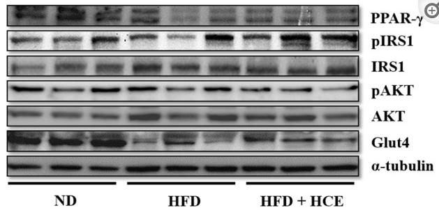
Application: WB    Species: Mice    Sample: epididymal white adipose tissue

Figure 5 Effect of djulis hull crude extract on the expression of proteins involved in glucose transportation in (A) epididymal white adipose tissue (eWAT) and (B) the liver of high-fat diet-induced hyperglycaemia. ND: normal diet; HFD: high-fat diet; HCE: high dosage of crude extract. Values represent the mean ± SEM (n = 6). The statistical methods used one-way ANOVA, and the values with different letters are significantly different at p < 0.05.

5). Black Sesame Seeds Ethanol Extract Ameliorates Hepatic Lipid Accumulation, Oxidative Stress, and Insulin Resistance in Fructose-Induced Nonalcoholic Fatty Liver Disease. JOURNAL OF AGRICULTURAL AND FOOD CHEMISTRY (PubMed: 30244573) [IF=6.1]

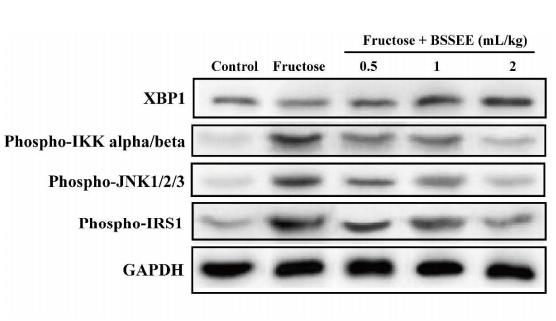
Application: WB    Species: mouse    Sample: Liver

Figure.6. |Effects of BSSEE (0.5, 1 and 2 mL/kg) on the expression of hepatic (A) XBP1, (B) phospho-IKK alpha/beta (Thr 183+Tyr 185), (C) phospho-JNK1/2/3 (Ser180/181) and (D) phospho-IRS1 (Ser 307).

6). Alleviation of Fufang Fanshiliu decoction on type II diabetes mellitus by reducing insulin resistance: A comprehensive network prediction and experimental validation. Journal of Ethnopharmacology (PubMed: 35568115) [IF=5.4]

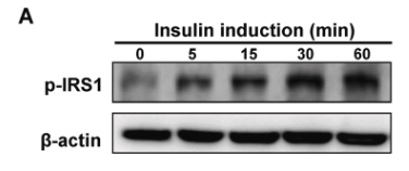
Application: WB    Species: Human    Sample: HepG2 cells

Fig. 7. FFSLD alleviated insulin resistance (IR) in the HepG2-IR cells model induced by high glucose (HG). (A) Western Blot detection and (B) quantitative analysis for the expression of p-IRS1 in HepG2 cells stimulated by insulin (1 μM) for 0, 5, 15, 30, 60 min, respectively. (C) Western Blot detection and (D) quantitative analysis for the expression of p-IRS1 in HepG2 cells incubated with 30 mM glucose for 0, 3, 6, 12, 18, 24 h, respectively, and followed by insulin stimulation (1 μM) for 30 min. (E) Cell viability of HepG2 cells treated by different concentrations of FFSLD for 24 h. (F) Western Blot detection and (G) quantitative analysis for the expression of p-IRS1 in HepG2 cells treated with different concentrations of FFSLD in 30 mM glucose of medium, followed by insulin stimulation (1 μM) for 30 min. Data (n = 3) are expressed as mean ± SEM. *p < 0.05, **p < 0.01, compared with the control group.

7). Silencing of ANGPTL8 Alleviates Insulin Resistance in Trophoblast Cells. Frontiers in Endocrinology (PubMed: 34163433) [IF=5.2]

Application: WB    Species: mouse    Sample: placenta

FIGURE 1 | Angiopoietin like-8 (ANGPTL8) was increased in serum and placenta tissues of gestational diabetes mellitus (GDM) mice.(J) Western blot was used to determine the levels of insulin signaling related molecules, p-IRb(Tyr1361), IRb, p-IRS-1(Ser307), p-IRS-1(Tyr896), IRS-1, p-Akt and Akt in placenta tissues.

Application: WB    Species: Mice    Sample: serum and placenta tissue

Figure 1 Angiopoietin like-8 (ANGPTL8) was increased in serum and placenta tissues of gestational diabetes mellitus (GDM) mice. (A) The mice were treated as described in the chart. (B) The body weight of mice in normal fat diet (NFD) and high fat diet (HFD) groups. (C) Oral glucose tolerance test (OGTT) was performed at gestational day (GD)0.5, 11.5 and 16.5. (D, E) Fasting blood glucose and insulin levels were measured at GD18.5. (F) Homeostasis model assessment insulin resistance (HOMA-IR) was calculated as follow: HOMA-IR= blood glucose (mM)×blood insulin (mU/l)/22.5. (G) The contents of triglyceride (TG), total cholesterol (TC), high density lipoprotein (HDL-C) and low density lipoprotein (LDL-C) in serum were detected. (H) HE staining was performed to detect the pathological changes in labyrinth zone of placenta tissues. (I) Periodic acid Schiff (PAS) staining was carried out to detect the glycogen accumulation in labyrinth zone of placenta tissues. (J) Western blot was used to determine the levels of insulin signaling related molecules, p-IRβ(Tyr1361), IRβ, p-IRS-1(Ser307), p-IRS-1(Tyr896), IRS-1, p-Akt and Akt in placenta tissues. (K) The expression levels of glucose transporter 1 (GLUT1) and GLUT4 in placenta tissues. (L) The serum level of ANGPTL8 in mice. (M, N) The mRNA and protein levels of ANGPTL8 in placenta tissues. (the scale bar represents 100 μm; **p < 0.01, ***p < 0.001 vs. NFD).

8). Hyperinsulinemia Can Cause Kidney Disease in the IGT Stage of OLETF Rats via the INS/IRS-1/PI3-K/Akt Signaling Pathway. Journal of Diabetes Research (PubMed: 31737684) [IF=4.3]

9). Curcumin attenuates BPA-induced insulin resistance in HepG2 cells through suppression of JNK/p38 pathways. TOXICOLOGY LETTERS (PubMed: 28300666) [IF=3.5]

Application: WB    Species: human    Sample: HepG2

Application: WB    Species: human    Sample: HepG2 cells

Figure 1| Effect of BPA on insulin resistance in HepG2 cells. (C) The levels of p-IR, p-IRS1 and p-AKT were measured by Western blot. Data are expressed as mean ± SD of five independent experiments. ∗P< 0.05 and ∗∗P < 0.01, significantly different as compared with the untreated control.

10). P53 modulates hepatic insulin sensitivity through NF-κB and p38/ERK MAPK pathways. BIOCHEMICAL AND BIOPHYSICAL RESEARCH COMMUNICATIONS (PubMed: 29258820) [IF=3.1]

Load more

Restrictive clause


Affinity Biosciences tests all products strictly. Citations are provided as a resource for additional applications that have not been validated by Affinity Biosciences. Please choose the appropriate format for each application and consult Materials and Methods sections for additional details about the use of any product in these publications.

For Research Use Only.
Not for use in diagnostic or therapeutic procedures. Not for resale. Not for distribution without written consent. Affinity Biosciences will not be held responsible for patent infringement or other violations that may occur with the use of our products. Affinity Biosciences, Affinity Biosciences Logo and all other trademarks are the property of Affinity Biosciences LTD.