ITGAM Antibody - #DF7553
, diluted 1/600 was used as secondary antibody.")
| Product: | ITGAM Antibody |
| Catalog: | DF7553 |
| Description: | Rabbit polyclonal antibody to ITGAM |
| Application: | WB IF/ICC |
| Cited expt.: | WB, IF/ICC |
| Reactivity: | Human, Mouse, Rat |
| Prediction: | Bovine, Horse, Sheep, Rabbit, Dog |
| Mol.Wt.: | 170 kDa(Observed); 127kD(Calculated). |
| Uniprot: | P11215 |
| RRID: | AB_2841048 |
Product Info
*The optimal dilutions should be determined by the end user. For optimal experimental results, antibody reuse is not recommended.
*Tips:
WB: For western blot detection of denatured protein samples. IHC: For immunohistochemical detection of paraffin sections (IHC-p) or frozen sections (IHC-f) of tissue samples. IF/ICC: For immunofluorescence detection of cell samples. ELISA(peptide): For ELISA detection of antigenic peptide.
Cite Format: Affinity Biosciences Cat# DF7553, RRID:AB_2841048.
Fold/Unfold
antigen CD11b (p170); Antigen CD11b p170; CD11 antigen like family member B; CD11 antigen-like family member B; CD11b; CD11b/CD18; CD49d; Cell surface glycoprotein MAC-1 subunit alpha; Complement component 3 receptor 3 subunit; Complement Component Receptor 3 Alpha; Complement receptor type 3, alpha subunit; CR 3 alpha chain (CR3A); CR 3 alpha chain; CR-3 alpha chain; CR3; CR3A; F730045J24Rik; Integrin Alpha M; Integrin alpha M chain; Integrin alpha-M; Integrin beta 2 alpha subunit; Integrin subunit alpha M; integrin, alpha M (complement component 3 receptor 3 subunit); ITAM_HUMAN; ITGAM; Leukocyte adhesion receptor MO1; Ly-40; MAC 1; Mac-1a; MAC1; Mac1, alpha subunit; MAC1A; Macrophage antigen alpha polypeptide; MGC117044; Mo1, alpha subunit; MO1A; Neutrophil adherence receptor alpha M subunit; Neutrophil adherence receptor; SLEB6;
Immunogens
A synthesized peptide derived from human ITGAM, corresponding to a region within the internal amino acids.
Predominantly expressed in monocytes and granulocytes (PubMed:1346576). Expressed in neutrophils (at protein level) (PubMed:21193407).
- P11215 ITAM_HUMAN:
- Protein BLAST With
- NCBI/
- ExPASy/
- Uniprot
MALRVLLLTALTLCHGFNLDTENAMTFQENARGFGQSVVQLQGSRVVVGAPQEIVAANQRGSLYQCDYSTGSCEPIRLQVPVEAVNMSLGLSLAATTSPPQLLACGPTVHQTCSENTYVKGLCFLFGSNLRQQPQKFPEALRGCPQEDSDIAFLIDGSGSIIPHDFRRMKEFVSTVMEQLKKSKTLFSLMQYSEEFRIHFTFKEFQNNPNPRSLVKPITQLLGRTHTATGIRKVVRELFNITNGARKNAFKILVVITDGEKFGDPLGYEDVIPEADREGVIRYVIGVGDAFRSEKSRQELNTIASKPPRDHVFQVNNFEALKTIQNQLREKIFAIEGTQTGSSSSFEHEMSQEGFSAAITSNGPLLSTVGSYDWAGGVFLYTSKEKSTFINMTRVDSDMNDAYLGYAAAIILRNRVQSLVLGAPRYQHIGLVAMFRQNTGMWESNANVKGTQIGAYFGASLCSVDVDSNGSTDLVLIGAPHYYEQTRGGQVSVCPLPRGRARWQCDAVLYGEQGQPWGRFGAALTVLGDVNGDKLTDVAIGAPGEEDNRGAVYLFHGTSGSGISPSHSQRIAGSKLSPRLQYFGQSLSGGQDLTMDGLVDLTVGAQGHVLLLRSQPVLRVKAIMEFNPREVARNVFECNDQVVKGKEAGEVRVCLHVQKSTRDRLREGQIQSVVTYDLALDSGRPHSRAVFNETKNSTRRQTQVLGLTQTCETLKLQLPNCIEDPVSPIVLRLNFSLVGTPLSAFGNLRPVLAEDAQRLFTALFPFEKNCGNDNICQDDLSITFSFMSLDCLVVGGPREFNVTVTVRNDGEDSYRTQVTFFFPLDLSYRKVSTLQNQRSQRSWRLACESASSTEVSGALKSTSCSINHPIFPENSEVTFNITFDVDSKASLGNKLLLKANVTSENNMPRTNKTEFQLELPVKYAVYMVVTSHGVSTKYLNFTASENTSRVMQHQYQVSNLGQRSLPISLVFLVPVRLNQTVIWDRPQVTFSENLSSTCHTKERLPSHSDFLAELRKAPVVNCSIAVCQRIQCDIPFFGIQEEFNATLKGNLSFDWYIKTSHNHLLIVSTAEILFNDSVFTLLPGQGAFVRSQTETKVEPFEVPNPLPLIVGSSVGGLLLLALITAALYKLGFFKRQYKDMMSEGGPPGAEPQ
Predictions
Score>80(red) has high confidence and is suggested to be used for WB detection. *The prediction model is mainly based on the alignment of immunogen sequences, the results are for reference only, not as the basis of quality assurance.
High(score>80) Medium(80>score>50) Low(score<50) No confidence
Research Backgrounds
Integrin ITGAM/ITGB2 is implicated in various adhesive interactions of monocytes, macrophages and granulocytes as well as in mediating the uptake of complement-coated particles and pathogens. It is identical with CR-3, the receptor for the iC3b fragment of the third complement component. It probably recognizes the R-G-D peptide in C3b. Integrin ITGAM/ITGB2 is also a receptor for fibrinogen, factor X and ICAM1. It recognizes P1 and P2 peptides of fibrinogen gamma chain. Regulates neutrophil migration. In association with beta subunit ITGB2/CD18, required for CD177-PRTN3-mediated activation of TNF primed neutrophils. May regulate phagocytosis-induced apoptosis in extravasated neutrophils (By similarity). May play a role in mast cell development (By similarity). Required with TYROBP/DAP12 in microglia to control production of microglial superoxide ions which promote the neuronal apoptosis that occurs during brain development (By similarity).
Cell membrane>Single-pass type I membrane protein. Membrane raft>Single-pass type I membrane protein.
Predominantly expressed in monocytes and granulocytes. Expressed in neutrophils (at protein level).
The integrin I-domain (insert) is a VWFA domain. Integrins with I-domains do not undergo protease cleavage.
Belongs to the integrin alpha chain family.
Research Fields
· Cellular Processes > Transport and catabolism > Phagosome. (View pathway)
· Cellular Processes > Cell motility > Regulation of actin cytoskeleton. (View pathway)
· Environmental Information Processing > Signal transduction > Rap1 signaling pathway. (View pathway)
· Environmental Information Processing > Signaling molecules and interaction > Cell adhesion molecules (CAMs). (View pathway)
· Human Diseases > Infectious diseases: Bacterial > Pertussis.
· Human Diseases > Infectious diseases: Bacterial > Legionellosis.
· Human Diseases > Infectious diseases: Parasitic > Leishmaniasis.
· Human Diseases > Infectious diseases: Parasitic > Amoebiasis.
· Human Diseases > Infectious diseases: Bacterial > Staphylococcus aureus infection.
· Human Diseases > Infectious diseases: Bacterial > Tuberculosis.
· Human Diseases > Cancers: Overview > Transcriptional misregulation in cancer.
· Human Diseases > Cancers: Specific types > Acute myeloid leukemia. (View pathway)
· Organismal Systems > Immune system > Complement and coagulation cascades. (View pathway)
· Organismal Systems > Immune system > Hematopoietic cell lineage. (View pathway)
· Organismal Systems > Immune system > Leukocyte transendothelial migration. (View pathway)
References
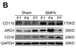
Application: WB Species: Rat Sample: spinal cord
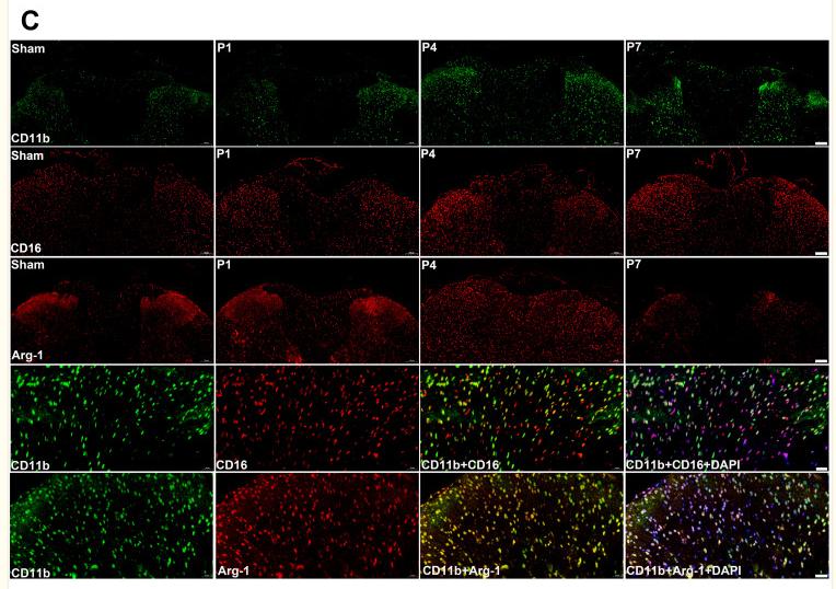
Application: IF/ICC Species: Rat Sample: spinal cord
Restrictive clause
Affinity Biosciences tests all products strictly. Citations are provided as a resource for additional applications that have not been validated by Affinity Biosciences. Please choose the appropriate format for each application and consult Materials and Methods sections for additional details about the use of any product in these publications.
For Research Use Only.
Not for use in diagnostic or therapeutic procedures. Not for resale. Not for distribution without written consent. Affinity Biosciences will not be held responsible for patent infringement or other violations that may occur with the use of our products. Affinity Biosciences, Affinity Biosciences Logo and all other trademarks are the property of Affinity Biosciences LTD.