MCP1 Antibody - #DF7554
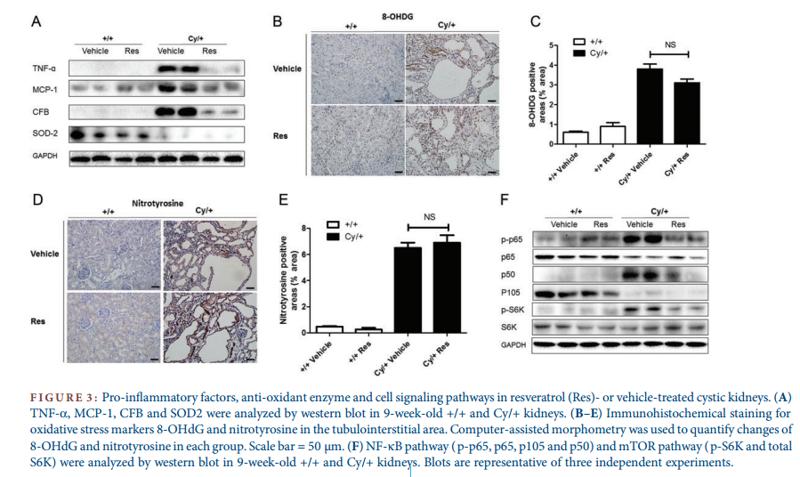
- or vehicle-treated cystic kidneys. (A) TNF-α, MCP-1, CFB and SOD2 were analyzed by western blot in 9-week-old +/+ and Cy/+ kidneys. (B–E) Immunohistochemical staining for oxidative stress markers 8-OHdG and nitrotyrosine in the tubulointerstitial area. Computer-assisted morphometry was used to quantify changes of 8-OHdG and nitrotyrosine in each group. Scale bar = 50 µm. (F) NF-κB pathway ( p-p65, p65, p105 and p50) and mTOR pathway ( p-S6K and total S6K) were analyzed by western blot in 9-week-old +/+ and Cy/+ kidneys. Blots are representative of three independent experiments.")
| Product: | MCP1 Antibody |
| Catalog: | DF7554 |
| Description: | Rabbit polyclonal antibody to MCP1 |
| Application: | WB IHC |
| Reactivity: | Human, Rat |
| Prediction: | Horse |
| Mol.Wt.: | 14 kDa; 11kD(Calculated). |
| Uniprot: | P13500 |
| RRID: | AB_2841049 |
Product Info
*The optimal dilutions should be determined by the end user.
*Tips:
WB: For western blot detection of denatured protein samples. IHC: For immunohistochemical detection of paraffin sections (IHC-p) or frozen sections (IHC-f) of tissue samples. IF/ICC: For immunofluorescence detection of cell samples. ELISA(peptide): For ELISA detection of antigenic peptide.
Cite Format: Affinity Biosciences Cat# DF7554, RRID:AB_2841049.
Fold/Unfold
C-C motif chemokine 2; CCL2; CCL2_HUMAN; Chemokine (C C motif) ligand 2; GDCF 2; GDCF-2; GDCF2; HC11; HSMCR30; JE; MCAF; MCP 1; MCP-1; MCP1; MGC9434; Monocyte chemoattractant protein 1; Monocyte chemotactic and activating factor; Monocyte chemotactic protein 1; Monocyte secretory protein JE; SCYA2; Small inducible cytokine A2 (monocyte chemotactic protein 1, homologous to mouse Sig je); Small inducible cytokine A2; Small inducible cytokine subfamily A (Cys Cys), member 2; Small-inducible cytokine A2; SMC CF; SMC-CF; SMCCF;
Immunogens
A synthesized peptide derived from human MCP1, corresponding to a region within the internal amino acids.
Expressed in the seminal plasma, endometrial fluid and follicular fluid (at protein level) (PubMed:23765988). Expressed in monocytes (PubMed:2513477).
- P13500 CCL2_HUMAN:
- Protein BLAST With
- NCBI/
- ExPASy/
- Uniprot
MKVSAALLCLLLIAATFIPQGLAQPDAINAPVTCCYNFTNRKISVQRLASYRRITSSKCPKEAVIFKTIVAKEICADPKQKWVQDSMDHLDKQTQTPKT
Predictions
Score>80(red) has high confidence and is suggested to be used for WB detection. *The prediction model is mainly based on the alignment of immunogen sequences, the results are for reference only, not as the basis of quality assurance.
High(score>80) Medium(80>score>50) Low(score<50) No confidence
Research Backgrounds
Acts as a ligand for C-C chemokine receptor CCR2. Signals through binding and activation of CCR2 and induces a strong chemotactic response and mobilization of intracellular calcium ions. Exhibits a chemotactic activity for monocytes and basophils but not neutrophils or eosinophils. May be involved in the recruitment of monocytes into the arterial wall during the disease process of atherosclerosis.
Processing at the N-terminus can regulate receptor and target cell selectivity. Deletion of the N-terminal residue converts it from an activator of basophil to an eosinophil chemoattractant.
N-Glycosylated.
Secreted.
Expressed in the seminal plasma, endometrial fluid and follicular fluid (at protein level). Expressed in monocytes.
Monomer or homodimer; in equilibrium. Is tethered on endothelial cells by glycosaminoglycan (GAG) side chains of proteoglycans.
Belongs to the intercrine beta (chemokine CC) family.
Research Fields
· Environmental Information Processing > Signaling molecules and interaction > Cytokine-cytokine receptor interaction. (View pathway)
· Environmental Information Processing > Signal transduction > TNF signaling pathway. (View pathway)
· Human Diseases > Infectious diseases: Parasitic > Chagas disease (American trypanosomiasis).
· Human Diseases > Infectious diseases: Parasitic > Malaria.
· Human Diseases > Infectious diseases: Viral > Influenza A.
· Human Diseases > Infectious diseases: Viral > Herpes simplex infection.
· Human Diseases > Immune diseases > Rheumatoid arthritis.
· Organismal Systems > Immune system > Chemokine signaling pathway. (View pathway)
· Organismal Systems > Immune system > NOD-like receptor signaling pathway. (View pathway)
· Organismal Systems > Immune system > IL-17 signaling pathway. (View pathway)
References
Application: WB Species: rat Sample:
Restrictive clause
Affinity Biosciences tests all products strictly. Citations are provided as a resource for additional applications that have not been validated by Affinity Biosciences. Please choose the appropriate format for each application and consult Materials and Methods sections for additional details about the use of any product in these publications.
For Research Use Only.
Not for use in diagnostic or therapeutic procedures. Not for resale. Not for distribution without written consent. Affinity Biosciences will not be held responsible for patent infringement or other violations that may occur with the use of our products. Affinity Biosciences, Affinity Biosciences Logo and all other trademarks are the property of Affinity Biosciences LTD.