MLC2 Antibody - #DF7911

| Product: | MLC2 Antibody |
| Catalog: | DF7911 |
| Description: | Rabbit polyclonal antibody to MLC2 |
| Application: | WB IHC IF/ICC |
| Reactivity: | Human, Mouse, Rat |
| Prediction: | Bovine, Horse, Sheep, Rabbit, Dog, Chicken, Xenopus |
| Mol.Wt.: | 19 kDa; 19kD(Calculated). |
| Uniprot: | P10916 |
| RRID: | AB_2841328 |
Related Downloads
Protocols
Product Info
*The optimal dilutions should be determined by the end user.
*Tips:
WB: For western blot detection of denatured protein samples. IHC: For immunohistochemical detection of paraffin sections (IHC-p) or frozen sections (IHC-f) of tissue samples. IF/ICC: For immunofluorescence detection of cell samples. ELISA(peptide): For ELISA detection of antigenic peptide.
Cite Format: Affinity Biosciences Cat# DF7911, RRID:AB_2841328.
Fold/Unfold
Cardiac myosin light chain-2; Cardiac ventricular myosin light chain 2; CMH10; MLC 2v; MLC-2; MLC-2v; MLC2; MLRV_HUMAN; MYL 2; MYL2; Myosin light chain 2 regulatory cardiac slow; Myosin light polypeptide 2 regulatory cardiac slow; Myosin regulatory light chain 2; Myosin regulatory light chain 2 ventricular/cardiac muscle isoform; Regulatory light chain of myosin; RLC of myosin; Slow cardiac myosin regulatory light chain 2; ventricular/cardiac muscle isoform;
Immunogens
- P10916 MLRV_HUMAN:
- Protein BLAST With
- NCBI/
- ExPASy/
- Uniprot
MAPKKAKKRAGGANSNVFSMFEQTQIQEFKEAFTIMDQNRDGFIDKNDLRDTFAALGRVNVKNEEIDEMIKEAPGPINFTVFLTMFGEKLKGADPEETILNAFKVFDPEGKGVLKADYVREMLTTQAERFSKEEVDQMFAAFPPDVTGNLDYKNLVHIITHGEEKD
Predictions
Score>80(red) has high confidence and is suggested to be used for WB detection. *The prediction model is mainly based on the alignment of immunogen sequences, the results are for reference only, not as the basis of quality assurance.
High(score>80) Medium(80>score>50) Low(score<50) No confidence
PTMs - P10916 As Substrate
| Site | PTM Type | Enzyme | Source |
|---|---|---|---|
| S15 | Phosphorylation | Q15746 (MYLK) | Uniprot |
| S19 | Phosphorylation | Q5VT25 (CDC42BPA) | Uniprot |
| T80 | Phosphorylation | Uniprot | |
| T98 | Phosphorylation | Uniprot | |
| K104 | Acetylation | Uniprot | |
| K111 | Acetylation | Uniprot | |
| Y118 | Phosphorylation | Uniprot | |
| T124 | Phosphorylation | Uniprot | |
| Y152 | Phosphorylation | Uniprot | |
| K153 | Acetylation | Uniprot | |
| K165 | Acetylation | Uniprot |
Research Backgrounds
Contractile protein that plays a role in heart development and function (By similarity). Following phosphorylation, plays a role in cross-bridge cycling kinetics and cardiac muscle contraction by increasing myosin lever arm stiffness and promoting myosin head diffusion; as a consequence of the increase in maximum contraction force and calcium sensitivity of contraction force. These events altogether slow down myosin kinetics and prolong duty cycle resulting in accumulated myosins being cooperatively recruited to actin binding sites to sustain thin filament activation as a means to fine-tune myofilament calcium sensitivity to force (By similarity). During cardiogenesis plays an early role in cardiac contractility by promoting cardiac myofibril assembly (By similarity).
N-terminus is methylated by METTL11A/NTM1.
Phosphorylated by MYLK3 and MYLK2; promotes cardiac muscle contraction and function (By similarity). Dephosphorylated by PPP1CB complexed to PPP1R12B (By similarity). The phosphorylated form in adult is expressed as gradients across the heart from endocardium (low phosphorylation) to epicardium (high phosphorylation); regulates cardiac torsion and workload distribution (By similarity).
Cytoplasm>Myofibril>Sarcomere>A band.
Myosin is a hexamer of 2 heavy chains and 4 light chains. Interacts with MYOC.
References
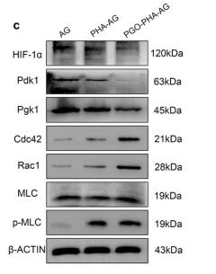
Application: WB Species: rat Sample: RAW 264.7 cells

Application: WB Species: Mice Sample: mRMVECs

Application: WB Species: Mice Sample: Colonic tissues
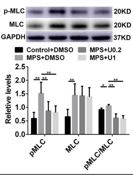
Application: WB Species: rat Sample: muscle
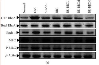
Application: WB Species: Mice Sample: colon tissue
Restrictive clause
Affinity Biosciences tests all products strictly. Citations are provided as a resource for additional applications that have not been validated by Affinity Biosciences. Please choose the appropriate format for each application and consult Materials and Methods sections for additional details about the use of any product in these publications.
For Research Use Only.
Not for use in diagnostic or therapeutic procedures. Not for resale. Not for distribution without written consent. Affinity Biosciences will not be held responsible for patent infringement or other violations that may occur with the use of our products. Affinity Biosciences, Affinity Biosciences Logo and all other trademarks are the property of Affinity Biosciences LTD.