Product: Phospho-MLKL (Ser358) Antibody
Catalog: AF7420
Description: Rabbit polyclonal antibody to Phospho-MLKL (Ser358)
Application: WB IHC
Reactivity: Human, Mouse, Rat
Mol.Wt.: 54kDa; 54kD(Calculated).
Uniprot: Q8NB16
RRID: AB_2843860

View similar products>>

   Size Price Inventory
 100ul $350 In stock
 200ul $450 In stock

Lead Time: Same day delivery

For pricing and ordering contact:
Local distributors

Product Info

Source:
Rabbit
Application:
WB 1:500-1:2000, IHC 1:50-1:200
*The optimal dilutions should be determined by the end user.
*Tips:

WB: For western blot detection of denatured protein samples. IHC: For immunohistochemical detection of paraffin sections (IHC-p) or frozen sections (IHC-f) of tissue samples. IF/ICC: For immunofluorescence detection of cell samples. ELISA(peptide): For ELISA detection of antigenic peptide.

Reactivity:
Human,Mouse,Rat
Clonality:
Polyclonal
Specificity:
Phospho-MLKL (Ser358) Antibody detects endogenous levels of MLKL only when phosphorylated at Ser358.
RRID:
AB_2843860
Cite Format: Affinity Biosciences Cat# AF7420, RRID:AB_2843860.
Conjugate:
Unconjugated.
Purification:
The antibody is from purified rabbit serum by affinity purification via sequential chromatography on phospho-peptide and non-phospho-peptide affinity columns.
Storage:
Rabbit IgG in phosphate buffered saline , pH 7.4, 150mM NaCl, 0.02% sodium azide and 50% glycerol. Store at -20 °C. Stable for 12 months from date of receipt.
Alias:

Fold/Unfold

9130019I15Rik; FLJ34389; hMLKL; Mixed lineage kinase domain like; Mixed lineage kinase domain like protein; Mixed lineage kinase domain like pseudokinase; Mixed lineage kinase domain-like protein; Mlkl; MLKL_HUMAN;

Immunogens

Immunogen:
Uniprot:
Gene(ID):
Sequence:
MENLKHIITLGQVIHKRCEEMKYCKKQCRRLGHRVLGLIKPLEMLQDQGKRSVPSEKLTTAMNRFKAALEEANGEIEKFSNRSNICRFLTASQDKILFKDVNRKLSDVWKELSLLLQVEQRMPVSPISQGASWAQEDQQDADEDRRAFQMLRRDNEKIEASLRRLEINMKEIKETLRQYLPPKCMQEIPQEQIKEIKKEQLSGSPWILLRENEVSTLYKGEYHRAPVAIKVFKKLQAGSIAIVRQTFNKEIKTMKKFESPNILRIFGICIDETVTPPQFSIVMEYCELGTLRELLDREKDLTLGKRMVLVLGAARGLYRLHHSEAPELHGKIRSSNFLVTQGYQVKLAGFELRKTQTSMSLGTTREKTDRVKSTAYLSPQELEDVFYQYDVKSEIYSFGIVLWEIATGDIPFQGCNSEKIRKLVAVKRQQEPLGEDCPSELREIIDECRAHDPSVRPSVDEILKKLSTFSK

PTMs - Q8NB16 As Substrate

Site PTM Type Enzyme
K40 Ubiquitination
K50 Ubiquitination
S52 Phosphorylation
K57 Ubiquitination
T59 Phosphorylation
K66 Ubiquitination
K78 Ubiquitination
S92 Phosphorylation
S106 Phosphorylation
S125 Phosphorylation
S128 Phosphorylation
K157 Ubiquitination
S161 Phosphorylation
K173 Ubiquitination
K183 Ubiquitination
K198 Ubiquitination
K219 Ubiquitination
K230 Ubiquitination
T246 Phosphorylation
K249 Ubiquitination
T302 Phosphorylation
K331 Ubiquitination
S334 Phosphorylation
K354 Acetylation
K354 Ubiquitination
T357 Phosphorylation Q9Y572 (RIPK3)
S358 Phosphorylation Q9Y572 (RIPK3)
T364 Phosphorylation
K372 Ubiquitination
S373 Phosphorylation
T374 Phosphorylation
S393 Phosphorylation
S417 Phosphorylation
S467 Phosphorylation

Research Backgrounds

Function:

Pseudokinase that plays a key role in TNF-induced necroptosis, a programmed cell death process. Activated following phosphorylation by RIPK3, leading to homotrimerization, localization to the plasma membrane and execution of programmed necrosis characterized by calcium influx and plasma membrane damage. Does not have protein kinase activity. Binds to highly phosphorylated inositol phosphates such as inositolhexakisphosphate (InsP6) which is essential for its necroptotic function.

PTMs:

Phosphorylation by RIPK3 induces a conformational switch that is required for necroptosis. It also induces homotrimerization and localization to the plasma membrane.

Subcellular Location:

Cytoplasm. Cell membrane.
Note: Localizes to the cytoplasm and translocates to the plasma membrane on necroptosis induction.

Extracellular region or secreted Cytosol Plasma membrane Cytoskeleton Lysosome Endosome Peroxisome ER Golgi apparatus Nucleus Mitochondrion Manual annotation Automatic computational assertionSubcellular location
Subunit Structure:

Homooligomer. Homotrimer; forms homotrimers on necroptosis induction. Interacts with RIPK3; the interaction is direct. Upon TNF-induced necrosis, forms in complex with PGAM5, RIPK1 and RIPK3. Within this complex, may play a role in the proper targeting of RIPK1/RIPK3 to its downstream effector PGAM5.

Family&Domains:

The protein kinase domain is catalytically inactive but contains an unusual pseudoactive site with an interaction between Lys-230 and Gln-356 residues. Upon phosphorylation by RIPK3, undergoes an active conformation (By similarity).

The coiled coil region 2 is responsible for homotrimerization.

Belongs to the protein kinase superfamily.

Research Fields

· Cellular Processes > Cell growth and death > Necroptosis.   (View pathway)

· Environmental Information Processing > Signal transduction > TNF signaling pathway.   (View pathway)

References

1). Polysaccharide from Strongylocentrotus nudus eggs regulates intestinal epithelial autophagy through CD36/PI3K-Akt pathway to ameliorate inflammatory bowel disease. International Journal of Biological Macromolecules (PubMed: 37327932) [IF=8.2]

2). Pterocephin A, a Novel Triterpenoid Saponin from Pterocephalus hookeri Induced Liver Injury by Activation of Necroptosis. PHYTOMEDICINE (PubMed: 33831690) [IF=7.9]

3). Protective Effect of Calpain Inhibition During Cold Ischemia on Ischemia–reperfusion Injury After Lung Transplantation. Transplantation (PubMed: 36648297) [IF=6.2]

4). Inhibition of extracellular signal-regulated kinase/calpain-2 pathway reduces neuroinflammation and necroptosis after cerebral ischemia-reperfusion injury in a rat model of cardiac arrest. International Immunopharmacology (PubMed: 33517223) [IF=5.6]

5). Cisatracurium besylate rescues Mycobacterium Tuberculosis-infected macrophages from necroptosis and enhances the bactericidal effect of isoniazid. International Immunopharmacology (PubMed: 37182451) [IF=5.6]

6). Inhibiting Heat Shock Protein 90 Protects Nucleus Pulposus-Derived Stem/Progenitor Cells From Compression-Induced Necroptosis and Apoptosis. Frontiers in Cell and Developmental Biology (PubMed: 32850811) [IF=5.5]

Application: WB    Species: human    Sample: NPSCs

FIGURE 2 | RIPK1/RIPK3/MLKL-mediated necroptosis was involved in compression-induced death of NPSCs. (A) Representative WB graphs of the expression of RIPK1, P-RIPK1, RIPK3, P-RIPK3, MLKL, and P-MLKL. (B) Quantitation of the expression levels of RIPK1, P-RIPK1, RIPK3, P-RIPK3, MLKL, and P-MLKL. (C) The effects of different concentrations of Nec-1, GSK0872 and NSA on cell viability of NPSCs exposed to 0, 24, 36, and 48 h compression measured by CCK-8 assays. (D) Representative dot plots of PI staining obtained from flow cytometry analysis of NPSCs. (E) The statistical analysis of PI positive ratio of NPSCs. The data were expressed as mean ± SD from at least three independent experiments, and they were analyzed by a two-tailed t-test. (*P < 0.05, **P < 0.01, ***P < 0.001 vs. 0 h, 0 µM or control, NS, not significant)

7). Sacubitril Ameliorates Cardiac Fibrosis Through Inhibiting TRPM7 Channel. Frontiers in Cell and Developmental Biology (PubMed: 34778271) [IF=5.5]

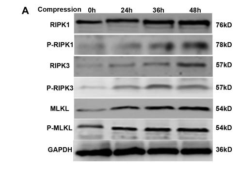
Application: WB    Species: Rat    Sample: H9C2 cells

FIGURE 4 LBQ657 reduced hypoxia-induced necrosis by inhibiting TRPM7-mediated Ca2+ influx in cardiomyocytes. (A) Comparison of TRPM7 protein levels in MEF and H9C2 cells. (B) Fluorescence intensity of Fluo-4-AM fluorescent dye in 488 nm excitation light of normoxic H9C2, hypoxic H9C2 and hypoxic H9C2 pre-treated with LBQ657. (C) Protein levels of caspase3, cleaved caspase3, LC3B-I and LC3B-II in hypoxic H9C2 and hypoxic H9C2 pre-treated with LBQ657. (D) Protein levels of RIPK1/RIPK3/MLKL/p-MLKL in hypoxic H9C2 and hypoxic H9C2 pre-treated with LBQ657. (E) Protein levels of RIPK1/RIPK3/MLKL/p-MLKL in normoxic H9C2 and normoxic H9C2 treated with LBQ657. (F) Protein levels of RIPK1/RIPK3/MLKL/p-MLKL in hypoxic H9C2 and hypoxic H9C2 pre-treated with BAPTA-AM. The data presented are mean ± SD. Each group contained the results of three independent repeated trials. β-actin was used as the internal reference protein for normalization and statistical analysis. ∗∗∗∗P < 0.0001, ∗∗∗P < 0.001, ∗∗P < 0.01, and ∗P < 0.05.

8). Autophagy-related LC3 accumulation interacted directly with LIR containing RIPK1 and RIPK3, stimulating necroptosis in hypoxic cardiomyocytes. Frontiers in Cell and Developmental Biology (PubMed: 34368130) [IF=5.5]

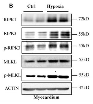
Application: WB    Species: mouse    Sample: myocardium

FIGURE 1 | Necroptosis mediates cardiac dysfunction caused by hypoxia. (B,C) Representative bands of western blotting and statistical analysis, which were performed to detect RIPK1, RIPK3, p-RIPK3, MLKL, and p-MLKL levels after hypoxia treatment in the myocardium, Means ± SEM, n = 5.*p < 0.05, **p < 0.01, and ***p < 0.001 versus the control group.

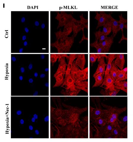
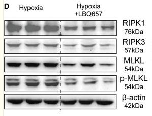
Application: IF/ICC    Species: mouse    Sample: myocardium

FIGURE 1 | Necroptosis mediates cardiac dysfunction caused by hypoxia. (I,J) Representative confocal images and statistical analysis of p-MLKL after hypoxia treatment for 9 h.Scale bar, 10 µm. n = 3, Mean ± SEM. ***p < 0.001 versus the control group, ###p < 0.001 versus the hypoxia group.

9). Adenosine kinase inhibition prevents severe acute pancreatitis via suppressing inflammation and acinar cell necroptosis. Frontiers in Cell and Developmental Biology (PubMed: 35281076) [IF=5.5]

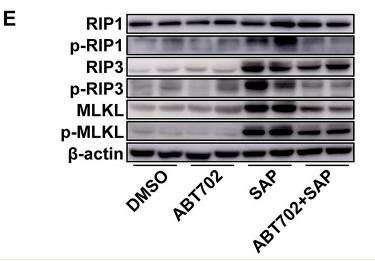
Application: WB    Species: Mice    Sample: pancreas

FIGURE 2 Effects of ADK inhibition on inflammation and pancreatic cell necrosis. (A), Neutrophils were immunochemically staining using anti-CD11b antibody. (n = 10). Scale bar = 50 μm. (B), Macrophages were visualized using immunofluorescence staining with anti-MOMA-2 antibody. (n = 10). Scale bar = 50 μm. (C). NF-κB-P65 and the phosphorylation of NF-κB-P65 were immunoblotted. (n = 5). (D), Pancreatic necrosis was visualized by double staining of EBD and anti-amylase. (n = 10). (E), The critical molecules of the necroptotic pathway were immunoblotted. (n = 5).

10). Tanshinone I exerts cardiovascular protective effects in vivo and in vitro through inhibiting necroptosis via Akt/Nrf2 signaling pathway. Chinese Medicine (PubMed: 34183021) [IF=4.9]

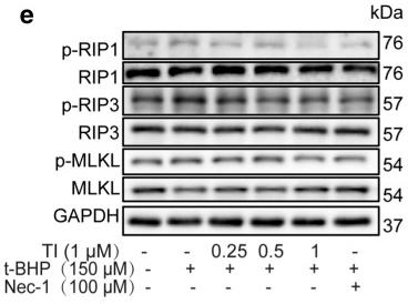
Application: WB    Species: Rat    Sample: H9c2 cells

Fig. 2 TI ameliorated t‑BHP induced cell necroptosis via RIP1/RIP3/MLKL pathway. a H9c2 cells were cultured with Nec-1 for 12 h. b Cells were exposed to t-BHP (150 μM) for 10 h after treated with Nec-1 for 2 h. c Cells were exposed to t-BHP (150 μM) for 10 h after treated with TI (1 μM) or Nec-1 (100 μM) for 2 h. MTT was employed to detect cell viability. d Cells were treated with t-BHP (150 μM) for 10 h when pretreated with TI (1 μM) or Nec-1 (100 μM) for 2 h, the LDH level was monitored by LDH kit. e–h Pretreated with TI (0.25, 0.5, and 1 μM) or Nec-1 (100 μM) for 2 h respectively, then exposed to t-BHP (150 μM) for 4 h, the protein expression was determined by western blotting. n = 3. *p < 0.05, **p < 0.01, ***p < 0.001 vs. t-BHP group

Load more

Restrictive clause


Affinity Biosciences tests all products strictly. Citations are provided as a resource for additional applications that have not been validated by Affinity Biosciences. Please choose the appropriate format for each application and consult Materials and Methods sections for additional details about the use of any product in these publications.

For Research Use Only.
Not for use in diagnostic or therapeutic procedures. Not for resale. Not for distribution without written consent. Affinity Biosciences will not be held responsible for patent infringement or other violations that may occur with the use of our products. Affinity Biosciences, Affinity Biosciences Logo and all other trademarks are the property of Affinity Biosciences LTD.