Product: Phospho-VE-Cadherin (Tyr731) Antibody
Catalog: AF3265
Description: Rabbit polyclonal antibody to Phospho-VE-Cadherin (Tyr731)
Application: WB IHC IF/ICC
Reactivity: Human, Mouse, Rat
Prediction: Pig, Zebrafish, Bovine, Horse, Sheep, Dog, Chicken
Mol.Wt.: 130kDa; 88kD(Calculated).
Uniprot: P33151
RRID: AB_2834691

View similar products>>

   Size Price Inventory
 100ul $280 In stock
 200ul $350 In stock

Lead Time: Same day delivery

For pricing and ordering contact:
Local distributors

Product Info

Source:
Rabbit
Application:
WB 1:500-1:2000, IHC 1:50-1:200, IF/ICC 1:100-1:500
*The optimal dilutions should be determined by the end user.
*Tips:

WB: For western blot detection of denatured protein samples. IHC: For immunohistochemical detection of paraffin sections (IHC-p) or frozen sections (IHC-f) of tissue samples. IF/ICC: For immunofluorescence detection of cell samples. ELISA(peptide): For ELISA detection of antigenic peptide.

Reactivity:
Human,Mouse,Rat
Prediction:
Pig(83%), Zebrafish(92%), Bovine(92%), Horse(100%), Sheep(88%), Dog(92%), Chicken(92%)
Clonality:
Polyclonal
Specificity:
Phospho-VE-Cadherin (Tyr731) Antibody detects endogenous levels of VE-Cadherin only when phosphorylated at Tyrosine 731.
RRID:
AB_2834691
Cite Format: Affinity Biosciences Cat# AF3265, RRID:AB_2834691.
Conjugate:
Unconjugated.
Purification:
The antibody is from purified rabbit serum by affinity purification via sequential chromatography on phospho-peptide and non-phospho-peptide affinity columns.
Storage:
Rabbit IgG in phosphate buffered saline , pH 7.4, 150mM NaCl, 0.02% sodium azide and 50% glycerol. Store at -20 °C. Stable for 12 months from date of receipt.
Alias:

Fold/Unfold

7B 4; 7B4; 7B4 antigen; CADH5_HUMAN; Cadherin 5; Cadherin 5 type 2; Cadherin 5, type 2 (vascular endothelium); Cadherin 5, type 2, VE cadherin (vascular epithelium); cadherin, vascular endothelial; cadherin, vascular endothelial, 1; Cadherin-5; Cadherin5; CD 144; CD144; CD144 antigen; CDH 5; CDH5; CDH5 protein; Endothelial specific cadherin; FLJ17376; OTTHUMP00000174777; Vascular endothelial cadherin; Vascular epithelium cadherin; VE Cad; VE-cadherin; VEC;

Immunogens

Immunogen:
Uniprot:
Gene(ID):
Expression:
P33151 CADH5_HUMAN:

Endothelial tissues and brain.

Description:
This gene is a classical cadherin from the cadherin superfamily and is located in a six-cadherin cluster in a region on the long arm of chromosome 16 that is involved in loss of heterozygosity events in breast and prostate cancer.
Sequence:
MQRLMMLLATSGACLGLLAVAAVAAAGANPAQRDTHSLLPTHRRQKRDWIWNQMHIDEEKNTSLPHHVGKIKSSVSRKNAKYLLKGEYVGKVFRVDAETGDVFAIERLDRENISEYHLTAVIVDKDTGENLETPSSFTIKVHDVNDNWPVFTHRLFNASVPESSAVGTSVISVTAVDADDPTVGDHASVMYQILKGKEYFAIDNSGRIITITKSLDREKQARYEIVVEARDAQGLRGDSGTATVLVTLQDINDNFPFFTQTKYTFVVPEDTRVGTSVGSLFVEDPDEPQNRMTKYSILRGDYQDAFTIETNPAHNEGIIKPMKPLDYEYIQQYSFIVEATDPTIDLRYMSPPAGNRAQVIINITDVDEPPIFQQPFYHFQLKENQKKPLIGTVLAMDPDAARHSIGYSIRRTSDKGQFFRVTKKGDIYNEKELDREVYPWYNLTVEAKELDSTGTPTGKESIVQVHIEVLDENDNAPEFAKPYQPKVCENAVHGQLVLQISAIDKDITPRNVKFKFILNTENNFTLTDNHDNTANITVKYGQFDREHTKVHFLPVVISDNGMPSRTGTSTLTVAVCKCNEQGEFTFCEDMAAQVGVSIQAVVAILLCILTITVITLLIFLRRRLRKQARAHGKSVPEIHEQLVTYDEEGGGEMDTTSYDVSVLNSVRRGGAKPPRPALDARPSLYAQVQKPPRHAPGAHGGPGEMAAMIEVKKDEADHDGDGPPYDTLHIYGYEGSESIAESLSSLGTDSSDSDVDYDFLNDWGPRFKMLAELYGSDPREELLY

Predictions

Predictions:

Score>80(red) has high confidence and is suggested to be used for WB detection. *The prediction model is mainly based on the alignment of immunogen sequences, the results are for reference only, not as the basis of quality assurance.

Species
Results
Score
Horse
100
Bovine
92
Dog
92
Zebrafish
92
Chicken
92
Sheep
88
Pig
83
Xenopus
0
Rabbit
0
Model Confidence:
High(score>80) Medium(80>score>50) Low(score<50) No confidence

PTMs - P33151 As Substrate

Site PTM Type Enzyme
N61 N-Glycosylation
T62 Phosphorylation
S63 Phosphorylation
Y82 Phosphorylation
N112 N-Glycosylation
N157 N-Glycosylation
T212 Phosphorylation
Y223 Phosphorylation
N362 N-Glycosylation
T392 Phosphorylation
N442 N-Glycosylation
N523 N-Glycosylation
N535 N-Glycosylation
Y658 Phosphorylation P12931 (SRC)
S665 Phosphorylation
S683 Phosphorylation
Y685 Phosphorylation P12931 (SRC)
Y725 Phosphorylation
Y731 Phosphorylation P12931 (SRC)
Y733 Phosphorylation
Y774 Phosphorylation
S776 Phosphorylation
Y784 Phosphorylation

Research Backgrounds

Function:

Cadherins are calcium-dependent cell adhesion proteins (By similarity). They preferentially interact with themselves in a homophilic manner in connecting cells; cadherins may thus contribute to the sorting of heterogeneous cell types. This cadherin may play a important role in endothelial cell biology through control of the cohesion and organization of the intercellular junctions (By similarity). It associates with alpha-catenin forming a link to the cytoskeleton. Acts in concert with KRIT1 and MPP5 to establish and maintain correct endothelial cell polarity and vascular lumen (By similarity). These effects are mediated by recruitment and activation of the Par polarity complex and RAP1B. Required for activation of PRKCZ and for the localization of phosphorylated PRKCZ, PARD3, TIAM1 and RAP1B to the cell junction.

PTMs:

Phosphorylated on tyrosine residues by KDR/VEGFR-2. Dephosphorylated by PTPRB (By similarity).

O-glycosylated.

Subcellular Location:

Cell junction. Cell membrane>Single-pass type I membrane protein.
Note: Found at cell-cell boundaries and probably at cell-matrix boundaries. KRIT1 and CDH5 reciprocally regulate their localization to endothelial cell-cell junctions.

Extracellular region or secreted Cytosol Plasma membrane Cytoskeleton Lysosome Endosome Peroxisome ER Golgi apparatus Nucleus Mitochondrion Manual annotation Automatic computational assertionSubcellular location
Tissue Specificity:

Endothelial tissues and brain.

Subunit Structure:

Interacts (via cadherin 5 domain) with PTPRB (By similarity). Interacts with TRPC4. Interacts with KRIT1. Interacts with PARD3 (By similarity). Interacts with RTN4 (isoform B). Interacts with MPP5; the interaction promotes MPP5 localization to cell junctions and is required for CDH5-mediated vascular lumen formation and endothelial cell.

Family&Domains:

Three calcium ions are usually bound at the interface of each cadherin domain and rigidify the connections, imparting a strong curvature to the full-length ectodomain.

Research Fields

· Environmental Information Processing > Signaling molecules and interaction > Cell adhesion molecules (CAMs).   (View pathway)

· Organismal Systems > Immune system > Leukocyte transendothelial migration.   (View pathway)

References

1). Robo4 inhibits gamma radiation-induced permeability of a murine microvascular endothelial cell by regulating the junctions. Cellular & Molecular Biology Letters (PubMed: 36647012) [IF=8.3]

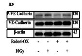
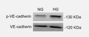
Application: WB    Species: Human    Sample: ECs

Fig. 4 Overexpression of Robo4 improves IR-induced downregulation of VE-cadherin and decreases the permeability of the EC monolayer. A Confirmation of Robo4 overexpression after establishing a stable cell line using western blot and qPCR analysis, respectively. B Effect of Robo4 overexpression and irradiation on cell survival and proliferation of ECs. C and E Functional analysis of Robo4 overexpression by FITC-dextran (70 kDa) permeability assay and EC monolayer crystal violet staining after irradiation. D Western blot analysis of total and pY-731 VE-cadherin after Robo4 overexpression and irradiation; the relative protein expression of phosphorylated Y-731 and total VE-cadherin normalized with endogenous beta-actin protein; The ratio of pY-731 VE-cadherin to total VE-cadherin. Cycloheximide treatment and immunoblotting were conducted as in (F). The CD144 band intensity was normalized to actin and then normalized to the t = 0 controls. G Confocal microscopic images of VE-cadherin (red) and DAPI (blue). H Western blot analysis showing connexin 43 expression levels in Robo4 overexpression microvascular ECs after gamma radiation treatment. J Confocal images show immunofluorescence of connexin 43 (red) in Robo4 overexpression microvascular ECs, with or without irradiation. Nuclei appear in blue. I Flow cytometric analysis to determine EC GJ coupling ability in vitro. For all experiments, scale bars = 20 µm, n ≥ 3, and error bars represent std.

2). Stealthy nanoparticles protect endothelial barrier from leakiness by resisting the absorption of VE-cadherin. Nanoscale (PubMed: 34259298) [IF=6.7]

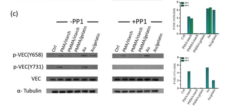
Application: WB    Species: Human    Sample: HMVEC cells

Fig. 4 (a) Schematic showing the protein pull down experimental setup. Immunoblotting (left panel) and its semi-quantitative analysis (right panel) of (b) VEC, SOD1, claudin-5 and α-tublin from whole cell lysate and pulled down by different NPs. (c) Y658 (p-VEC(Y658)), Y731 (p-VEC(Y731), VEC and α-tublin from the whole cell lysate with/without the Src kinase inhibitor, PP1.

3). YAP promotes endothelial barrier repair by repressing STAT3/VEGF signaling. LIFE SCIENCES (PubMed: 32502546) [IF=6.1]

Application: WB    Species: Human    Sample: HUVECs

Fig. 3. YAP inhibits VEGF expression to mediate endothelial barrier repair. (A–E) HUVECs were transfected with control scrambled (scr) or YAP shRNA and stable cell lines were screened. Cells were treated with TNF-α for 24 h. VEGF (A,B) and VE-cadherin (D,E) protein levels were analyzed by western blotting. Relative levels of VEGF (C) mRNA were determined by qRT-PCR. (F–I) HUVECs were transfected with GFP or YAP, and stable cell lines were screened. Cells were treated with TNF-α for 24 h. VEGF (F,G) and VE-cadherin (F,H) protein levels were assessed by western blot analysis. Relative levels of VEGF (I) mRNA were determined by qRT-PCR. All experiments were repeated three times (n = 3), and data are presented as means ± S.E. *p < 0.05, **p < 0.01, ***p < 0.001.

4). Skeletal muscle-derived FSTL1 starting up angiogenesis by regulating endothelial junction via activating Src pathway can be upregulated by hydrogen sulfide. American journal of physiology. Cell physiology (PubMed: 37694287) [IF=5.5]

5). Losartan protects against myocardial ischemia and reperfusion injury via vascular integrity preservation. FASEB JOURNAL (PubMed: 30991833) [IF=4.8]

6). Orai–vascular endothelial-cadherin signaling complex regulates high-glucose exposure-induced increased permeability of mouse aortic endothelial cells. BMJ Open Diabetes Research & Care (PubMed: 33888544) [IF=4.1]

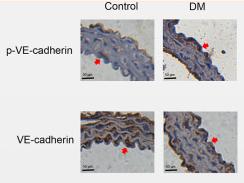
Application: IHC    Species: Mouse    Sample: aortic endothelial cells

Figure 2 Phosphorylated vascular endothelial (p-VE)-cadherin protein expression level is markedly increased in mouse aortic endothelial cells (MAECs) cultured in high glucose (HG) for 7 days and in thoracic aorta endothelial cells of streptozotocin (STZ)-induced mice with diabetes with no change in VE-cadherin expression level; HG exposure promotes intercellular permeability in MAECs. Representative western blot analysis images (A) and summary data (B) showing p-VE-cadherin and VE-cadherin expression levels in MAECs cultured in normal glucose (NG) or HG medium. (C, E) MAECs were cultured in HG medium for 7 days and then fixed and incubated with anti-p-VE-cadherin antibody (red), anti-VE-cadherin antibody (red) and 4',6-diamidino-2-phenylindole (DAPI) (blue; cell nuclei) and then imaged with a confocal microscope. Representative confocal microscopy images and the final merged images are shown (C, E). (D, F) Fluorescence intensity profiles and summary data for anti-p-VE-cadherin antibody and anti-VE-cadherin antibody in the regions delineated by the corresponding yellow line shown in (C) and (E). (G) Representative images of thoracic aorta endothelium immunostaining (brown; eg, red arrowheads) for expression levels of p-VE-cadherin and VE-cadherin in a mouse model of diabetes mellitus (DM) and control mice. Magnification, ×200. (H) Quantification of p-VE-cadherin and VE-cadherin expression levels in the thoracic aorta endothelium of a mouse model of diabetes and control mice. IOD, integrated optical density. (I) HG-induced transendothelial electrical resistance (TER) was examined in vitro after MAECs were cultured in HG medium for 7 days. (J) FD-20 permeability was tested in a monolayer of aortic endothelial cells using a transwell permeability assay. OD, optical density; values, means±SEM (n=4–8 samples). *P<0.05, ***p<0.001 compared with NG-cultured cells or control groups.

Application: WB    Species: Mouse    Sample: aortic endothelial cells

Figure 2 Phosphorylated vascular endothelial (p-VE)-cadherin protein expression level is markedly increased in mouse aortic endothelial cells (MAECs) cultured in high glucose (HG) for 7 days and in thoracic aorta endothelial cells of streptozotocin (STZ)-induced mice with diabetes with no change in VE-cadherin expression level; HG exposure promotes intercellular permeability in MAECs. Representative western blot analysis images (A) and summary data (B) showing p-VE-cadherin and VE-cadherin expression levels in MAECs cultured in normal glucose (NG) or HG medium. (C, E) MAECs were cultured in HG medium for 7 days and then fixed and incubated with anti-p-VE-cadherin antibody (red), anti-VE-cadherin antibody (red) and 4',6-diamidino-2-phenylindole (DAPI) (blue; cell nuclei) and then imaged with a confocal microscope. Representative confocal microscopy images and the final merged images are shown (C, E). (D, F) Fluorescence intensity profiles and summary data for anti-p-VE-cadherin antibody and anti-VE-cadherin antibody in the regions delineated by the corresponding yellow line shown in (C) and (E). (G) Representative images of thoracic aorta endothelium immunostaining (brown; eg, red arrowheads) for expression levels of p-VE-cadherin and VE-cadherin in a mouse model of diabetes mellitus (DM) and control mice. Magnification, ×200. (H) Quantification of p-VE-cadherin and VE-cadherin expression levels in the thoracic aorta endothelium of a mouse model of diabetes and control mice. IOD, integrated optical density. (I) HG-induced transendothelial electrical resistance (TER) was examined in vitro after MAECs were cultured in HG medium for 7 days. (J) FD-20 permeability was tested in a monolayer of aortic endothelial cells using a transwell permeability assay. OD, optical density; values, means±SEM (n=4–8 samples). *P<0.05, ***p<0.001 compared with NG-cultured cells or control groups.

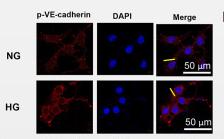
Application: IF/ICC    Species: Mouse    Sample: aortic endothelial cells

Figure 2 Phosphorylated vascular endothelial (p-VE)-cadherin protein expression level is markedly increased in mouse aortic endothelial cells (MAECs) cultured in high glucose (HG) for 7 days and in thoracic aorta endothelial cells of streptozotocin (STZ)-induced mice with diabetes with no change in VE-cadherin expression level; HG exposure promotes intercellular permeability in MAECs. Representative western blot analysis images (A) and summary data (B) showing p-VE-cadherin and VE-cadherin expression levels in MAECs cultured in normal glucose (NG) or HG medium. (C, E) MAECs were cultured in HG medium for 7 days and then fixed and incubated with anti-p-VE-cadherin antibody (red), anti-VE-cadherin antibody (red) and 4',6-diamidino-2-phenylindole (DAPI) (blue; cell nuclei) and then imaged with a confocal microscope. Representative confocal microscopy images and the final merged images are shown (C, E). (D, F) Fluorescence intensity profiles and summary data for anti-p-VE-cadherin antibody and anti-VE-cadherin antibody in the regions delineated by the corresponding yellow line shown in (C) and (E). (G) Representative images of thoracic aorta endothelium immunostaining (brown; eg, red arrowheads) for expression levels of p-VE-cadherin and VE-cadherin in a mouse model of diabetes mellitus (DM) and control mice. Magnification, ×200. (H) Quantification of p-VE-cadherin and VE-cadherin expression levels in the thoracic aorta endothelium of a mouse model of diabetes and control mice. IOD, integrated optical density. (I) HG-induced transendothelial electrical resistance (TER) was examined in vitro after MAECs were cultured in HG medium for 7 days. (J) FD-20 permeability was tested in a monolayer of aortic endothelial cells using a transwell permeability assay. OD, optical density; values, means±SEM (n=4–8 samples). *P<0.05, ***p<0.001 compared with NG-cultured cells or control groups.

7). Zanthoxylum nitidum extract attenuates BMP-2 induced inflammation and hyperpermeability. BIOSCIENCE REPORTS (PubMed: 33030503) [IF=4.0]

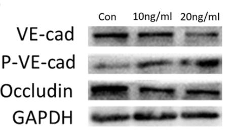
Application: WB    Species: human    Sample: HUVECs

Figure 3. |BMP-2 increased the HUVECs permeability(A and B) VE-cad, p-VE-cad, and Occludin protein levels in HUVECs were determined (n=5).

Restrictive clause


Affinity Biosciences tests all products strictly. Citations are provided as a resource for additional applications that have not been validated by Affinity Biosciences. Please choose the appropriate format for each application and consult Materials and Methods sections for additional details about the use of any product in these publications.

For Research Use Only.
Not for use in diagnostic or therapeutic procedures. Not for resale. Not for distribution without written consent. Affinity Biosciences will not be held responsible for patent infringement or other violations that may occur with the use of our products. Affinity Biosciences, Affinity Biosciences Logo and all other trademarks are the property of Affinity Biosciences LTD.