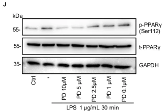
Product: Phospho-PPAR gamma (Ser112) Antibody
Catalog: AF3284
Description: Rabbit polyclonal antibody to Phospho-PPAR gamma (Ser112)
Application: WB IHC IF/ICC
Reactivity: Human, Mouse, Rat
Prediction: Pig, Bovine, Horse, Sheep, Rabbit, Dog
Mol.Wt.: 57kDa; 58kD(Calculated).
Uniprot: P37231
RRID: AB_2834705

View similar products>>

   Size Price Inventory
 100ul $280 In stock
 200ul $350 In stock

Lead Time: Same day delivery

For pricing and ordering contact:
Local distributors

Product Info

Source:
Rabbit
Application:
WB 1:500-1:2000, IHC 1:50-1:200, IF/ICC 1:200
*The optimal dilutions should be determined by the end user.
*Tips:

WB: For western blot detection of denatured protein samples. IHC: For immunohistochemical detection of paraffin sections (IHC-p) or frozen sections (IHC-f) of tissue samples. IF/ICC: For immunofluorescence detection of cell samples. ELISA(peptide): For ELISA detection of antigenic peptide.

Reactivity:
Human,Mouse,Rat
Prediction:
Pig(92%), Bovine(92%), Horse(100%), Sheep(92%), Rabbit(100%), Dog(100%)
Clonality:
Polyclonal
Specificity:
Phospho-PPAR gamma (Ser112) Antibody detects endogenous levels of PPAR gamma only when phosphorylated at Serine 112.
RRID:
AB_2834705
Cite Format: Affinity Biosciences Cat# AF3284, RRID:AB_2834705.
Conjugate:
Unconjugated.
Purification:
The antibody is from purified rabbit serum by affinity purification via sequential chromatography on phospho-peptide and non-phospho-peptide affinity columns.
Storage:
PBS, pH 7.4,50% glycerol. Store at -20 °C. Stable for 12 months from date of receipt.
Alias:

Fold/Unfold

CIMT1; GLM1; NR1C3; Nuclear receptor subfamily 1 group C member 3; OTTHUMP00000185032; OTTHUMP00000185036; Peroxisome proliferator activated nuclear receptor gamma variant 1; Peroxisome proliferator activated receptor gamma 1; Peroxisome Proliferator Activated Receptor gamma; Peroxisome proliferator-activated receptor gamma; PPAR gamma; PPAR-gamma; PPARG; PPARG_HUMAN; PPARG1; PPARG2; PPARgamma;

Immunogens

Immunogen:
Uniprot:
Gene(ID):
Expression:
P37231 PPARG_HUMAN:

Highest expression in adipose tissue. Lower in skeletal muscle, spleen, heart and liver. Also detectable in placenta, lung and ovary.

Description:
The protein encoded by this gene is a member of the peroxisome proliferator-activated receptor (PPAR) subfamily of nuclear receptors. PPARs form heterodimers with retinoid X receptors (RXRs) and these heterodimers regulate transcription of various genes. Three subtypes of PPARs are known: PPAR-alpha, PPAR-delta, and PPAR-gamma.
Sequence:
MGETLGDSPIDPESDSFTDTLSANISQEMTMVDTEMPFWPTNFGISSVDLSVMEDHSHSFDIKPFTTVDFSSISTPHYEDIPFTRTDPVVADYKYDLKLQEYQSAIKVEPASPPYYSEKTQLYNKPHEEPSNSLMAIECRVCGDKASGFHYGVHACEGCKGFFRRTIRLKLIYDRCDLNCRIHKKSRNKCQYCRFQKCLAVGMSHNAIRFGRMPQAEKEKLLAEISSDIDQLNPESADLRALAKHLYDSYIKSFPLTKAKARAILTGKTTDKSPFVIYDMNSLMMGEDKIKFKHITPLQEQSKEVAIRIFQGCQFRSVEAVQEITEYAKSIPGFVNLDLNDQVTLLKYGVHEIIYTMLASLMNKDGVLISEGQGFMTREFLKSLRKPFGDFMEPKFEFAVKFNALELDDSDLAIFIAVIILSGDRPGLLNVKPIEDIQDNLLQALELQLKLNHPESSQLFAKLLQKMTDLRQIVTEHVQLLQVIKKTETDMSLHPLLQEIYKDLY

Predictions

Predictions:

Score>80(red) has high confidence and is suggested to be used for WB detection. *The prediction model is mainly based on the alignment of immunogen sequences, the results are for reference only, not as the basis of quality assurance.

Species
Results
Score
Horse
100
Dog
100
Rabbit
100
Pig
92
Bovine
92
Sheep
92
Xenopus
69
Zebrafish
62
Chicken
0
Model Confidence:
High(score>80) Medium(80>score>50) Low(score<50) No confidence

PTMs - P37231 As Substrate

Site PTM Type Enzyme
Y78 Phosphorylation
Y95 Phosphorylation
Y102 Phosphorylation
K107 Sumoylation
S112 Phosphorylation P50750 (CDK9) , P45983 (MAPK8) , P50613 (CDK7) , Q02750 (MAP2K1) , P27361 (MAPK3) , P28482 (MAPK1)
K184 Ubiquitination
K185 Ubiquitination
T269 Phosphorylation
S273 Phosphorylation Q00535 (CDK5)
K395 Sumoylation

Research Backgrounds

Function:

Nuclear receptor that binds peroxisome proliferators such as hypolipidemic drugs and fatty acids. Once activated by a ligand, the nuclear receptor binds to DNA specific PPAR response elements (PPRE) and modulates the transcription of its target genes, such as acyl-CoA oxidase. It therefore controls the peroxisomal beta-oxidation pathway of fatty acids. Key regulator of adipocyte differentiation and glucose homeostasis. ARF6 acts as a key regulator of the tissue-specific adipocyte P2 (aP2) enhancer. Acts as a critical regulator of gut homeostasis by suppressing NF-kappa-B-mediated proinflammatory responses. Plays a role in the regulation of cardiovascular circadian rhythms by regulating the transcription of ARNTL/BMAL1 in the blood vessels (By similarity).

(Microbial infection) Upon treatment with M.tuberculosis or its lipoprotein LpqH, phosphorylation of MAPK p38 and IL-6 production are modulated, probably via this protein.

PTMs:

O-GlcNAcylation at Thr-84 reduces transcriptional activity in adipocytes.

Phosphorylated in basal conditions and dephosphorylated when treated with the ligand. May be dephosphorylated by PPP5C. The phosphorylated form may be inactive and dephosphorylation at Ser-112 induces adipogenic activity (By similarity).

Subcellular Location:

Nucleus. Cytoplasm.
Note: Redistributed from the nucleus to the cytosol through a MAP2K1/MEK1-dependent manner. NOCT enhances its nuclear translocation.

Extracellular region or secreted Cytosol Plasma membrane Cytoskeleton Lysosome Endosome Peroxisome ER Golgi apparatus Nucleus Mitochondrion Manual annotation Automatic computational assertionSubcellular location
Tissue Specificity:

Highest expression in adipose tissue. Lower in skeletal muscle, spleen, heart and liver. Also detectable in placenta, lung and ovary.

Subunit Structure:

Interacts with FOXO1 (acetylated form) (By similarity). Heterodimer with other nuclear receptors, such as RXRA. The heterodimer with the retinoic acid receptor RXRA is called adipocyte-specific transcription factor ARF6. Interacts with NCOA6 coactivator, leading to a strong increase in transcription of target genes. Interacts with coactivator PPARBP, leading to a mild increase in transcription of target genes. Interacts with NOCA7 in a ligand-inducible manner. Interacts with NCOA1 and NCOA2 LXXLL motifs. Interacts with ASXL1, ASXL2, DNTTIP2, FAM120B, MAP2K1/MEK1, NR0B2, PDPK1, PRDM16, PRMT2 and TGFB1I1. Interacts (when activated by agonist) with PPP5C. Interacts with HELZ2 and THRAP3; the interaction stimulates the transcriptional activity of PPARG. Interacts with PER2, the interaction is ligand dependent and blocks PPARG recruitment to target promoters. Interacts with NOCT. Interacts with ACTN4. Interacts (when in the liganded conformation) with GPS2 (By similarity). Interacts with CRY1 and CRY2 in a ligand-dependent manner (By similarity). In the absence of hormonal ligand, interacts with TACC1.

Family&Domains:

The 9aaTAD motif is a transactivation domain present in a large number of yeast and animal transcription factors.

Belongs to the nuclear hormone receptor family. NR1 subfamily.

Research Fields

· Environmental Information Processing > Signal transduction > AMPK signaling pathway.   (View pathway)

· Human Diseases > Neurodegenerative diseases > Huntington's disease.

· Human Diseases > Cancers: Overview > Pathways in cancer.   (View pathway)

· Human Diseases > Cancers: Overview > Transcriptional misregulation in cancer.

· Human Diseases > Cancers: Specific types > Thyroid cancer.   (View pathway)

· Organismal Systems > Endocrine system > PPAR signaling pathway.

· Organismal Systems > Aging > Longevity regulating pathway.   (View pathway)

· Organismal Systems > Development > Osteoclast differentiation.   (View pathway)

References

1). Pioglitazone alleviates maternal sleep deprivation-induced cognitive deficits in male rat offspring by enhancing microglia-mediated neurogenesis. BRAIN BEHAVIOR AND IMMUNITY (PubMed: 32032783) [IF=15.1]

Application: WB    Species: rat    Sample: dorsal hippocampus

Fig. 3. Effects of PPARγ activation on microglial phenotype switching. Levels of mRNAs 23 encoding (A) PPARα, (B) PPAR β/δ or (C) PPARγ in hippocampus of rat offspring. (D) Representative western blots and (E-F) quantitation of phospho- and non-phospho-PPARγ protein in hippocampus of rat offspring (n = 5 offspring per group). (G) Immunofluorescence staining of PPARγ located in microglia. Scale bars: 20 μm. (H) Expression of PPARγ in primary microglia after poly(I:C) stimulation. (I) Heat map of mRNA expression of M2 microglia markers Arg1, IL-4, and IL-10 as well as M1 microglia markers IL-6, TNFα, and IL- 1β (n = 5 replicates per group). Statistical analysis is shown in Table S4. *p < 0.05, **p < 0.01, ***p < 0.001. Abbreviations: Ctrl, control; MSD, maternal sleep deprivation; Veh(Vehicle), treatment with normal saline; Pio, pioglitazone.

2). Emodin attenuates severe acute pancreatitis-associated acute lung injury by suppressing pancreatic exosome-mediated alveolar macrophage activation. Acta Pharmaceutica Sinica B (PubMed: 36213542) [IF=14.5]

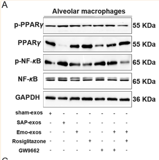
Application: WB    Species: Rat    Sample: macrophages and lung tissues

Figure 7 Effect of emodin on PPARγ-NF-κB pathway in macrophages and lung tissues. (A) Western blot analysis of p-PPARγ, PPARγ, p-NF-κB, and NF-κB in alveolar macrophages exposed to plasma exosomes from sham rats, SAP rats, and emodin-treated SAP rats and subsequently treated with the PPARγ agonist rosiglitazone or the antagonist GW9662. GAPDH was used as the loading control. (B) Quantitative analysis of p-PPARγ, PPARγ, and p-NF-κB expression in alveolar macrophages (n = 3). (C) Levels of the pro-inflammatory factors NO, TNF-α, and MIP-2 in culture media as detected by ELISA (n = 3). (D) Western blot analysis of p-PPARγ, PPARγ, p-NF-κB, and NF-κB in the lung tissues of healthy rats administered plasma exosomes from sham rats, SAP rats, and emodin-treated SAP rats and subsequently treated with the PPARγ agonist rosiglitazone or the antagonist GW9662. GAPDH was used as the loading control. (E) Quantitative analysis of p-PPARγ, PPARγ, and p-NF-κB expression in the lung tissues (n = 3). (F) Levels of the pro-inflammatory factors NO, TNF-α, and MIP-2 in the lung tissues as detected by ELISA (n = 3). All experiments were independently performed three times. All data were expressed as mean ± SD. ∗P < 0.05, ∗∗P < 0.01, ∗∗∗P < 0.001, ∗∗∗∗P < 0.0001 vs. the sham group, ∗P < 0.05, ∗∗P < 0.01, ∗∗∗P < 0.001, ∗∗∗∗P < 0.0001 vs. the Emo-exos-AMs or Emo-exos-lung; #P < 0.05, ##P < 0.01, ###P < 0.001 and ####P < 0.0001 vs. the SAP group, by one-way ANOVA followed by Tukey tests.

3). EGFR tyrosine kinase activity and Rab GTPases coordinate EGFR trafficking to regulate macrophage activation in sepsis. Cell Death & Disease (PubMed: 36344490) [IF=9.0]

Application: WB    Species: Mouse    Sample: RAW264.7 cells

Fig. 7: Inhibition of EGFR phosphorylation promote M2 polarization by regulating glutamine metabolism through activation of PPARγ. A–F RAW264.7 macrophages were treated with LPS (1 μg/mL) for 24 h, with or without Erlotinib (20 μM) pretreatment for 30 min. A RT-qPCR analysis of mRNA expression of M2-related genes Mcr1 (n = 3). B RT-qPCR analysis of mRNA expression of M2-related genes Ym1 (n = 3). C Representative western blot of Arg1. D Flow cytometry analysis showing the level of M2 macrophage-associated markers CD206. E Percentage of CD206-positive RAW264.7 is shown (n = 3). I Mean fluorescence intensity (MFI) is shown (n = 3). G–I Macrophages were collected from bronchoalveolar lavage fluid of C57BL/6 mice subjected to CLP and were divided into sham-operated, CLP and CLP plus Erlotinib (100 mg/kg, gavage) pretreatmend for 2 h, and alveolar macrophages were identified with CD45 + CD11b + F4/80high. G CD206 expression on the surface of alveolar macrophage was analyzed by flow cytometry. H Percentage of CD206-positive alveolar macrophage is shown (n = 9). I Mean fluorescence intensity (MFI) is shown (n = 9). J Immunoblot analysis of p-PPARγ (Ser112), t-PPARγ in RAW264.7 cells treated with LPS (1 μg/mL) for 30 min with or without indicated concentration of PD168393 (10 μM) pretreatment for 30 min. K Fluorescence images depicting PPARγ translocation (left panel, scale bar, 50 μm; right panel, scale bar, 5 μm). L–N RAW264.7 cells were treated with LPS (1 μg/mL) for 24 h with or without Erlotinib (10 μM) or Rosiglitazone (Rosi (20 μM) pretreatment. L Cell surface CD206 were analyzed by flow cytometry. M Percentage of CD206-positive RAW264.7 is shown (n = 3). N Mean fluorescence intensity (MFI) is shown (n = 3). O–Q Macrophages were collected from bronchoalveolar lavage fluid of C57BL/6 mice subjected to CLP and were divided into Sham-operated, CLP and CLP plus Erlotinib (100 mg/kg, gavage) pretreatmend for 2 h, and alveolar macrophages were identified with CD45 + CD11b + F4/80high. O CD206 expression on the surface of alveolar macrophage was analyzed by flow cytometry. P Percentage of CD206-positive alveolar macrophage is shown (n = 9). Q Mean fluorescence intensity (MFI) is shown (n = 9). R–V RAW264.7 cells were treated with LPS (1 μg/mL) for 30 min with or without PD168393 (10 μM) pretreatment for 30 min. R Flow cytometry analysis of JC-1 for the detecting the change of mitochondrial membrane potential (ΔΨm) (left panel, JC-1 aggregates; right panel, JC-1 monomers). S The ratio of JC-1 aggregates /JC-1 monomers was calculated as Δψm. T Total cellular ATP level was detected (n = 3). U Immunoblot analysis of IRG1, ATP5A, SDHA, Tubulin as a loading control. V Immunoblot analysis of SDHA and IRG1 in Control or IRG1 silenced RAW264.7 cells, Tubulin as a loading control. The graphs depict mean ± SD based on three independent experiments.

4). Aerobic exercise and metformin on intermuscular adipose tissue (IMAT): insights from multimodal MRI and histological changes in prediabetic rats. Diabetology & metabolic syndrome (PubMed: 37899436) [IF=4.8]

Application: WB    Species: Rat    Sample: intermuscular adipose tissues (IMATs)

Fig. 6 Characterization of lipid and glucose metabolism in intermuscular adipose tissues (IMATs). Representative western blots (A, C, E, G, I) and quantification of the gene-expression levels of peroxisome proliferators-activated receptor-γ (PPAR-γ) (B), phosphorylated PPAR-γ (p-PPAR-γ; Ser112) (F), nuclear respiratory factor-1 (NRF-1) (J), glucose transporter-4 (GLUT-4) (D), glucose transporter-1 (GLUT-1) (H), and perilipin-5 (Plin-5) (K). The data represent the mean ± the standard error of the mean (n = 5–6/group). CON control, EMA combined therapies + compound-c, EMC combined therapies, EXE moderate exercise, GAPDH glyceraldehyde-3-phosphate dehydrogenase, MET metformin, PRE prediabetes. ap 

5). HuoXueTongFu Formula Alleviates Intraperitoneal Adhesion by Regulating Macrophage Polarization and the SOCS/JAK2/STAT/PPAR-γ Signalling Pathway. MEDIATORS OF INFLAMMATION (PubMed: 31772499) [IF=4.6]

6). Autologous decellularized extracellular matrix promotes adipogenic differentiation of adipose derived stem cells in low serum culture system by regulating the ERK1/2-PPARγ pathway. Adipocyte (PubMed: 33825675) [IF=3.3]

Application: WB    Species: Human    Sample: ADSCs

Figure 7. Effects of d-ECM on the ERK1/2-PPARγ pathway and the expression of adipocyte secreting factors ADIPOQ and aP2 in the fully differentiated ADSCs. After 3-days treatments and 14-days adipogenic induction, ADSCs at the 5th passage were collected and used for Western blotting analysis (a). Protein levels of ERK1/2 (b), p-ERK1/2 (c), PPARγ (d), p-PPARγ (e), ADIPOQ (f) and aP2 (g) were examined. GAPDH demonstrated the equal loading of protein samples. N = 3. *, p < 0.05, vs. 10% FBS group; **, p < 0.01, vs. 10% FBS group; #, p < 0.05, vs. 2% FBS group; ##, p < 0.01, vs. 2% FBS group; $, p < 0.05, vs. 2% FBS + d-ECM group; $$, p < 0.01, vs. 2% FBS + d-ECM group. ADIPOQ, adiponectin; aP2, adipocyte fatty-acid binding protein

7). Cholecystokinin Octapeptide Promotes ANP Secretion through Activation of NOX4–PGC-1α–PPARα/PPARγ Signaling in Isolated Beating Rat Atria. Oxidative Medicine and Cellular Longevity (PubMed: 35770043)

Restrictive clause


Affinity Biosciences tests all products strictly. Citations are provided as a resource for additional applications that have not been validated by Affinity Biosciences. Please choose the appropriate format for each application and consult Materials and Methods sections for additional details about the use of any product in these publications.

For Research Use Only.
Not for use in diagnostic or therapeutic procedures. Not for resale. Not for distribution without written consent. Affinity Biosciences will not be held responsible for patent infringement or other violations that may occur with the use of our products. Affinity Biosciences, Affinity Biosciences Logo and all other trademarks are the property of Affinity Biosciences LTD.