Parkin Antibody - #AF0235

Related Downloads
Protocols
Product Info
*The optimal dilutions should be determined by the end user.
*Tips:
WB: For western blot detection of denatured protein samples. IHC: For immunohistochemical detection of paraffin sections (IHC-p) or frozen sections (IHC-f) of tissue samples. IF/ICC: For immunofluorescence detection of cell samples. ELISA(peptide): For ELISA detection of antigenic peptide.
Cite Format: Affinity Biosciences Cat# AF0235, RRID:AB_2833410.
Fold/Unfold
AR JP; E3 ubiquitin ligase; E3 ubiquitin protein ligase parkin; E3 ubiquitin-protein ligase parkin; FRA6E; LPRS 2; LPRS2; PARK 2; Park2; Parkin 2; Parkinson disease (autosomal recessive juvenile) 2; Parkinson disease (autosomal recessive, juvenile) 2, parkin; Parkinson disease protein 2; Parkinson juvenile disease protein 2; Parkinson protein 2 E3 ubiquitin protein ligase; Parkinson protein 2, E3 ubiquitin protein ligase (parkin); PDJ; PRKN 2; PRKN; PRKN2; PRKN2_HUMAN; Ubiquitin E3 ligase PRKN;
Immunogens
Highly expressed in the brain including the substantia nigra. Expressed in heart, testis and skeletal muscle. Expression is down-regulated or absent in tumor biopsies, and absent in the brain of PARK2 patients. Overexpression protects dopamine neurons from kainate-mediated apoptosis. Found in serum (at protein level).
- O60260 PRKN_HUMAN:
- Protein BLAST With
- NCBI/
- ExPASy/
- Uniprot
MIVFVRFNSSHGFPVEVDSDTSIFQLKEVVAKRQGVPADQLRVIFAGKELRNDWTVQNCDLDQQSIVHIVQRPWRKGQEMNATGGDDPRNAAGGCEREPQSLTRVDLSSSVLPGDSVGLAVILHTDSRKDSPPAGSPAGRSIYNSFYVYCKGPCQRVQPGKLRVQCSTCRQATLTLTQGPSCWDDVLIPNRMSGECQSPHCPGTSAEFFFKCGAHPTSDKETSVALHLIATNSRNITCITCTDVRSPVLVFQCNSRHVICLDCFHLYCVTRLNDRQFVHDPQLGYSLPCVAGCPNSLIKELHHFRILGEEQYNRYQQYGAEECVLQMGGVLCPRPGCGAGLLPEPDQRKVTCEGGNGLGCGFAFCRECKEAYHEGECSAVFEASGTTTQAYRVDERAAEQARWEAASKETIKKTTKPCPRCHVPVEKNGGCMHMKCPQPQCRLEWCWNCGCEWNRVCMGDHWFDV
PTMs - O60260 As Substrate
| Site | PTM Type | Enzyme | Source |
|---|---|---|---|
| Ubiquitination | Uniprot | ||
| S9 | Phosphorylation | Uniprot | |
| S19 | Phosphorylation | Uniprot | |
| K27 | Ubiquitination | Uniprot | |
| K48 | Ubiquitination | Uniprot | |
| S65 | Phosphorylation | Q9BXM7 (PINK1) | Uniprot |
| K76 | Ubiquitination | Uniprot | |
| S101 | Phosphorylation | P48729 (CSNK1A1) | Uniprot |
| S108 | Phosphorylation | Uniprot | |
| S116 | Phosphorylation | Uniprot | |
| S131 | Phosphorylation | Q00535 (CDK5) | Uniprot |
| S136 | Phosphorylation | Uniprot | |
| Y143 | Phosphorylation | P00519 (ABL1) | Uniprot |
| S193 | Phosphorylation | Uniprot | |
| S198 | Phosphorylation | Uniprot | |
| T204 | Phosphorylation | Uniprot | |
| S296 | Phosphorylation | Uniprot | |
| Y372 | Phosphorylation | Uniprot | |
| S378 | Phosphorylation | P48729 (CSNK1A1) , P53350 (PLK1) | Uniprot |
| T388 | Phosphorylation | Uniprot | |
| Y391 | Phosphorylation | Uniprot |
Research Backgrounds
Functions within a multiprotein E3 ubiquitin ligase complex, catalyzing the covalent attachment of ubiquitin moieties onto substrate proteins, such as BCL2, SYT11, CCNE1, GPR37, RHOT1/MIRO1, MFN1, MFN2, STUB1, SNCAIP, SEPTIN5, TOMM20, USP30, ZNF746 and AIMP2. Mediates monoubiquitination as well as 'Lys-6', 'Lys-11', 'Lys-48'-linked and 'Lys-63'-linked polyubiquitination of substrates depending on the context. Participates in the removal and/or detoxification of abnormally folded or damaged protein by mediating 'Lys-63'-linked polyubiquitination of misfolded proteins such as PARK7: 'Lys-63'-linked polyubiquitinated misfolded proteins are then recognized by HDAC6, leading to their recruitment to aggresomes, followed by degradation. Mediates 'Lys-63'-linked polyubiquitination of a 22 kDa O-linked glycosylated isoform of SNCAIP, possibly playing a role in Lewy-body formation. Mediates monoubiquitination of BCL2, thereby acting as a positive regulator of autophagy. Promotes the autophagic degradation of dysfunctional depolarized mitochondria (mitophagy) by promoting the ubiquitination of mitochondrial proteins such as TOMM20, RHOT1/MIRO1 and USP30. Preferentially assembles 'Lys-6'-, 'Lys-11'- and 'Lys-63'-linked polyubiquitin chains following mitochondrial damage, leading to mitophagy. Mediates 'Lys-48'-linked polyubiquitination of ZNF746, followed by degradation of ZNF746 by the proteasome; possibly playing a role in the regulation of neuron death. Limits the production of reactive oxygen species (ROS). Regulates cyclin-E during neuronal apoptosis. In collaboration with CHPF isoform 2, may enhance cell viability and protect cells from oxidative stress. Independently of its ubiquitin ligase activity, protects from apoptosis by the transcriptional repression of p53/TP53. May protect neurons against alpha synuclein toxicity, proteasomal dysfunction, GPR37 accumulation, and kainate-induced excitotoxicity. May play a role in controlling neurotransmitter trafficking at the presynaptic terminal and in calcium-dependent exocytosis. May represent a tumor suppressor gene.
Auto-ubiquitinates in an E2-dependent manner leading to its own degradation. Also polyubiquitinated by RNF41 for proteasomal degradation.
S-nitrosylated. The inhibition of PRKN ubiquitin E3 ligase activity by S-nitrosylation could contribute to the degenerative process in PD by impairing the ubiquitination of PRKN substrates.
Phosphorylation at Ser-65 by PINK1 contributes to activate PRKN activity. It is however not sufficient and requires binding to phosphorylated ubiquitin as well.
Cytoplasm>Cytosol. Nucleus. Endoplasmic reticulum. Mitochondrion.
Note: Mainly localizes in the cytosol. Co-localizes with SYT11 in neutrites. Co-localizes with SNCAIP in brainstem Lewy bodies. Mitochondrial localization gradually increases with cellular growth. Also relocates to dysfunctional mitochondria that have lost the mitochondrial membrane potential; recruitment to mitochondria is PINK1-dependent.
Highly expressed in the brain including the substantia nigra. Expressed in heart, testis and skeletal muscle. Expression is down-regulated or absent in tumor biopsies, and absent in the brain of PARK2 patients. Overexpression protects dopamine neurons from kainate-mediated apoptosis. Found in serum (at protein level).
Forms an E3 ubiquitin ligase complex with UBE2L3 or UBE2L6. Mediates 'Lys-63'-linked polyubiquitination by associating with UBE2V1. Part of a SCF-like complex, consisting of PRKN, CUL1 and FBXW7. Interacts with SNCAIP. Binds to the C2A and C2B domains of SYT11. Interacts and regulates the turnover of SEPTIN5. Part of a complex, including STUB1, HSP70 and GPR37. The amount of STUB1 in the complex increases during ER stress. STUB1 promotes the dissociation of HSP70 from PRKN and GPR37, thus facilitating PRKN-mediated GPR37 ubiquitination. HSP70 transiently associates with unfolded GPR37 and inhibits the E3 activity of PRKN, whereas, STUB1 enhances the E3 activity of PRKN through promotion of dissociation of HSP70 from PRKN-GPR37 complexes. Interacts with PSMD4 and PACRG. Interacts with LRRK2. Interacts with RANBP2. Interacts with SUMO1 but not SUMO2, which promotes nuclear localization and autoubiquitination. Interacts (via first RING-type domain) with AIMP2 (via N-terminus). Interacts with PSMA7 and RNF41. Interacts with PINK1. Interacts with CHPF, the interaction with isoform 2 may facilitate PRKN transport into the mitochondria. Interacts with MFN2 (phosphorylated), promotes PRKN localization in dysfunctional depolarized mitochondria. Interacts with FBXO7; this promotes translocation to dysfunctional depolarized mitochondria. Interacts with heat shock protein 70 family members, including HSPA1L, HSPA1A and HSPA8; interaction HSPA1L promotes translocation to damaged mitochondria. Interacts with BAG4 and, to a lesser extent, BAG5; interaction with BAG4 inhibits translocation to damaged mitochondria. Forms a complex with PINK1 and PARK7.
The ubiquitin-like domain binds the PSMD4 subunit of 26S proteasomes.
The RING-type 1 zinc finger domain is required to repress p53/TP53 transcription.
Members of the RBR family are atypical E3 ligases. They interact with the E2 conjugating enzyme UBE2L3 and function like HECT-type E3 enzymes: they bind E2s via the first RING domain, but require an obligate trans-thiolation step during the ubiquitin transfer, requiring a conserved cysteine residue in the second RING domain (PubMed:21532592).
Belongs to the RBR family. Parkin subfamily.
Research Fields
· Genetic Information Processing > Folding, sorting and degradation > Ubiquitin mediated proteolysis. (View pathway)
· Genetic Information Processing > Folding, sorting and degradation > Protein processing in endoplasmic reticulum. (View pathway)
· Human Diseases > Neurodegenerative diseases > Parkinson's disease.
References
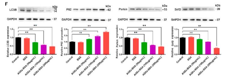
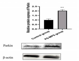
Application: WB Species: mice Sample: bone marrow mesenchymal stem (BMSCs)
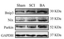
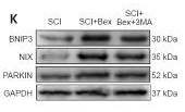
Application: WB Species: Mice Sample: spinal cords
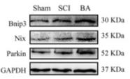
Application: WB Species: Mice Sample: spinal cords
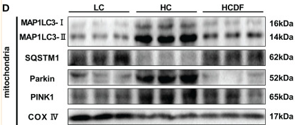
Application: WB Species: Hu Sheep Sample: liver tissue
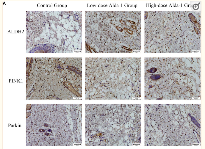
Application: IHC Species: Mouse Sample:
Application: WB Species: mouse Sample: GC-2 cells
Application: WB Species: Mouse Sample:
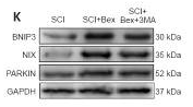
Application: WB Species: Mouse Sample: spinal cord
Restrictive clause
Affinity Biosciences tests all products strictly. Citations are provided as a resource for additional applications that have not been validated by Affinity Biosciences. Please choose the appropriate format for each application and consult Materials and Methods sections for additional details about the use of any product in these publications.
For Research Use Only.
Not for use in diagnostic or therapeutic procedures. Not for resale. Not for distribution without written consent. Affinity Biosciences will not be held responsible for patent infringement or other violations that may occur with the use of our products. Affinity Biosciences, Affinity Biosciences Logo and all other trademarks are the property of Affinity Biosciences LTD.