Phospho-Smad3 (Ser425) Antibody - #AF3362
 Antibody.
Lane 1:mouse brain tissue,
Lane 2:rat brain tissue,
Lane 3:mouse brain with blocking peptide.")


 by IF/ICC. The samples were fixed with PFA and permeabilized in 0.1% Triton X-100,then blocked in 10% serum for 45 minutes at 25°C. Samples were then incubated with primary Ab(AF3362 1:200) and mouse anti-beta tubulin Ab(T0023 1:200) for 1 hour at 37°C. An AlexaFluor594 conjugated goat anti-rabbit IgG(H+L) Ab(Red) and an AlexaFluor488 conjugated goat anti-mouse IgG(H+L) Ab(Green) were used as the secondary antibody.
The nuclear counter stain is DAPI(blue).")
 and mouse anti-beta tubulin Ab(T0023) for 1 hour at 37°C. An AlexaFluor594 conjugated goat anti-rabbit IgG(H+L) Ab(Red) and an AlexaFluor488 conjugated goat anti-mouse IgG(H+L) Ab(Green) were used as the secondary antibody.
The nuclear counter stain is DAPI(blue).")

 and pCX43 (C) showed a significant decrease but TGF-β1 (D), Smad3 (E), and pSmad3 (F) showed a significant increase in the bladder detrusor after SCI. Those changes were more significant in transection than in hemisection of sacral spine cord. CX45 was not changed among three groups.")
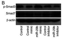
 and Smad3 (B) as well as total Smad7 (C) in sham, RDP, UUO and RDP+UUO groups. Data are presented as mean ±SEM (n = 3 in each group). #P < 0.05, ##P < 0.01, UUO group vs. sham group; *P <0.05, **P < 0.01, UUO + RDP group vs. UUO group.")
 for 48 h before being exposed to H/R. D-F, Western blot analysis for the protein expression of ALK5, Smad2, Smad3, p-Smad2, and Smad3 in the indicated groups and quantitative analysis of ALK5, p-Smad2, and p-Smad3, n = 3.")
 Typical images of p-Smad 2 and p-Smad 3 afer immunohistochemical staining in the control and NTP groups. Te image in the lower lef corner is a magnifed view of the blue square. Bar = 200 μm/20 μm.")
 Luciferase assays of CAGA-NIH3T3 cells. Cells were pretreated with Remdesivir (0–50 μM) for 30 min and then incubated with TGF-β1 (5 ng ml−1) for 24 h, then analyzed with luciferase assay. SB431542 is a TGF-β1/Smad pathway inhibitor and serves as a positive control (B) NIH-3T3 cells were co-treated with TGF-β1 (5 ng ml−1) and Remdesivir (12.5, 25, 50 μM) for 1 h. P-Smad3 and Smad3 were assessed using western blot. GAPDH was used as the internal control (C) PPF cells were co-treated with TGF-β1 (5 ng ml−1) and Remdesivir (12.5, 25, 50 μM) for 1 h. P-Smad3 and Smad3 were assessed using western blot. GAPDH was used as the internal control (D) NIH-3T3 cells were co-treated with TGF-β1 (5 ng ml−1) and Remdesivir (12.5, 25, 50 μM) for 1 h and the phosphorylation levels of P-38, JNK, ERK and Akt were analyzed by Western blot. β-tubulin was used as a loading control in grayscale analysis. Scale bar = 60 μm. Data was presented as the means ± SD, n = 3. * p < 0.05, ** p < 0.01, *** p < 0.001, **** p < 0.0001.")
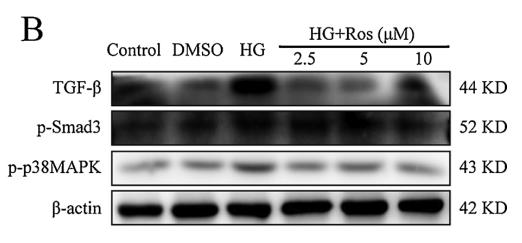
 Western blot analysis of the protein levels of p-Akt, p-Smad3, p-p38 and p-Erk1/2 expression.")
 Western blot results of TGF-β1, Smad2, p-Smad2, Smad3, p-Smad3 and Smad7 protein expression levels. n = 3 rats per group.")
 The representative image of flow-cytometry of CD206 expression and CD86 expression in THP-1 macrophages with different stimulations. (B) The quantification of CD206 expression in THP-1 macrophages with different stimulations. (C) The quantification of CD86 expression in THP-1 macrophages with different stimulations. (D) The expression of Smad3 and p-Smad3 in THP-1 macrophages with different stimulations, and related quantification (E and F). N=7. ***p<0.001.")
 for 24 h, followed by exposure to colchicine (4 μg/ml) or GZFL (8 or 10 mg/ml) for another 24 h. (a, b) TGF-β1, TGF-βR2, CUGBP1, p-STAT1, p-Smad2, p-Smad3, Smad7,α-SMA and Collagen I expressions in LX-2 cells were detected by western blot assay.")

 RT-PCR assay demonstrated that the I-125 seed radiation downregulate the expression of TGF-β1, Smad2, and Snail. (b). WB assay demonstrated that the I-125 seed radiation downregulate the expression of TGF-β1, p-Smad2, and p-Smad3. (c) By immunofluorescence staining, the I-125 seed irradiation downregulates the expression of TGF-β1 in PLC and Huh7 cells, respectively (data of the immunofluorescence for Smad2, p-Smad2, Smad3, and p-Smad3 not shown, bar: 20 μm). (d) Relative protein expression of the genes in PLC. (e) Relative protein expression of the genes in Huh7. Data presented as the mean ± standard error of the mean (n = 3). ∗P < 0.05 vs. the control group.")
 and invasion (b) of PLC and Huh7. TGF-β1 activator reverses the roles in the EMT and TGF-β1 signaling pathway-related markers by WB assay (c) and immunofluorescence assay for PLC (d) and for Huh7 (e) and their relative IOD (integrated optical density) per cell were plotted, respectively (f). The average number of invasive cells was calculated by counting then number of cells in 5 fields per chamber. Data was expressed as mean ± SD (n = 3). The difference was of statistical significance (P < 0.05).")

 Western blot analysis of SMAD3 and p-SMAD3 protein expression levels in the kidneys of mice in the sham, ALD, ALD + Exo-NC and ALD + Exo-miR-26a groups. (B) Western blot analysis of SMAD3 and p-SMAD3 protein expression levels in mTECs co-transfected with oe-CTGF or oe-NC and miR-26a mimic or NC mimic for 6 h, and then treated with ALD (1×10⁻⁶ M) for 48 h. (C) Western blot analysis of SMAD3 and p-SMAD3 protein levels in mTECs co-transfected with si-CTGF or si-NC and miR-26a inhibitor or NC inhibitor for 6 h, and then treated with ALD (1×10⁻⁶ M) for 48 h. Data are presented as mean ± SD; Data are presented as the mean ± SD; *P<0.05, **P<0.01, ***P<0.001; #P<0.05, ##P<0.01. ALD, aldosterone; CTGF, connective tissue growth factor; Exo, exosome encapsulated; miR, microRNA; mTEC, mouse tubular epithelial cells; NC, negative control; p-phosphorylated.")
 for 48 h. MLFs were cultured with 5 ng/ml TGF-β1 with or without IL-22 (1 ng/ml, 5 ng/ml) for 48 h. Western blotting was used to detect proteins (n = 3). Relative intensity of each band was normalized to GAPDH protein. The relevant gels and blots were cropped. (B) MLFs were cultured with 5 ng/ml TGF-β1 with or without IL-22(1 ng/ml, 5 ng/ml, 10 ng/ml) for 48 h. Western blotting was used to detect proteins as indicated (n = 3). Relative intensity of each band was normalized to GAPDH protein. The relevant gels and blots were cropped. C A549 cells, NIH/3T3 cells, and MLFs were cultured with gradient doses of IL-22 for 24 h, 48 h, and 72 h in the presence of 5 ng/ml TGF-β1. Cell viability was measured using the CCK-8 assay (n = 6). Viability of cells without IL-22 treatment was set as 100%. D NIH/3T3 cells without TGF-β1 were co-cultured with T cells of WT or IL-22KO mice for 24 h with or without IL-22(10 ng/ml), and T cells were activated with CD3 and CD28. Relative intensity of each band was normalized to GAPDH protein. The relevant gels and blots were cropped. Data are mean ± SEM, compared using one-way ANOVA test.")
 Western blotting was used to detect Smad3 and p-Smad3 expression in nasal polyp tissues and normal tissues. (C, D) Quantitative real-time polymerase chain reaction (qRT-PCR) and representative images of immunohistochemical staining were used to detect Smad3 expression in nasal polyp tissues and normal tissues. Scale bar=100 μm (left) and 20 μm (right). (E) Assessment of Smad3 transfection efficiency through qRT-PCR. (F, G) Assessment of Smad3 transfection efficiency through Western blotting. (H-J) qRT-PCR was employed to assess the mRNA expression of α-smooth muscle actin (SMA), N-cadherin, and vimentin. (K-N) Western blotting was employed to assess the expression of E-cadherin, N-cadherin, and vimentin. (O) Diagram of the mechanism of nasal polyp formation. All data are shown as mean±standard deviation. n = 3 per group. LV, lentivirus vector; TGF, transforming growth factor. *P")
. All experiments were repeated three times.")
 GSEA pathway enrichment analysis of TGF β signaling pathway. (B) Heat map of expression of genes related to TGF β signaling pathway. (C) The protein expression levels of TGFβ and P-SMAD3 were detected using Western blot analysis. (D) The expression level of TGFβ was detected by immunohistochemistry. Compared with platycodin D 0 μM group. Abbreviation: ns, not significant. ***P")

| Product: | Phospho-Smad3 (Ser425) Antibody |
| Catalog: | AF3362 |
| Description: | Rabbit polyclonal antibody to Phospho-Smad3 (Ser425) |
| Application: | WB IHC IF/ICC |
| Cited expt.: | WB, IHC, IF/ICC |
| Reactivity: | Human, Mouse, Rat |
| Prediction: | Pig, Bovine, Horse, Sheep, Rabbit, Dog, Chicken, Xenopus |
| Mol.Wt.: | 58kDa; 48kD(Calculated). |
| Uniprot: | P84022 |
| RRID: | AB_2834777 |
Product Info
*The optimal dilutions should be determined by the end user. For optimal experimental results, antibody reuse is not recommended.
*Tips:
WB: For western blot detection of denatured protein samples. IHC: For immunohistochemical detection of paraffin sections (IHC-p) or frozen sections (IHC-f) of tissue samples. IF/ICC: For immunofluorescence detection of cell samples. ELISA(peptide): For ELISA detection of antigenic peptide.
Cite Format: Affinity Biosciences Cat# AF3362, RRID:AB_2834777.
Fold/Unfold
DKFZP586N0721; DKFZp686J10186; hMAD 3; hMAD-3; hSMAD3; HSPC193; HST17436; JV15 2; JV15-2; JV152; LDS1C; LDS3; MAD (mothers against decapentaplegic Drosophila) homolog 3; MAD homolog 3; Mad homolog JV15 2; Mad protein homolog; MAD, mothers against decapentaplegic homolog 3; Mad3; MADH 3; MADH3; MGC60396; Mothers against decapentaplegic homolog 3; Mothers against DPP homolog 3; SMA and MAD related protein 3; SMAD 3; SMAD; SMAD family member 3; SMAD, mothers against DPP homolog 3; Smad3; SMAD3_HUMAN;
Immunogens
A synthesized peptide derived from human Smad3 around the phosphorylation site of Ser425.
- P84022 SMAD3_HUMAN:
- Protein BLAST With
- NCBI/
- ExPASy/
- Uniprot
MSSILPFTPPIVKRLLGWKKGEQNGQEEKWCEKAVKSLVKKLKKTGQLDELEKAITTQNVNTKCITIPRSLDGRLQVSHRKGLPHVIYCRLWRWPDLHSHHELRAMELCEFAFNMKKDEVCVNPYHYQRVETPVLPPVLVPRHTEIPAEFPPLDDYSHSIPENTNFPAGIEPQSNIPETPPPGYLSEDGETSDHQMNHSMDAGSPNLSPNPMSPAHNNLDLQPVTYCEPAFWCSISYYELNQRVGETFHASQPSMTVDGFTDPSNSERFCLGLLSNVNRNAAVELTRRHIGRGVRLYYIGGEVFAECLSDSAIFVQSPNCNQRYGWHPATVCKIPPGCNLKIFNNQEFAALLAQSVNQGFEAVYQLTRMCTIRMSFVKGWGAEYRRQTVTSTPCWIELHLNGPLQWLDKVLTQMGSPSIRCSSVS
Predictions
Score>80(red) has high confidence and is suggested to be used for WB detection. *The prediction model is mainly based on the alignment of immunogen sequences, the results are for reference only, not as the basis of quality assurance.
High(score>80) Medium(80>score>50) Low(score<50) No confidence
Research Backgrounds
Receptor-regulated SMAD (R-SMAD) that is an intracellular signal transducer and transcriptional modulator activated by TGF-beta (transforming growth factor) and activin type 1 receptor kinases. Binds the TRE element in the promoter region of many genes that are regulated by TGF-beta and, on formation of the SMAD3/SMAD4 complex, activates transcription. Also can form a SMAD3/SMAD4/JUN/FOS complex at the AP-1/SMAD site to regulate TGF-beta-mediated transcription. Has an inhibitory effect on wound healing probably by modulating both growth and migration of primary keratinocytes and by altering the TGF-mediated chemotaxis of monocytes. This effect on wound healing appears to be hormone-sensitive. Regulator of chondrogenesis and osteogenesis and inhibits early healing of bone fractures. Positively regulates PDPK1 kinase activity by stimulating its dissociation from the 14-3-3 protein YWHAQ which acts as a negative regulator.
Phosphorylated on serine and threonine residues. Enhanced phosphorylation in the linker region on Thr-179, Ser-204 and Ser-208 on EGF and TGF-beta treatment. Ser-208 is the main site of MAPK-mediated phosphorylation. CDK-mediated phosphorylation occurs in a cell-cycle dependent manner and inhibits both the transcriptional activity and antiproliferative functions of SMAD3. This phosphorylation is inhibited by flavopiridol. Maximum phosphorylation at the G(1)/S junction. Also phosphorylated on serine residues in the C-terminal SXS motif by TGFBR1 and ACVR1. TGFBR1-mediated phosphorylation at these C-terminal sites is required for interaction with SMAD4, nuclear location and transactivational activity, and appears to be a prerequisite for the TGF-beta mediated phosphorylation in the linker region. Dephosphorylated in the C-terminal SXS motif by PPM1A. This dephosphorylation disrupts the interaction with SMAD4, promotes nuclear export and terminates TGF-beta-mediated signaling. Phosphorylation at Ser-418 by CSNK1G2/CK1 promotes ligand-dependent ubiquitination and subsequent proteasome degradation, thus inhibiting SMAD3-mediated TGF-beta responses. Phosphorylated by PDPK1.
Acetylation in the nucleus by EP300 in the MH2 domain regulates positively its transcriptional activity and is enhanced by TGF-beta.
Poly-ADP-ribosylated by PARP1 and PARP2. ADP-ribosylation negatively regulates SMAD3 transcriptional responses during the course of TGF-beta signaling.
Ubiquitinated. Monoubiquitinated, leading to prevent DNA-binding. Deubiquitination by USP15 alleviates inhibition and promotes activation of TGF-beta target genes. Ubiquitinated by RNF111, leading to its degradation: only SMAD3 proteins that are 'in use' are targeted by RNF111, RNF111 playing a key role in activating SMAD3 and regulating its turnover (By similarity). Undergoes STUB1-mediated ubiquitination and degradation.
Cytoplasm. Nucleus.
Note: Cytoplasmic and nuclear in the absence of TGF-beta. On TGF-beta stimulation, migrates to the nucleus when complexed with SMAD4 (PubMed:15799969). Through the action of the phosphatase PPM1A, released from the SMAD2/SMAD4 complex, and exported out of the nucleus by interaction with RANBP1 (PubMed:16751101, PubMed:19289081). Co-localizes with LEMD3 at the nucleus inner membrane (PubMed:15601644). MAPK-mediated phosphorylation appears to have no effect on nuclear import (PubMed:19218245). PDPK1 prevents its nuclear translocation in response to TGF-beta (PubMed:17327236).
The MH1 domain is required for DNA binding. Also binds zinc ions which are necessary for the DNA binding.
The MH2 domain is required for both homomeric and heteromeric interactions and for transcriptional regulation. Sufficient for nuclear import.
The linker region is required for the TGFbeta-mediated transcriptional activity and acts synergistically with the MH2 domain.
Belongs to the dwarfin/SMAD family.
Research Fields
· Cellular Processes > Cell growth and death > Cell cycle. (View pathway)
· Cellular Processes > Transport and catabolism > Endocytosis. (View pathway)
· Cellular Processes > Cell growth and death > Cellular senescence. (View pathway)
· Cellular Processes > Cellular community - eukaryotes > Adherens junction. (View pathway)
· Cellular Processes > Cellular community - eukaryotes > Signaling pathways regulating pluripotency of stem cells. (View pathway)
· Environmental Information Processing > Signal transduction > FoxO signaling pathway. (View pathway)
· Environmental Information Processing > Signal transduction > Wnt signaling pathway. (View pathway)
· Environmental Information Processing > Signal transduction > TGF-beta signaling pathway. (View pathway)
· Environmental Information Processing > Signal transduction > Apelin signaling pathway. (View pathway)
· Environmental Information Processing > Signal transduction > Hippo signaling pathway. (View pathway)
· Human Diseases > Infectious diseases: Parasitic > Chagas disease (American trypanosomiasis).
· Human Diseases > Infectious diseases: Viral > Hepatitis B.
· Human Diseases > Infectious diseases: Viral > HTLV-I infection.
· Human Diseases > Cancers: Overview > Pathways in cancer. (View pathway)
· Human Diseases > Cancers: Specific types > Colorectal cancer. (View pathway)
· Human Diseases > Cancers: Specific types > Pancreatic cancer. (View pathway)
· Human Diseases > Cancers: Specific types > Chronic myeloid leukemia. (View pathway)
· Human Diseases > Cancers: Specific types > Hepatocellular carcinoma. (View pathway)
· Human Diseases > Cancers: Specific types > Gastric cancer. (View pathway)
· Human Diseases > Immune diseases > Inflammatory bowel disease (IBD).
· Organismal Systems > Immune system > Th17 cell differentiation. (View pathway)
· Organismal Systems > Endocrine system > Relaxin signaling pathway.
References
Application: WB Species: human Sample: HK2 cells
Application: WB Species: Mice Sample: H22 cells
Restrictive clause
Affinity Biosciences tests all products strictly. Citations are provided as a resource for additional applications that have not been validated by Affinity Biosciences. Please choose the appropriate format for each application and consult Materials and Methods sections for additional details about the use of any product in these publications.
For Research Use Only.
Not for use in diagnostic or therapeutic procedures. Not for resale. Not for distribution without written consent. Affinity Biosciences will not be held responsible for patent infringement or other violations that may occur with the use of our products. Affinity Biosciences, Affinity Biosciences Logo and all other trademarks are the property of Affinity Biosciences LTD.