Product: GSDMD Antibody
Catalog: DF12275
Description: Rabbit polyclonal antibody to GSDMD
Application: WB IHC IF/ICC
Reactivity: Human
Prediction: Pig, Bovine, Horse, Dog
Mol.Wt.: 50kDa; 53kD(Calculated).
Uniprot: P57764
RRID: AB_2845080

View similar products>>

   Size Price Inventory
 100ul $280 In stock
 200ul $350 In stock

Lead Time: Same day delivery

For pricing and ordering contact:
Local distributors

Product Info

Source:
Rabbit
Application:
WB 1:500-1:2000, IHC 1:50-1:200, IF/ICC 1:200
*The optimal dilutions should be determined by the end user.
*Tips:

WB: For western blot detection of denatured protein samples. IHC: For immunohistochemical detection of paraffin sections (IHC-p) or frozen sections (IHC-f) of tissue samples. IF/ICC: For immunofluorescence detection of cell samples. ELISA(peptide): For ELISA detection of antigenic peptide.

Reactivity:
Human
Prediction:
Pig(82%), Bovine(82%), Horse(82%), Dog(91%)
Clonality:
Polyclonal
Specificity:
GSDMD Antibody detects endogenous levels of total GSDMD.
RRID:
AB_2845080
Cite Format: Affinity Biosciences Cat# DF12275, RRID:AB_2845080.
Conjugate:
Unconjugated.
Purification:
The antiserum was purified by peptide affinity chromatography using SulfoLink™ Coupling Resin (Thermo Fisher Scientific).
Storage:
Rabbit IgG in phosphate buffered saline , pH 7.4, 150mM NaCl, 0.02% sodium azide and 50% glycerol. Store at -20 °C. Stable for 12 months from date of receipt.
Alias:

Fold/Unfold

1810036L03Rik; DF 5L; DF5L; DFNA 5L; DFNA5L; FKSG 10; FKSG10; FLJ12150; Gasdermin D; Gasdermin domain containing 1; Gasdermin domain containing protein 1; Gasdermin domain-containing protein 1; Gasdermin-D; GasderminD; GSDMD; GSDMD_HUMAN; GSDMDC 1; GSDMDC1;

Immunogens

Immunogen:
Uniprot:
Gene(ID):
Expression:
P57764 GSDMD_HUMAN:

Expressed in the suprabasal cells of esophagus, as well as in the isthmus/neck, pit, and gland of the stomach, suggesting preferential expression in differentiating cells.

Sequence:
MGSAFERVVRRVVQELDHGGEFIPVTSLQSSTGFQPYCLVVRKPSSSWFWKPRYKCVNLSIKDILEPDAAEPDVQRGRSFHFYDAMDGQIQGSVELAAPGQAKIAGGAAVSDSSSTSMNVYSLSVDPNTWQTLLHERHLRQPEHKVLQQLRSRGDNVYVVTEVLQTQKEVEVTRTHKREGSGRFSLPGATCLQGEGQGHLSQKKTVTIPSGSTLAFRVAQLVIDSDLDVLLFPDKKQRTFQPPATGHKRSTSEGAWPQLPSGLSMMRCLHNFLTDGVPAEGAFTEDFQGLRAEVETISKELELLDRELCQLLLEGLEGVLRDQLALRALEEALEQGQSLGPVEPLDGPAGAVLECLVLSSGMLVPELAIPVVYLLGALTMLSETQHKLLAEALESQTLLGPLELVGSLLEQSAPWQERSTMSLPPGLLGNSWGEGAPAWVLLDECGLELGEDTPHVCWEPQAQGRMCALYASLALLSGLSQEPH

Predictions

Predictions:

Score>80(red) has high confidence and is suggested to be used for WB detection. *The prediction model is mainly based on the alignment of immunogen sequences, the results are for reference only, not as the basis of quality assurance.

Species
Results
Score
Dog
91
Pig
82
Horse
82
Bovine
82
Rabbit
73
Sheep
40
Xenopus
0
Zebrafish
0
Chicken
0
Model Confidence:
High(score>80) Medium(80>score>50) Low(score<50) No confidence

PTMs - P57764 As Substrate

Site PTM Type Enzyme
Y37 Phosphorylation
K43 Ubiquitination
K51 Ubiquitination
K55 Ubiquitination
K62 Ubiquitination
S79 Phosphorylation
K145 Ubiquitination
S152 Phosphorylation
Y158 Phosphorylation
T161 Phosphorylation
K168 Ubiquitination
S181 Phosphorylation
S185 Phosphorylation
S201 Phosphorylation
K203 Ubiquitination
K204 Ubiquitination
K236 Ubiquitination
K248 Ubiquitination
S250 Phosphorylation
T251 Phosphorylation
S252 Phosphorylation
S261 Phosphorylation
K299 Ubiquitination

Research Backgrounds

Function:

Promotes pyroptosis in response to microbial infection and danger signals. Produced by the cleavage of gasdermin-D by inflammatory caspases CASP1 or CASP4 in response to canonical, as well as non-canonical (such as cytosolic LPS) inflammasome activators. After cleavage, moves to the plasma membrane where it strongly binds to inner leaflet lipids, including monophosphorylated phosphatidylinositols, such as phosphatidylinositol 4-phosphate, bisphosphorylated phosphatidylinositols, such as phosphatidylinositol (4,5)-bisphosphate, as well as phosphatidylinositol (3,4,5)-bisphosphate, and more weakly to phosphatidic acid and phosphatidylserine. Homooligomerizes within the membrane and forms pores of 10 - 15 nanometers (nm) of inner diameter, possibly allowing the release of mature IL1B and triggering pyroptosis. Exhibits bactericidal activity. Gasdermin-D, N-terminal released from pyroptotic cells into the extracellular milieu rapidly binds to and kills both Gram-negative and Gram-positive bacteria, without harming neighboring mammalian cells, as it does not disrupt the plasma membrane from the outside due to lipid-binding specificity. Under cell culture conditions, also active against intracellular bacteria, such as Listeria monocytogenes (By similarity). Strongly binds to bacterial and mitochondrial lipids, including cardiolipin. Does not bind to unphosphorylated phosphatidylinositol, phosphatidylethanolamine nor phosphatidylcholine.

PTMs:

Cleavage at Asp-275 by CASP1 (mature and uncleaved precursor forms) or CASP4 relieves autoinhibition and is sufficient to initiate pyroptosis. Cleavage at Asp-87 by CASP3.

Subcellular Location:

Cytoplasm>Cytosol. Inflammasome.
Note: In response to a canonical inflammasome stimulus, such as nigericin, recruited to NLRP3 inflammasone with similar kinetics to that of uncleaved CASP1 precursor.

Cell membrane. Secreted.
Note: Released in the extracellular milieu following pyroptosis.

Extracellular region or secreted Cytosol Plasma membrane Cytoskeleton Lysosome Endosome Peroxisome ER Golgi apparatus Nucleus Mitochondrion Manual annotation Automatic computational assertionSubcellular location
Tissue Specificity:

Expressed in the suprabasal cells of esophagus, as well as in the isthmus/neck, pit, and gland of the stomach, suggesting preferential expression in differentiating cells.

Subunit Structure:

In response to a canonical inflammasome stimulus, such as nigericin, recruited to NLRP3 inflammasone with similar kinetics to that of uncleaved CASP1 precursor. Although this recruitment is also observed in the absence of PYCARD, it is more efficient in its presence (By similarity). Gasdermin-D, N-terminal forms disulfide-linked homooligomers (16-mers) in a Ca(+2)-independent manner. Oligomerization occurs in the presence of membranes; cytosolic Gasdermin-D, N-terminal remains monomeric.

Family&Domains:

Intramolecular interactions between N- and C-terminal domains may be important for autoinhibition in the absence of cleavage by inflammatory caspases CASP1 or CASP4. The intrinsic pyroptosis-inducing activity is carried by gasdermin-D, N-terminal, that is released upon cleavage by inflammatory caspases.

Belongs to the gasdermin family.

Research Fields

· Organismal Systems > Immune system > NOD-like receptor signaling pathway.   (View pathway)

References

1). Elamipretide alleviates pyroptosis in traumatically injured spinal cord by inhibiting cPLA2-induced lysosomal membrane permeabilization. Journal of Neuroinflammation (PubMed: 36609266) [IF=9.3]

2). Shionone alleviates NLRP3 inflammasome mediated pyroptosis in interstitial cystitis injury. International Immunopharmacology (PubMed: 33223465) [IF=5.6]

3). Melatonin protects human nucleus pulposus cells from pyroptosis by regulating Nrf2 via melatonin membrane receptors. Bone & Joint Research (PubMed: 37051810) [IF=4.6]

4). Staphylococcus aureus mediates pyroptosis in bovine mammary epithelial cell via activation of NLRP3 inflammasome. Veterinary Research (PubMed: 35123552) [IF=4.4]

Application: WB    Species: bovine    Sample: MAC-T cells

Figure 1 GSDMD activation by S. aureus. A MAC-T cells were treated with S. aureus for the indicated times. GSDMD and GSDMD-N were detected by immunoblotting. B Cell death was measured by LDH release. C Subcellular localization of GSDMD-N or GSDMD in MAC-T cells incubated with or without S. aureus.

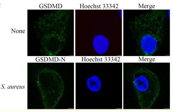
Application: IF/ICC    Species: bovine    Sample: MAC-T cells

Figure 1 GSDMD activation by S. aureus. A MAC-T cells were treated with S. aureus for the indicated times. GSDMD and GSDMD-N were detected by immunoblotting. B Cell death was measured by LDH release. C Subcellular localization of GSDMD-N or GSDMD in MAC-T cells incubated with or without S. aureus.

5). Dihydromyricetin inhibits African swine fever virus replication by downregulating toll-like receptor 4-dependent pyroptosis in vitro. Veterinary Research (PubMed: 37438783) [IF=4.4]

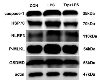
Application: WB    Species: pig    Sample:

Figure 7 DHM treatment inhibited ASFV-induced pyroptosis. PAMs were treated with A and B DHM or C disulfiram after ASFV infection; the cells were then collected at 24 h for Western blotting or RT-qPCR. D and E Western blotting of ASFV-infected PAMs were treated with DHM in the presence or absence of D polyphyllin VI or E RS 09 TFA; samples were collected after 24 h of treatment. *P 

6). Tryptophan alleviates lipopolysaccharide-induced liver injury and inflammation by modulating necroptosis and pyroptosis signaling pathways in piglets. Animal Biotechnology (PubMed: 37688392) [IF=3.7]

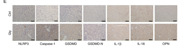
Application: WB    Species: piglets    Sample: liver

Figure 7. Effects of tryptophan on expression pyroptosis-related proteins in liver after 4h lipopolysaccharide (LPS) challenge in piglets. P values < 0.05 were considered significant. P values between 0.05 and 0.10 were regarded as a tendency. Values are presented as mean ± standard error, n = 4. CONTR: control group; LPS: piglets challenged with LPS; Trp + LPS: piglets fed with 0.2% tryptophan and challenged with LPS. NLRP3: NOD-like receptor family pyrin domain containing 3. GSDMD: gasdermin-D. MLKL: the mixed lineage kinase domain-like protein. HSP70: the 70-kDa heat shock proteins.

7). Oxalate‑induced renal pyroptotic injury and crystal formation mediated by NLRP3‑GSDMD signaling in vitro and in vivo. Molecular Medicine Reports (PubMed: 37732544) [IF=3.4]

Application: IHC    Species: Human    Sample: HK-2 cells

Figure 7. GSDMD mediates oxalate crystal-related pyroptosis in vivo. (A) Von Kossa staining revealed oxalate crystals in kidney of mice treated with Gly. (B) Haematoxylin and eosin staining demonstrated renal epithelial cell swelling, renal tubule edema and neutrophil infiltration in Gly treated mice. Scale bars, 50 µm. (C) Serum creatinine level of Gly-treated mice was examined by creatinine assay (n=6). (D) Western blot analysis of indicated proteins in NLRP3-GSDMD pathway in oxalated crystal mice. (E) The expression of same proteins was detected through immunohistochemical assays in mice. Scale bars, 50 µm. GSDMD, gasdermin D; Gly, glyoxylic acid; NLRP3, leucine-rich repeat-containing family pyrin domain-containing 3; OPN, osteopontin; Ctrl, control.

Application: WB    Species: Human    Sample: HK-2 cells

Figure 3. NLRP3 knockdown ameliorates oxalate-caused injury of HK-2 cells. (A) Western blot detection of NLRP3, caspase-1, GSDMD and GSDMD-N protein contents in HK-2 cells treated with 0.8 mM oxalate for 24 h after transfection with NLRP3-siRNA. (B) Relative lactate dehydrogenase levels in HK-2 cells transfected with NLRP3-siRNA upon 0.8 mM oxalate treatment for 24 h (n=6). (C) Images of two colors (blue and green) and merged colors in HK-2 cells with TUNEL staining were observed by confocal microscopy. HK-2 cells with NLRP3 silence were treated with 0.8 mM oxalate for 24 h followed by staining. Scale bars, 100 µm. (D) Images of three colors (blue, green and red) and merged colors in HK-2 cells with Calcein-AM/PI staining were recorded through confocal microscopy. HK-2 cells transfected with NLRP3-siRNA or scrambled-siRNA were dealt with 0.8 mM oxalate for 24 h. Scale bars, 100 µm. NLRP3, leucine-rich repeat-containing family pyrin domain-containing 3; GSDMD, gasdermin D; siRNA, small interfering RNA; Ctrl, control; NC, negative control.

8). LPS Enhances the Chemosensitivity of Oxaliplatin in HT29 Cells via GSDMD-Mediated Pyroptosis. Cancer Management and Research (PubMed: 33116894) [IF=3.3]

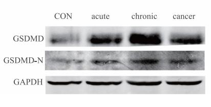
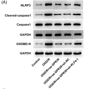
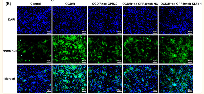
Application: WB    Species: mouse    Sample: colonials

Figure 1 |LPS improves anti-cancer effect of oxaliplatin in mice in vivo experiment.(D–F) Histopathology of colonials in different phases of model establishment. Western blot analyses and IHC of GSDMD and GSDMD-N in each group (n=6).

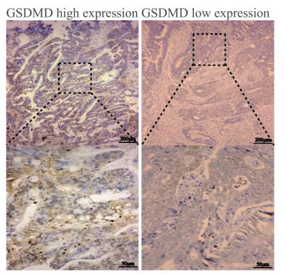
Application: IHC    Species: human    Sample: colorectal cancer tissues

Figure 2| The GSDMD expression is reduced in human colorectal cancer tissues and is significantly related to the overall survival of CRC patients.(C) Immunohistochemical analysis of GSDMD expression in human colorectal cancer tissues (n=244).

9). Immunohistochemical analysis of pyroptosis‐related protein expression in IgG4‐related sialadenitis. Journal of Oral Pathology & Medicine (PubMed: 35218237) [IF=3.3]

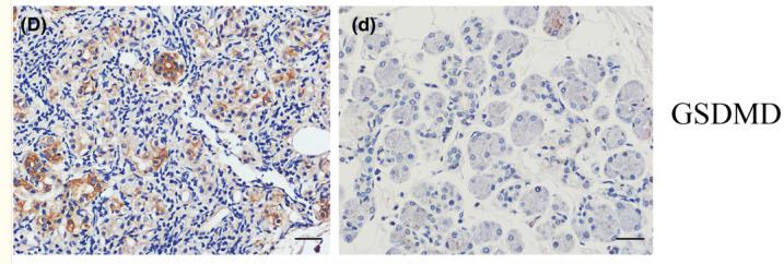
Application: IHC    Species: Human    Sample: IgG4‐RS tissues

FIGURE 1 Immunohistochemical (IHC) staining of pyroptosis‐related proteins in IgG4‐related sialadenitis (IgG4‐RS) tissues and control tissues (400×). IHC staining of IgG4‐RS tissues: (A) NLRP3 (NOD‐, LRR‐ and pyrin domain‐containing protein); (B) ASC (apoptosis‐associated speck‐like protein containing a CARD); (C) Caspase‐1; (D) GSDMD (gasdermin family members, including digestive dermatin D; (E) interleukin 1β (IL‐1β); (F) interleukin 18 (IL‐18). IHC staining of the control tissues: (a) NLRP3; (b) ASC; (c) Caspase‐1; (d) GSDMD; (e) IL‐1β; (f) IL‐18. Scale bars, 100 μm

10). Transcription factor Krüppel-like factor 4 upregulated G protein-coupled receptor 30 alleviates intestinal inflammation and apoptosis, and protects intestinal integrity from intestinal ischemia–reperfusion injury. Immunity, Inflammation and Disease (PubMed: 37506161) [IF=3.2]

Application: WB    Species: Human    Sample: Caco‐2 cells.

Figure 7 KLF4/GPR30 regulates NLRP3 inflammasome and pyroptosis in OGD/R‐exposed Caco‐2 cells. (A) Untransfected and transfected Caco‐2 cells were exposed to OGD/R, and protein expression of NLRP3, cleaved‐caspase1, caspase1, and GSDMD‐N was measured using western blot. (B) Immunofluorescence was conducted to examine GSDMD‐N expression. OGD/R, oxygen‐glucose deprivation/reoxygenation. ***p 

Application: IF/ICC    Species: Human    Sample: Caco‐2 cells.

Figure 7 KLF4/GPR30 regulates NLRP3 inflammasome and pyroptosis in OGD/R‐exposed Caco‐2 cells. (A) Untransfected and transfected Caco‐2 cells were exposed to OGD/R, and protein expression of NLRP3, cleaved‐caspase1, caspase1, and GSDMD‐N was measured using western blot. (B) Immunofluorescence was conducted to examine GSDMD‐N expression. OGD/R, oxygen‐glucose deprivation/reoxygenation. ***p 

Load more

Restrictive clause


Affinity Biosciences tests all products strictly. Citations are provided as a resource for additional applications that have not been validated by Affinity Biosciences. Please choose the appropriate format for each application and consult Materials and Methods sections for additional details about the use of any product in these publications.

For Research Use Only.
Not for use in diagnostic or therapeutic procedures. Not for resale. Not for distribution without written consent. Affinity Biosciences will not be held responsible for patent infringement or other violations that may occur with the use of our products. Affinity Biosciences, Affinity Biosciences Logo and all other trademarks are the property of Affinity Biosciences LTD.