Apelin Antibody - #DF13350

Related Downloads
Protocols
Product Info
*The optimal dilutions should be determined by the end user.
*Tips:
WB: For western blot detection of denatured protein samples. IHC: For immunohistochemical detection of paraffin sections (IHC-p) or frozen sections (IHC-f) of tissue samples. IF/ICC: For immunofluorescence detection of cell samples. ELISA(peptide): For ELISA detection of antigenic peptide.
Cite Format: Affinity Biosciences Cat# DF13350, RRID:AB_2846369.
Fold/Unfold
AGTRL1 ligand; APEL; APEL_HUMAN; Apelin-13; APJ endogenous ligand; Apln; XNPEP2;
Immunogens
Expressed in the brain with highest levels in the frontal cortex, thalamus, hypothalamus and midbrain (PubMed:10617103). Secreted by the mammary gland into the colostrum and the milk.
- Q9ULZ1 APEL_HUMAN:
- Protein BLAST With
- NCBI/
- ExPASy/
- Uniprot
MNLRLCVQALLLLWLSLTAVCGGSLMPLPDGNGLEDGNVRHLVQPRGSRNGPGPWQGGRRKFRRQRPRLSHKGPMPF
Predictions
Score>80(red) has high confidence and is suggested to be used for WB detection. *The prediction model is mainly based on the alignment of immunogen sequences, the results are for reference only, not as the basis of quality assurance.
High(score>80) Medium(80>score>50) Low(score<50) No confidence
Research Backgrounds
Endogenous ligand for the apelin receptor (APLNR). Drives internalization of the apelin receptor (By similarity). Apelin-36 dissociates more hardly than (pyroglu)apelin-13 from APLNR (By similarity). Hormone involved in the regulation of cardiac precursor cell movements during gastrulation and heart morphogenesis (By similarity). Has an inhibitory effect on cytokine production in response to T-cell receptor/CD3 cross-linking; the oral intake of apelin in the colostrum and the milk might therefore modulate immune responses in neonates (By similarity). Plays a role in early coronary blood vessels formation (By similarity). Mediates myocardial contractility in an ERK1/2-dependent manner (By similarity). May also have a role in the central control of body fluid homeostasis by influencing vasopressin release and drinking behavior (By similarity).
(Microbial infection) Endogenous ligand for the apelin receptor (APLNR), an alternative coreceptor with CD4 for HIV-1 infection. Inhibits HIV-1 entry in cells coexpressing CD4 and APLNR. Apelin-36 has a greater inhibitory activity on HIV infection than other synthetic apelin derivatives.
Several active peptides may be produced by proteolytic processing of the peptide precursor.
Secreted. Secreted>Extracellular space.
Note: Abundantly secreted in the colostrum. Lower level in milk. Decreases rapidly within several days after parturition in milk, but is still detectable even in commercial milk.
Expressed in the brain with highest levels in the frontal cortex, thalamus, hypothalamus and midbrain. Secreted by the mammary gland into the colostrum and the milk.
Belongs to the apelin family.
Research Fields
· Environmental Information Processing > Signal transduction > Apelin signaling pathway. (View pathway)
References
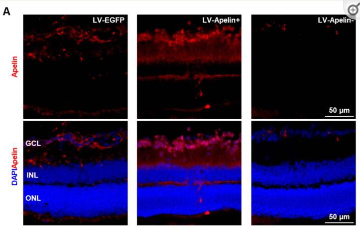
Application: IF/ICC Species: Mouse Sample:
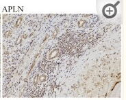
Application: IHC Species: Human Sample: AE samples
Restrictive clause
Affinity Biosciences tests all products strictly. Citations are provided as a resource for additional applications that have not been validated by Affinity Biosciences. Please choose the appropriate format for each application and consult Materials and Methods sections for additional details about the use of any product in these publications.
For Research Use Only.
Not for use in diagnostic or therapeutic procedures. Not for resale. Not for distribution without written consent. Affinity Biosciences will not be held responsible for patent infringement or other violations that may occur with the use of our products. Affinity Biosciences, Affinity Biosciences Logo and all other trademarks are the property of Affinity Biosciences LTD.