Product: nNOS Antibody
Catalog: AF6249
Description: Rabbit polyclonal antibody to nNOS
Application: WB IHC IF/ICC
Reactivity: Human, Mouse, Rat
Prediction: Pig, Zebrafish, Bovine, Horse, Rabbit, Dog, Chicken, Xenopus
Mol.Wt.: 120~160kDa; 161kD(Calculated).
Uniprot: P29475
RRID: AB_2835113

View similar products>>

   Size Price Inventory
 100ul $280 In stock
 200ul $350 In stock

Lead Time: Same day delivery

For pricing and ordering contact:
Local distributors

Product Info

Source:
Rabbit
Application:
WB 1:500-1:2000, IHC 1:50-1:200, IF/ICC 1:100-1:500
*The optimal dilutions should be determined by the end user.
*Tips:

WB: For western blot detection of denatured protein samples. IHC: For immunohistochemical detection of paraffin sections (IHC-p) or frozen sections (IHC-f) of tissue samples. IF/ICC: For immunofluorescence detection of cell samples. ELISA(peptide): For ELISA detection of antigenic peptide.

Reactivity:
Human,Mouse,Rat
Prediction:
Pig(100%), Zebrafish(100%), Bovine(100%), Horse(100%), Rabbit(100%), Dog(100%), Chicken(100%), Xenopus(85%)
Clonality:
Polyclonal
Specificity:
nNOS Antibody detects endogenous levels of total nNOS.
RRID:
AB_2835113
Cite Format: Affinity Biosciences Cat# AF6249, RRID:AB_2835113.
Conjugate:
Unconjugated.
Purification:
The antiserum was purified by peptide affinity chromatography using SulfoLink™ Coupling Resin (Thermo Fisher Scientific).
Storage:
Rabbit IgG in phosphate buffered saline , pH 7.4, 150mM NaCl, 0.02% sodium azide and 50% glycerol. Store at -20 °C. Stable for 12 months from date of receipt.
Alias:

Fold/Unfold

2310005C01Rik; BNOS; Constitutive NOS; EC 1.14.13.39; IHPS 1; IHPS1; N-NOS; NC-NOS; neuronal Nitric Oxide Synthase; Neuronal NOS; Nitric oxide synthase , neuronal, included; Nitric oxide synthase 1 (neuronal); Nitric oxide synthase 1; Nitric oxide synthase, brain; Nitric oxide synthase, penile neuronal, included; NNOS; NO; NOS 1; NOS; NOS type I; NOS-I; NOS1; NOS1_HUMAN; Peptidyl-cysteine S-nitrosylase NOS1;

Immunogens

Immunogen:
Uniprot:
Gene(ID):
Expression:
P29475 NOS1_HUMAN:

Isoform 1 is ubiquitously expressed: detected in skeletal muscle and brain, also in testis, lung and kidney, and at low levels in heart, adrenal gland and retina. Not detected in the platelets. Isoform 3 is expressed only in testis. Isoform 4 is detected in testis, skeletal muscle, lung, and kidney, at low levels in the brain, but not in the heart and adrenal gland.

Description:
nNOS nitric oxide synthase 1, neuronal form. Produces nitric oxide which is a messenger molecule with diverse functions and displays many properties of a neurotransmitter in the brain and peripheral nervous system. May be an effector enzyme for the dystrophin complex.
Sequence:
MEDHMFGVQQIQPNVISVRLFKRKVGGLGFLVKERVSKPPVIISDLIRGGAAEQSGLIQAGDIILAVNGRPLVDLSYDSALEVLRGIASETHVVLILRGPEGFTTHLETTFTGDGTPKTIRVTQPLGPPTKAVDLSHQPPAGKEQPLAVDGASGPGNGPQHAYDDGQEAGSLPHANGLAPRPPGQDPAKKATRVSLQGRGENNELLKEIEPVLSLLTSGSRGVKGGAPAKAEMKDMGIQVDRDLDGKSHKPLPLGVENDRVFNDLWGKGNVPVVLNNPYSEKEQPPTSGKQSPTKNGSPSKCPRFLKVKNWETEVVLTDTLHLKSTLETGCTEYICMGSIMHPSQHARRPEDVRTKGQLFPLAKEFIDQYYSSIKRFGSKAHMERLEEVNKEIDTTSTYQLKDTELIYGAKHAWRNASRCVGRIQWSKLQVFDARDCTTAHGMFNYICNHVKYATNKGNLRSAITIFPQRTDGKHDFRVWNSQLIRYAGYKQPDGSTLGDPANVQFTEICIQQGWKPPRGRFDVLPLLLQANGNDPELFQIPPELVLEVPIRHPKFEWFKDLGLKWYGLPAVSNMLLEIGGLEFSACPFSGWYMGTEIGVRDYCDNSRYNILEEVAKKMNLDMRKTSSLWKDQALVEINIAVLYSFQSDKVTIVDHHSATESFIKHMENEYRCRGGCPADWVWIVPPMSGSITPVFHQEMLNYRLTPSFEYQPDPWNTHVWKGTNGTPTKRRAIGFKKLAEAVKFSAKLMGQAMAKRVKATILYATETGKSQAYAKTLCEIFKHAFDAKVMSMEEYDIVHLEHETLVLVVTSTFGNGDPPENGEKFGCALMEMRHPNSVQEERKSYKVRFNSVSSYSDSQKSSGDGPDLRDNFESAGPLANVRFSVFGLGSRAYPHFCAFGHAVDTLLEELGGERILKMREGDELCGQEEAFRTWAKKVFKAACDVFCVGDDVNIEKANNSLISNDRSWKRNKFRLTFVAEAPELTQGLSNVHKKRVSAARLLSRQNLQSPKSSRSTIFVRLHTNGSQELQYQPGDHLGVFPGNHEDLVNALIERLEDAPPVNQMVKVELLEERNTALGVISNWTDELRLPPCTIFQAFKYYLDITTPPTPLQLQQFASLATSEKEKQRLLVLSKGLQEYEEWKWGKNPTIVEVLEEFPSIQMPATLLLTQLSLLQPRYYSISSSPDMYPDEVHLTVAIVSYRTRDGEGPIHHGVCSSWLNRIQADELVPCFVRGAPSFHLPRNPQVPCILVGPGTGIAPFRSFWQQRQFDIQHKGMNPCPMVLVFGCRQSKIDHIYREETLQAKNKGVFRELYTAYSREPDKPKKYVQDILQEQLAESVYRALKEQGGHIYVCGDVTMAADVLKAIQRIMTQQGKLSAEDAGVFISRMRDDNRYHEDIFGVTLRTYEVTNRLRSESIAFIEESKKDTDEVFSS

Predictions

Predictions:

Score>80(red) has high confidence and is suggested to be used for WB detection. *The prediction model is mainly based on the alignment of immunogen sequences, the results are for reference only, not as the basis of quality assurance.

Species
Results
Score
Pig
100
Horse
100
Bovine
100
Dog
100
Zebrafish
100
Chicken
100
Rabbit
100
Xenopus
85
Sheep
0
Model Confidence:
High(score>80) Medium(80>score>50) Low(score<50) No confidence

PTMs - P29475 As Substrate

Site PTM Type Enzyme
S292 Phosphorylation
T395 Phosphorylation
T396 Phosphorylation
S397 Phosphorylation
Y453 Phosphorylation
Y609 Phosphorylation
T724 Phosphorylation
S746 Phosphorylation Q14012 (CAMK1)
T766 Phosphorylation
T768 Phosphorylation
S852 Phosphorylation Q9UQM7 (CAMK2A)
S857 Phosphorylation
S859 Phosphorylation
S885 Phosphorylation
T1358 Phosphorylation
S1378 Phosphorylation
S1387 Phosphorylation
S1417 Phosphorylation

Research Backgrounds

Function:

Produces nitric oxide (NO) which is a messenger molecule with diverse functions throughout the body. In the brain and peripheral nervous system, NO displays many properties of a neurotransmitter. Probably has nitrosylase activity and mediates cysteine S-nitrosylation of cytoplasmic target proteins such SRR.

PTMs:

Ubiquitinated; mediated by STUB1/CHIP in the presence of Hsp70 and Hsp40 (in vitro).

Subcellular Location:

Cell membrane>Sarcolemma>Peripheral membrane protein. Cell projection>Dendritic spine.
Note: In skeletal muscle, it is localized beneath the sarcolemma of fast-twitch muscle fiber by associating with the dystrophin glycoprotein complex. In neurons, enriched in dendritic spines (By similarity).

Extracellular region or secreted Cytosol Plasma membrane Cytoskeleton Lysosome Endosome Peroxisome ER Golgi apparatus Nucleus Mitochondrion Manual annotation Automatic computational assertionSubcellular location
Tissue Specificity:

Isoform 1 is ubiquitously expressed: detected in skeletal muscle and brain, also in testis, lung and kidney, and at low levels in heart, adrenal gland and retina. Not detected in the platelets. Isoform 3 is expressed only in testis. Isoform 4 is detected in testis, skeletal muscle, lung, and kidney, at low levels in the brain, but not in the heart and adrenal gland.

Subunit Structure:

Homodimer. Interacts with DLG4; the interaction possibly being prevented by the association between NOS1 and CAPON. Forms a ternary complex with CAPON and RASD1. Forms a ternary complex with CAPON and SYN1. Interacts with ZDHHC23. Interacts with NOSIP; which may impair its synaptic location (By similarity). Interacts with HTR4. Interacts with VAC14 (By similarity). Interacts with SLC6A4 (By similarity). Interacts (via N-terminal domain) with DLG4 (via N-terminal tandem pair of PDZ domains) (By similarity).

Family&Domains:

The PDZ domain in the N-terminal part of the neuronal isoform participates in protein-protein interaction, and is responsible for targeting nNos to synaptic membranes in muscles. Mediates interaction with VAC14 (By similarity).

Belongs to the NOS family.

Research Fields

· Cellular Processes > Transport and catabolism > Phagosome.   (View pathway)

· Environmental Information Processing > Signal transduction > Calcium signaling pathway.   (View pathway)

· Environmental Information Processing > Signal transduction > Apelin signaling pathway.   (View pathway)

· Human Diseases > Neurodegenerative diseases > Alzheimer's disease.

· Human Diseases > Neurodegenerative diseases > Amyotrophic lateral sclerosis (ALS).

· Metabolism > Amino acid metabolism > Arginine biosynthesis.

· Metabolism > Amino acid metabolism > Arginine and proline metabolism.

· Metabolism > Global and overview maps > Metabolic pathways.

· Organismal Systems > Environmental adaptation > Circadian entrainment.

· Organismal Systems > Nervous system > Long-term depression.

· Organismal Systems > Endocrine system > Relaxin signaling pathway.

· Organismal Systems > Digestive system > Salivary secretion.

References

1). Human umbilical cord mesenchymal stem cells ameliorate erectile dysfunction in rats with diabetes mellitus through the attenuation of ferroptosis. Stem Cell Research & Therapy (PubMed: 36064453) [IF=7.5]

2). Gastroprotective mechanism of modified lvdou gancao decoction on ethanol-induced gastric lesions in mice: Involvement of Nrf-2/HO-1/NF-κB signaling pathway. Frontiers in Pharmacology (PubMed: 36120337) [IF=5.6]

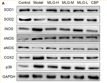
Application: WB    Species: Mouse    Sample: gastric tissues

FIGURE 7 Effect of MLG on some protein levels in ethanol-induced gastric lesions mice. Some representative western blot bands (A). Protein levels of SOD1/GAPDH (B), SOD2/GAPDH (C), iNOS/GAPDH (D), nNOS/GAPDH (E), eNOS/GAPDH (F), COX2/GAPDH (G), p38/GAPDH (H) in mice gastric tissues. SOD1, Superoxide dismutase one or Cu/Zn-superoxide dismutase; SOD2/Mn SOD, superoxide dismutase 2. Control: the group administered zero ethanol; Model: the group administered ethanol intragastrically (13.25 ml/kg BW); MLG-L: low-dose (5 g/kg body weight) MLG-treated group; MLG-M: medium-dose (10 g/kg body weight) MLG-treated group; MLG-H: high-dose (20 g/kg body weight) MLG-treated group; CBP: the group receiving Colloid Bismuth Pectin (57 mg/kg body weight). Each group’s data was expressed as mean ± standard deviation (SD), n = 6. By one-way analysis of variance (ANOVA) test, *p < 0.05, **p < 0.01, ***p < 0.001, ****p < 0.0001 vs. each group. Whole page of western blot can be found in Supplementary Figures S1, S3, S7, S8, S10, S14, S15, respectively.

3). Exosomes derived from smooth muscle cells ameliorate diabetes‐induced erectile dysfunction by inhibiting fibrosis and modulating the NO/cGMP pathway. Journal of Cellular and Molecular Medicine (PubMed: 33009701) [IF=5.3]

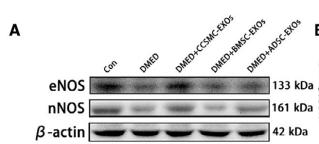
Application: WB    Species: Rat    Sample: corpus cavernosum

FIGURE 6 eNOS and nNOS expression with the NO and cGMP concentration in the corpus cavernosum. (A) Representative results of Western blot analysis of eNOS and nNOS in the rats of all five groups. (B) Expression of eNOS in all five groups with β-actin as the loading control is presented as a bar graph. (C) Expression of nNOS in all five groups with β-actin as the loading control is presented as a bar graph. (D) The black rectangle indicates the area of the corpus cavernosum selected for comparison. (E) The eNOS level is defined as eNOS IOD/area according to the immunofluorescence results. (F) The nNOS level is defined as nNOS IOD/area according to the immunofluorescence results. (G) Representative results of immunofluorescence analysis of eNOS and nNOS in all five groups. Scale bars = 50 μm. (H) Representative results of immunohistochemistry analysis of eNOS and nNOS in all five groups. The arrows indicate eNOS and nNOS expression (Because the expression levels of eNOS and nNOS in DMED group were very low, the coloring was very light). Scale bars = 50 μm. (I) The NO production in all five groups was determined with an assay kit. (J) The cGMP concentration in all five groups was determined with an assay kit. Data are expressed as the mean ± standard deviation. &P < .05 compared with the Con group. # P < .05 compared with the DMED group. * P < .05 compared with the DMED + CCSMC-EXO group. CCSMC-EXOs: exosomes derived from corpus cavernosum smooth muscle cells; BMSC-EXOs: exosomes derived from bone marrow stem cells; ADSC-EXOs: exosomes derived from adipose-derived stem cells; DMED: diabetes mellitus-induced erectile dysfunction; DAPI: 4',6-diamidino-2-phenylindole; eNOS: endothelial nitric oxide synthase; nNOS: neuronal nitric oxide synthase; NO: nitric oxide; cGMP: cyclic guanosine monophosphate; IOD: integral optical density; IOD/area: mean optical density

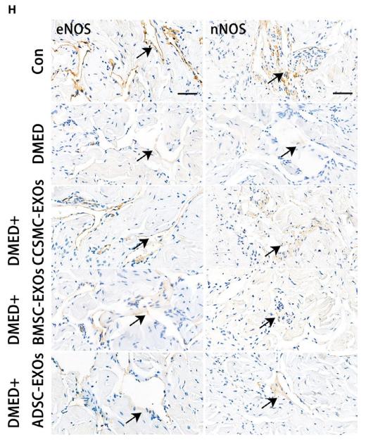
Application: IHC    Species: Rat    Sample: corpus cavernosum

FIGURE 6 eNOS and nNOS expression with the NO and cGMP concentration in the corpus cavernosum. (A) Representative results of Western blot analysis of eNOS and nNOS in the rats of all five groups. (B) Expression of eNOS in all five groups with β-actin as the loading control is presented as a bar graph. (C) Expression of nNOS in all five groups with β-actin as the loading control is presented as a bar graph. (D) The black rectangle indicates the area of the corpus cavernosum selected for comparison. (E) The eNOS level is defined as eNOS IOD/area according to the immunofluorescence results. (F) The nNOS level is defined as nNOS IOD/area according to the immunofluorescence results. (G) Representative results of immunofluorescence analysis of eNOS and nNOS in all five groups. Scale bars = 50 μm. (H) Representative results of immunohistochemistry analysis of eNOS and nNOS in all five groups. The arrows indicate eNOS and nNOS expression (Because the expression levels of eNOS and nNOS in DMED group were very low, the coloring was very light). Scale bars = 50 μm. (I) The NO production in all five groups was determined with an assay kit. (J) The cGMP concentration in all five groups was determined with an assay kit. Data are expressed as the mean ± standard deviation. &P < .05 compared with the Con group. # P < .05 compared with the DMED group. * P < .05 compared with the DMED + CCSMC-EXO group. CCSMC-EXOs: exosomes derived from corpus cavernosum smooth muscle cells; BMSC-EXOs: exosomes derived from bone marrow stem cells; ADSC-EXOs: exosomes derived from adipose-derived stem cells; DMED: diabetes mellitus-induced erectile dysfunction; DAPI: 4',6-diamidino-2-phenylindole; eNOS: endothelial nitric oxide synthase; nNOS: neuronal nitric oxide synthase; NO: nitric oxide; cGMP: cyclic guanosine monophosphate; IOD: integral optical density; IOD/area: mean optical density

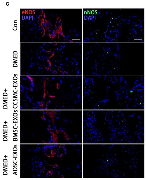
Application: IF/ICC    Species: Rat    Sample: corpus cavernosum

FIGURE 6 eNOS and nNOS expression with the NO and cGMP concentration in the corpus cavernosum. (A) Representative results of Western blot analysis of eNOS and nNOS in the rats of all five groups. (B) Expression of eNOS in all five groups with β-actin as the loading control is presented as a bar graph. (C) Expression of nNOS in all five groups with β-actin as the loading control is presented as a bar graph. (D) The black rectangle indicates the area of the corpus cavernosum selected for comparison. (E) The eNOS level is defined as eNOS IOD/area according to the immunofluorescence results. (F) The nNOS level is defined as nNOS IOD/area according to the immunofluorescence results. (G) Representative results of immunofluorescence analysis of eNOS and nNOS in all five groups. Scale bars = 50 μm. (H) Representative results of immunohistochemistry analysis of eNOS and nNOS in all five groups. The arrows indicate eNOS and nNOS expression (Because the expression levels of eNOS and nNOS in DMED group were very low, the coloring was very light). Scale bars = 50 μm. (I) The NO production in all five groups was determined with an assay kit. (J) The cGMP concentration in all five groups was determined with an assay kit. Data are expressed as the mean ± standard deviation. &P < .05 compared with the Con group. # P < .05 compared with the DMED group. * P < .05 compared with the DMED + CCSMC-EXO group. CCSMC-EXOs: exosomes derived from corpus cavernosum smooth muscle cells; BMSC-EXOs: exosomes derived from bone marrow stem cells; ADSC-EXOs: exosomes derived from adipose-derived stem cells; DMED: diabetes mellitus-induced erectile dysfunction; DAPI: 4',6-diamidino-2-phenylindole; eNOS: endothelial nitric oxide synthase; nNOS: neuronal nitric oxide synthase; NO: nitric oxide; cGMP: cyclic guanosine monophosphate; IOD: integral optical density; IOD/area: mean optical density

4). Acetyl‐L‐carnitine improves erectile function in bilateral cavernous nerve injury rats via promoting cavernous nerve regeneration. Andrology (PubMed: 35420721) [IF=4.5]

5). Neuronal NO synthase mediates plenylephrine induced cardiomyocyte hypertrophy through facilitation of NFAT-dependent transcriptional activity. Biochemistry and Biophysics Reports (PubMed: 30899802) [IF=2.7]

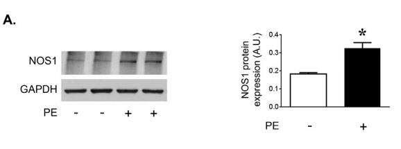
Application: WB    Species: rat    Sample: cardiomyocyte

Fig. 1. |Selective neuronal nitric oxide synthase inhibition blocks cardiomyocyte hypertrophy in vitro. A,Representative immunoblot showing NOS1 expression in response to PE stimulation. Values are expressed as mean ± SEM of triplicates from three independent experiments.

Restrictive clause


Affinity Biosciences tests all products strictly. Citations are provided as a resource for additional applications that have not been validated by Affinity Biosciences. Please choose the appropriate format for each application and consult Materials and Methods sections for additional details about the use of any product in these publications.

For Research Use Only.
Not for use in diagnostic or therapeutic procedures. Not for resale. Not for distribution without written consent. Affinity Biosciences will not be held responsible for patent infringement or other violations that may occur with the use of our products. Affinity Biosciences, Affinity Biosciences Logo and all other trademarks are the property of Affinity Biosciences LTD.