Product: DR4 Antibody
Catalog: AF0304
Description: Rabbit polyclonal antibody to DR4
Application: WB IHC IF/ICC
Reactivity: Human, Mouse, Rat
Mol.Wt.: 50kDa; 50kD(Calculated).
Uniprot: O00220
RRID: AB_2833468

View similar products>>

   Size Price Inventory
 100ul $280 In stock
 200ul $350 In stock

Lead Time: Same day delivery

For pricing and ordering contact:
Local distributors

Product Info

Source:
Rabbit
Application:
WB 1:500-1:3000, IF/ICC 1:100-1:500, IHC 1:50-1:200
*The optimal dilutions should be determined by the end user.
*Tips:

WB: For western blot detection of denatured protein samples. IHC: For immunohistochemical detection of paraffin sections (IHC-p) or frozen sections (IHC-f) of tissue samples. IF/ICC: For immunofluorescence detection of cell samples. ELISA(peptide): For ELISA detection of antigenic peptide.

Reactivity:
Human,Mouse,Rat
Clonality:
Polyclonal
Specificity:
DR4 Antibody detects endogenous levels of total DR4.
RRID:
AB_2833468
Cite Format: Affinity Biosciences Cat# AF0304, RRID:AB_2833468.
Conjugate:
Unconjugated.
Purification:
The antiserum was purified by peptide affinity chromatography using SulfoLink™ Coupling Resin (Thermo Fisher Scientific).
Storage:
Rabbit IgG in phosphate buffered saline , pH 7.4, 150mM NaCl, 0.02% sodium azide and 50% glycerol. Store at -20 °C. Stable for 12 months from date of receipt.
Alias:

Fold/Unfold

Apo2; CD261; Cytotoxic TRAIL receptor; Death receptor 4; DR4; NF related apoptosis-inducing ligand receptor 1; TNF receptor superfamily member 10a; TNF-related apoptosis-inducing ligand receptor 1; TNFRSF10A; TR10A_HUMAN; TRAIL receptor 1; TRAIL-R1; TRAILR 1; TRAILR1; Tumor necrosis factor receptor superfamily member 10A; Tumor necrosis factor receptor superfamily member 10a variant 2; Tumor necrosis factor receptor superfamily, member 10a;

Immunogens

Immunogen:
Uniprot:
Gene(ID):
Expression:
O00220 TR10A_HUMAN:

Widely expressed. High levels are found in spleen, peripheral blood leukocytes, small intestine and thymus, but also in K-562 erythroleukemia cells, MCF-7 breast carcinoma cells and activated T-cells.

Description:
TRAIL-R1 Receptor for the cytotoxic ligand TNFSF10/TRAIL. The adapter molecule FADD recruits caspase-8 to the activated receptor. The resulting death-inducing signaling complex (DISC) performs caspase-8 proteolytic activation which initiates the subsequent cascade of caspases (aspartate-specific cysteine proteases) mediating apoptosis. Promotes the activation of NF- kappa-B.
Sequence:
MAPPPARVHLGAFLAVTPNPGSAASGTEAAAATPSKVWGSSAGRIEPRGGGRGALPTSMGQHGPSARARAGRAPGPRPAREASPRLRVHKTFKFVVVGVLLQVVPSSAATIKLHDQSIGTQQWEHSPLGELCPPGSHRSEHPGACNRCTEGVGYTNASNNLFACLPCTACKSDEEERSPCTTTRNTACQCKPGTFRNDNSAEMCRKCSRGCPRGMVKVKDCTPWSDIECVHKESGNGHNIWVILVVTLVVPLLLVAVLIVCCCIGSGCGGDPKCMDRVCFWRLGLLRGPGAEDNAHNEILSNADSLSTFVSEQQMESQEPADLTGVTVQSPGEAQCLLGPAEAEGSQRRRLLVPANGADPTETLMLFFDKFANIVPFDSWDQLMRQLDLTKNEIDVVRAGTAGPGDALYAMLMKWVNKTGRNASIHTLLDALERMEERHAREKIQDLLVDSGKFIYLEDGTGSAVSLE

PTMs - O00220 As Substrate

Site PTM Type Enzyme
K36 Ubiquitination
R52 Methylation
S83 Phosphorylation
S178 Phosphorylation
K219 Ubiquitination
K273 Ubiquitination
C336 S-Nitrosylation
K391 Ubiquitination
S424 Phosphorylation
K443 Ubiquitination
K453 Ubiquitination
Y456 Phosphorylation
T461 Phosphorylation
S463 Phosphorylation
S466 Phosphorylation

Research Backgrounds

Function:

Receptor for the cytotoxic ligand TNFSF10/TRAIL. The adapter molecule FADD recruits caspase-8 to the activated receptor. The resulting death-inducing signaling complex (DISC) performs caspase-8 proteolytic activation which initiates the subsequent cascade of caspases (aspartate-specific cysteine proteases) mediating apoptosis. Promotes the activation of NF-kappa-B.

Subcellular Location:

Membrane>Single-pass type I membrane protein.

Extracellular region or secreted Cytosol Plasma membrane Cytoskeleton Lysosome Endosome Peroxisome ER Golgi apparatus Nucleus Mitochondrion Manual annotation Automatic computational assertionSubcellular location
Tissue Specificity:

Widely expressed. High levels are found in spleen, peripheral blood leukocytes, small intestine and thymus, but also in K-562 erythroleukemia cells, MCF-7 breast carcinoma cells and activated T-cells.

Subunit Structure:

Monomer. Three TNFRSF10A molecules interact with the TNFSF10 homotrimer. Can interact with TRADD and RIPK1. Interacts with ARAP1. Interacts with HCMV protein UL141; this interaction prevents TNFRSF10A cell surface expression. In the absence of stimulation, interacts with BIRC2, DDX3X and GSK3B. The interaction with BIRC2 and DDX3X is further enhanced upon receptor stimulation and accompanied by DDX3X and BIRC2 cleavage.

Research Fields

· Cellular Processes > Cell growth and death > Apoptosis.   (View pathway)

· Cellular Processes > Cell growth and death > Necroptosis.   (View pathway)

· Environmental Information Processing > Signaling molecules and interaction > Cytokine-cytokine receptor interaction.   (View pathway)

· Human Diseases > Infectious diseases: Viral > Measles.

· Human Diseases > Infectious diseases: Viral > Influenza A.

· Organismal Systems > Immune system > Natural killer cell mediated cytotoxicity.   (View pathway)

References

1). Sodium fluoride activates the extrinsic apoptosis via regulating NOX4/ROS-mediated p53/DR5 signaling pathway in lung cells both in vitro and in vivo. Free Radical Biology and Medicine (PubMed: 33857626) [IF=7.4]

Application: WB    Species: Human    Sample: BEAS-2B cells

Fig. 3. DR5 upregulation is critical for NaF-induced caspase-8 activation in BEAS-2B cells. (A–C) BEAS-2B cells were stimulated with 2 mM NaF for the indicated time. The mRNA and protein levels of DR4 and DR5 were tested using qRT-PCR and Western blot, respectively. (D) Cells were treated with or without 2 mM NaF for 9 h and cell surface expression of DR5 was determined by flow cytometry using an FITC-labeled anti-DR5 antibody. (E) The BEAS-2B cells transfected with scramble siRNA or DR5 siRNA were further stimulated with 2 mM NaF for 9 h, Western blotting was performed to detect the expression of DR5. BEAS-2B cells were transfected with scramble or DR5 siRNA (48 h), treated with 2 mM NaF (24 h). Enzymatic activity assays for caspase-8 (F) and caspase-3 (G). (H) Percentages of cells that underwent apoptosis for BEAS-2B cells treated with NaF. (I) Sum of Annexin V+PI− (Q4 quadrant) and Annexin V+PI+ (Q2 quadrant) populations is represented as the percentage of Annexin V+ cells. Results are expressed as means ± S.D and are representative of three independent experiments. **p < 0.01.

2). Akkermansia muciniphila Aspartic Protease Amuc_1434* Inhibits Human Colorectal Cancer LS174T Cell Viability via TRAIL-Mediated Apoptosis Pathway. INTERNATIONAL JOURNAL OF MOLECULAR SCIENCES (PubMed: 32403433) [IF=5.6]

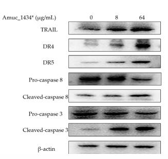
Application: WB    Species: human    Sample: LS174T cells

Figure 5. | Amuc_1434* mediated the activation of the apoptosis pathway in LS174T cells. (A) The expression of death receptor 4 (DR4), death receptor 5 (DR5), cysteinyl aspartate specific proteinase 8(caspase 8) and cysteinyl aspartate specific proteinase 3 (caspase 3) induced by Amuc_1434* in LS174T cells was dependent on tumor necrosis factor-related apoptosis-inducing ligand (TRAIL). (a) LS174T cells were treated with 8 and 64 µg/mL Amuc_1434* for 24 h, respectively. The cell lysates were analyzed by Western blot.

3). Bisimidazolium Salt Glycosyltransferase Inhibitors Suppress Hepatocellular Carcinoma Progression In Vitro and In Vivo. Pharmaceuticals (PubMed: 35745636) [IF=4.6]

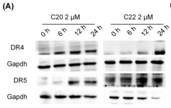
Application: WB    Species: Human    Sample: HepG2 cells

Figure 5 C20/C22 increased the sensitivity of HepG2 cells to TRAIL-induced apoptosis. (A,B) WB analysis of the expression of DR4 and 5 in HepG2 cells that were treated with 2 μM C20/C22 for 0, 6, 12, and 24 h. ImageJ software was used to calculate the gray value (n = 3). (C,D) The immunofluorescence assays used to determine the intracellular and cell surface distribution of DR4/5. (E,F) Annexin V/PI double staining was used to detect the apoptotic cells after the indicated treatments (n = 3). (G,H) WB analysis of the expression of Fas in HepG2 cells treated with C20/C22 for 0, 6, 12, and 24 h. (I) The immunofluorescence assays used to determine the intracellular and cell surface distribution of Fas. (J,K) Flow cytometry analysis of the apoptotic cells after the indicated treatments (n = 3). Scale bars, 10 µm. Data shown correspond to the mean ± SD of three independent experiments. The p-value for all datasets was analyzed by one-way ANOVA followed by Tukey’s test using GraphPad Prism version 8.00, except the data in (B) whose p-value was analyzed by nonparametric Dunnett’s test using GraphPad Prism version 8.00. * p < 0.05, ** p < 0.01, *** p < 0.001, **** p < 0.0001, and ns nonsignificant vs. 0 h/control group.

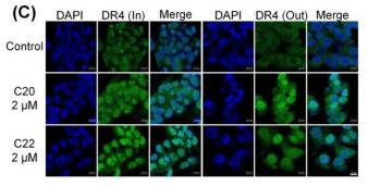
Application: IF/ICC    Species: Human    Sample: HepG2 cells

Figure 5 C20/C22 increased the sensitivity of HepG2 cells to TRAIL-induced apoptosis. (A,B) WB analysis of the expression of DR4 and 5 in HepG2 cells that were treated with 2 μM C20/C22 for 0, 6, 12, and 24 h. ImageJ software was used to calculate the gray value (n = 3). (C,D) The immunofluorescence assays used to determine the intracellular and cell surface distribution of DR4/5. (E,F) Annexin V/PI double staining was used to detect the apoptotic cells after the indicated treatments (n = 3). (G,H) WB analysis of the expression of Fas in HepG2 cells treated with C20/C22 for 0, 6, 12, and 24 h. (I) The immunofluorescence assays used to determine the intracellular and cell surface distribution of Fas. (J,K) Flow cytometry analysis of the apoptotic cells after the indicated treatments (n = 3). Scale bars, 10 µm. Data shown correspond to the mean ± SD of three independent experiments. The p-value for all datasets was analyzed by one-way ANOVA followed by Tukey’s test using GraphPad Prism version 8.00, except the data in (B) whose p-value was analyzed by nonparametric Dunnett’s test using GraphPad Prism version 8.00. * p < 0.05, ** p < 0.01, *** p < 0.001, **** p < 0.0001, and ns nonsignificant vs. 0 h/control group.

4). Sphingomyelin synthase 2 overexpression promotes cisplatin-induced apoptosis of HepG2 cells. Oncology Letters (PubMed: 29375716) [IF=2.9]

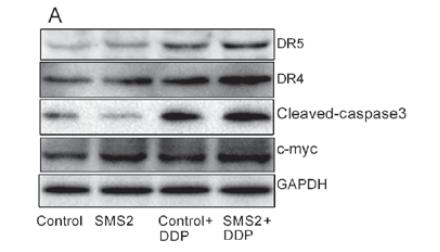
Application: WB    Species: human    Sample: HepG2 cells

Figure 2.| Expression of DR4, DR5, cleaved caspase‑3 and c‑Myc in HepG2 cells. (A) The protein expression of DR4, DR5, cleaved caspase‑3 and c‑Myc was investigated using western blotting. Quantification and statistical analysis was performed on (B) DR4, (C) DR5, (D) cleaved caspase‑3 and (E) c‑Myc. n=3, *P<0.05, **P<0.001 vs. control group; #P<0.05, ##P<0.001 vs. control + DDP group. DR. death receptor; DDP, cisplatin; SMS2, sphyngomyelin synthase 2; c‑Myc, avian myelocytomatosis viral oncogene homolog.

Restrictive clause


Affinity Biosciences tests all products strictly. Citations are provided as a resource for additional applications that have not been validated by Affinity Biosciences. Please choose the appropriate format for each application and consult Materials and Methods sections for additional details about the use of any product in these publications.

For Research Use Only.
Not for use in diagnostic or therapeutic procedures. Not for resale. Not for distribution without written consent. Affinity Biosciences will not be held responsible for patent infringement or other violations that may occur with the use of our products. Affinity Biosciences, Affinity Biosciences Logo and all other trademarks are the property of Affinity Biosciences LTD.