CLCN7 Antibody - #DF3932

Related Downloads
Protocols
Product Info
*The optimal dilutions should be determined by the end user.
*Tips:
WB: For western blot detection of denatured protein samples. IHC: For immunohistochemical detection of paraffin sections (IHC-p) or frozen sections (IHC-f) of tissue samples. IF/ICC: For immunofluorescence detection of cell samples. ELISA(peptide): For ELISA detection of antigenic peptide.
Cite Format: Affinity Biosciences Cat# DF3932, RRID:AB_2836285.
Fold/Unfold
Chloride channel protein 7; CLC 7; ClC-7; ClC7; CLCN7; CLCN7_HUMAN; FLJ26686; FLJ39644; FLJ46423; H(+)/Cl(-) exchange transporter 7; OPTA2; OPTB4;
Immunogens
- P51798 CLCN7_HUMAN:
- Protein BLAST With
- NCBI/
- ExPASy/
- Uniprot
MANVSKKVSWSGRDRDDEEAAPLLRRTARPGGGTPLLNGAGPGAARQSPRSALFRVGHMSSVELDDELLDPDMDPPHPFPKEIPHNEKLLSLKYESLDYDNSENQLFLEEERRINHTAFRTVEIKRWVICALIGILTGLVACFIDIVVENLAGLKYRVIKGNIDKFTEKGGLSFSLLLWATLNAAFVLVGSVIVAFIEPVAAGSGIPQIKCFLNGVKIPHVVRLKTLVIKVSGVILSVVGGLAVGKEGPMIHSGSVIAAGISQGRSTSLKRDFKIFEYFRRDTEKRDFVSAGAAAGVSAAFGAPVGGVLFSLEEGASFWNQFLTWRIFFASMISTFTLNFVLSIYHGNMWDLSSPGLINFGRFDSEKMAYTIHEIPVFIAMGVVGGVLGAVFNALNYWLTMFRIRYIHRPCLQVIEAVLVAAVTATVAFVLIYSSRDCQPLQGGSMSYPLQLFCADGEYNSMAAAFFNTPEKSVVSLFHDPPGSYNPLTLGLFTLVYFFLACWTYGLTVSAGVFIPSLLIGAAWGRLFGISLSYLTGAAIWADPGKYALMGAAAQLGGIVRMTLSLTVIMMEATSNVTYGFPIMLVLMTAKIVGDVFIEGLYDMHIQLQSVPFLHWEAPVTSHSLTAREVMSTPVTCLRRREKVGVIVDVLSDTASNHNGFPVVEHADDTQPARLQGLILRSQLIVLLKHKVFVERSNLGLVQRRLRLKDFRDAYPRFPPIQSIHVSQDERECTMDLSEFMNPSPYTVPQEASLPRVFKLFRALGLRHLVVVDNRNQVVGLVTRKDLARYRLGKRGLEELSLAQT
Predictions
Score>80(red) has high confidence and is suggested to be used for WB detection. *The prediction model is mainly based on the alignment of immunogen sequences, the results are for reference only, not as the basis of quality assurance.
High(score>80) Medium(80>score>50) Low(score<50) No confidence
PTMs - P51798 As Substrate
| Site | PTM Type | Enzyme | Source |
|---|---|---|---|
| K6 | Ubiquitination | Uniprot | |
| K7 | Ubiquitination | Uniprot | |
| S9 | Phosphorylation | Uniprot | |
| S11 | Phosphorylation | Uniprot | |
| T34 | Phosphorylation | Uniprot | |
| S48 | Phosphorylation | Uniprot | |
| S60 | Phosphorylation | Uniprot | |
| S61 | Phosphorylation | Uniprot | |
| K81 | Ubiquitination | Uniprot | |
| K88 | Ubiquitination | Uniprot | |
| K93 | Sumoylation | Uniprot | |
| K93 | Ubiquitination | Uniprot | |
| Y99 | Phosphorylation | Uniprot | |
| T137 | Phosphorylation | Uniprot | |
| K160 | Ubiquitination | Uniprot | |
| S237 | Phosphorylation | Uniprot | |
| K274 | Ubiquitination | Uniprot | |
| Y278 | Phosphorylation | Uniprot | |
| T633 | Phosphorylation | Uniprot | |
| T636 | Phosphorylation | Uniprot | |
| S682 | Phosphorylation | Uniprot | |
| S801 | Phosphorylation | Uniprot | |
| T805 | Phosphorylation | Uniprot |
Research Backgrounds
Slowly voltage-gated channel mediating the exchange of chloride ions against protons. Functions as antiporter and contributes to the acidification of the lysosome lumen.
Lysosome membrane>Multi-pass membrane protein.
Brain, testis, muscle and kidney.
Chloride channel 7 are heteromers of alpha (CLCN7) and beta (OSTM1) subunits.
Belongs to the chloride channel (TC 2.A.49) family. ClC-7/CLCN7 subfamily.
References
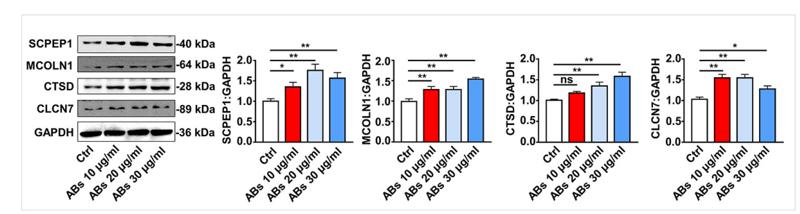
Application: WB Species: human Sample: human umbilical vein endothelial cells (HUVECs)
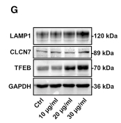
Application: WB Species: Human Sample: endothelial cells
Application: WB Species: Mouse Sample:
Restrictive clause
Affinity Biosciences tests all products strictly. Citations are provided as a resource for additional applications that have not been validated by Affinity Biosciences. Please choose the appropriate format for each application and consult Materials and Methods sections for additional details about the use of any product in these publications.
For Research Use Only.
Not for use in diagnostic or therapeutic procedures. Not for resale. Not for distribution without written consent. Affinity Biosciences will not be held responsible for patent infringement or other violations that may occur with the use of our products. Affinity Biosciences, Affinity Biosciences Logo and all other trademarks are the property of Affinity Biosciences LTD.