Product: AFP Antibody
Catalog: AF5134
Description: Rabbit polyclonal antibody to AFP
Application: WB IHC
Reactivity: Human, Mouse, Rat
Prediction: Pig, Bovine, Horse, Sheep, Rabbit, Dog
Mol.Wt.: 62-65 kDa; 69kD(Calculated).
Uniprot: P02771
RRID: AB_2837620

View similar products>>

   Size Price Inventory
 100ul $280 In stock
 200ul $350 In stock

Lead Time: Same day delivery

For pricing and ordering contact:
Local distributors

Product Info

Source:
Rabbit
Application:
WB 1:500-1:2000, IHC 1:50-1:200
*The optimal dilutions should be determined by the end user.
*Tips:

WB: For western blot detection of denatured protein samples. IHC: For immunohistochemical detection of paraffin sections (IHC-p) or frozen sections (IHC-f) of tissue samples. IF/ICC: For immunofluorescence detection of cell samples. ELISA(peptide): For ELISA detection of antigenic peptide.

Reactivity:
Human,Mouse,Rat
Prediction:
Pig(100%), Bovine(92%), Horse(100%), Sheep(92%), Rabbit(92%), Dog(100%)
Clonality:
Polyclonal
Specificity:
AFP Antibody detects endogenous levels of total AFP.
RRID:
AB_2837620
Cite Format: Affinity Biosciences Cat# AF5134, RRID:AB_2837620.
Conjugate:
Unconjugated.
Purification:
The antiserum was purified by peptide affinity chromatography using SulfoLink™ Coupling Resin (Thermo Fisher Scientific).
Storage:
Rabbit IgG in phosphate buffered saline , pH 7.4, 150mM NaCl, 0.02% sodium azide and 50% glycerol. Store at -20 °C. Stable for 12 months from date of receipt.
Alias:

Fold/Unfold

Afp; AFPD; Alpha fetoglobulin; Alpha fetoprotein; Alpha fetoprotein precursor; Alpha-1-fetoprotein; Alpha-fetoglobulin; Alpha-fetoprotein; alpha-fetoprotein, Hereditary persistence of, included; FETA; FETA_HUMAN; Hereditary persistence of alpha fetoprotein; HPAFP;

Immunogens

Immunogen:
Uniprot:
Gene(ID):
Expression:
P02771 FETA_HUMAN:

Plasma. Synthesized by the fetal liver and yolk sac.

Description:
Binds copper, nickel, and fatty acids as well as, and bilirubin less well than, serum albumin. Only a small percentage (less than 2%) of the human AFP shows estrogen-binding properties.
Sequence:
MKWVESIFLIFLLNFTESRTLHRNEYGIASILDSYQCTAEISLADLATIFFAQFVQEATYKEVSKMVKDALTAIEKPTGDEQSSGCLENQLPAFLEELCHEKEILEKYGHSDCCSQSEEGRHNCFLAHKKPTPASIPLFQVPEPVTSCEAYEEDRETFMNKFIYEIARRHPFLYAPTILLWAARYDKIIPSCCKAENAVECFQTKAATVTKELRESSLLNQHACAVMKNFGTRTFQAITVTKLSQKFTKVNFTEIQKLVLDVAHVHEHCCRGDVLDCLQDGEKIMSYICSQQDTLSNKITECCKLTTLERGQCIIHAENDEKPEGLSPNLNRFLGDRDFNQFSSGEKNIFLASFVHEYSRRHPQLAVSVILRVAKGYQELLEKCFQTENPLECQDKGEEELQKYIQESQALAKRSCGLFQKLGEYYLQNAFLVAYTKKAPQLTSSELMAITRKMAATAATCCQLSEDKLLACGEGAADIIIGHLCIRHEMTPVNPGVGQCCTSSYANRRPCFSSLVVDETYVPPAFSDDKFIFHKDLCQAQGVALQTMKQEFLINLVKQKPQITEEQLEAVIADFSGLLEKCCQGQEQEVCFAEEGQKLISKTRAALGV

Predictions

Predictions:

Score>80(red) has high confidence and is suggested to be used for WB detection. *The prediction model is mainly based on the alignment of immunogen sequences, the results are for reference only, not as the basis of quality assurance.

Species
Results
Score
Pig
100
Horse
100
Dog
100
Bovine
92
Sheep
92
Rabbit
92
Chicken
55
Xenopus
0
Zebrafish
0
Model Confidence:
High(score>80) Medium(80>score>50) Low(score<50) No confidence

PTMs - P02771 As Substrate

Site PTM Type Enzyme
S111 Phosphorylation
S115 Phosphorylation
S117 Phosphorylation
T248 Phosphorylation
S344 Phosphorylation
S368 Phosphorylation
K413 Acetylation
S415 Phosphorylation
S444 Phosphorylation
S445 Phosphorylation

Research Backgrounds

Function:

Binds copper, nickel, and fatty acids as well as, and bilirubin less well than, serum albumin. Only a small percentage (less than 2%) of the human AFP shows estrogen-binding properties.

PTMs:

Independent studies suggest heterogeneity of the N-terminal sequence of the mature protein and of the cleavage site of the signal sequence.

Sulfated.

Subcellular Location:

Secreted.

Extracellular region or secreted Cytosol Plasma membrane Cytoskeleton Lysosome Endosome Peroxisome ER Golgi apparatus Nucleus Mitochondrion Manual annotation Automatic computational assertionSubcellular location
Tissue Specificity:

Plasma. Synthesized by the fetal liver and yolk sac.

Subunit Structure:

Dimeric and trimeric forms have been found in addition to the monomeric form.

Family&Domains:

Belongs to the ALB/AFP/VDB family.

Research Fields

· Environmental Information Processing > Signal transduction > Hippo signaling pathway.   (View pathway)

References

1). Human umbilical cord-derived mesenchymal stem cells improve the function of liver in rats with acute-on-chronic liver failure via downregulating Notch and Stat1/Stat3 signaling. Stem Cell Research & Therapy (PubMed: 34256837) [IF=7.5]

Application: IHC    Species: Rat    Sample: liver tissue

Fig. 7 The effects of hUC-MSC transplantation on hepatocyte regeneration in ACLF rats. Liver sections from ACLF rats 12 and 24 h post-hUC-MSC transplantation or 0.9% sodium chloride injection as a control were used for immunohistochemical staining of AFP, CK18, HGF, and PCNA and microscopic examination (n = 4/group). Representative photographs are shown (a). Positive staining was quantified and is presented as the mean ± SD (b, c). d Serum levels of HGF were detected by ELISA (4h, n = 3/group; 12h and 24h, n = 4/group). Data are presented as the mean ± SD. *P < 0.05, **P < 0.01, ***P < 0.001

2). Enhanced hepatic differentiation of rat bone marrow-derived mesenchymal stem cells in spheroidal aggregate culture on a decellularized liver scaffold. INTERNATIONAL JOURNAL OF MOLECULAR MEDICINE (PubMed: 27314916) [IF=5.4]

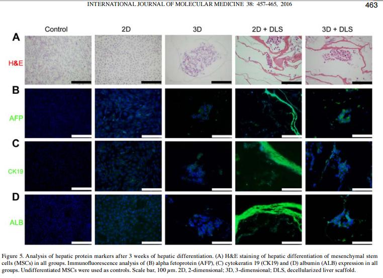
Application: IF/ICC    Species: rat    Sample:

Figure 5. Analysis of hepatic protein markers after 3 weeks of hepatic differentiation. (A) H&E staining of hepatic differentiation of mesenchymal stem cells (MSCs) in all groups. Immunofluorescence analysis of (B) alpha fetoprotein (AFP), (C) cytokeratin 19 (CK19) and (D) albumin (ALB) expression in all groups. Undifferentiated MSCs were used as controls. Scale bar, 100 µm. 2D, 2-dimensional; 3D, 3-dimensional; DLS, decellularized liver scaffold.

3). Glycyrrhizic Acid-Induced Differentiation Repressed Stemness in Hepatocellular Carcinoma by Targeting c-Jun N-Terminal Kinase 1. Frontiers in Oncology (PubMed: 31998631) [IF=4.7]

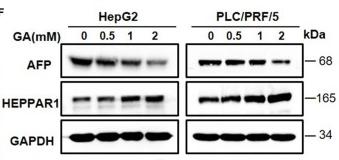
Application: WB    Species: Mice    Sample: hepatic cancer cells

Figure 1 GA reduces stemness and induces differentiation of hepatic cancer cells. (A) Survival of HepG2 and PLC/PRF/5 cells incubated with the indicated amounts of GA for 48 h. (B) Cell proliferation was assessed through a colony formation assay. The number of colonies was compared (one-way ANOVA; *P < 0.05, **P < 0.01, ***P < 0.001). (C) Images of sphere formation assay of HepG2 and PLC/PRF/5. The number of spheres was compared (one-way ANOVA; *P < 0.05, **P < 0.01, ***P < 0.001). (D) Expression of representative CSC markers (SOX2 and OCT4) was analyzed by Western blot assay (one-way ANOVA; *P < 0.05, **P < 0.01, ***P < 0.001). (E) Images of HepG2 and PLC/PRF/5 cells were taken after incubation with different GA concentrations. (F) Expressions of representative differentiation markers (AFP and HEPPAR1) were analyzed by Western blot assay (one-way ANOVA; *P < 0.05, **P < 0.01). Scale bars in C and E are at 200 and 20 μm, respectively.

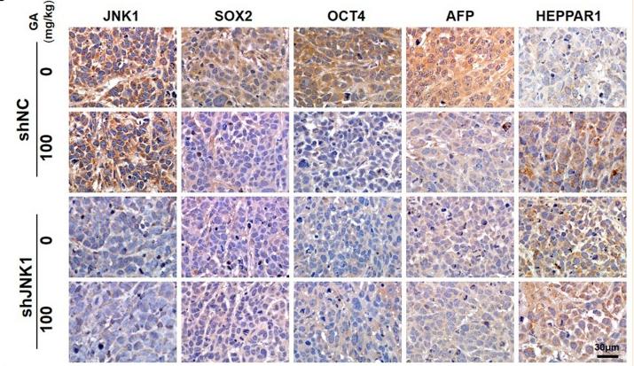
Application: IHC    Species: Mice    Sample: tumors tissue

Figure 6 GA suppresses tumor growth and enhances the antitumor effect of sorafenib. Images of tumors (A), tumor volumes (B), and body weight (C) of PLC/PRF/5 transfected with shNC or shJNK1 in BALB/c nu/nu mice treated with 100 mg/kg GA or saline as control. (D) IHC staining indicates the expression of CSC markers (SOX2 and OCT4) and differentiation markers (AFP and HEPPAR1) in tumors. (E) IHC analysis of SOX2, OCT4, AFP, and HEPPAR1 in tumors (one-way ANOVA; ***P < 0.001). Scale bars in (D), 30 μm. Images of tumors (F), tumor volumes (G) and body weight (H) of PLC/PRF/5-bearing BALB/c nu/nu mice. GA or sorafenib treatment obviously inhibited tumor growth. Furthermore, GA could enhance the anti-tumor effect of sorafenib.

4). Transient Inhibition of mTORC1 Signaling Ameliorates Irradiation-Induced Liver Damage. Frontiers in Physiology (PubMed: 30984007) [IF=4.0]

Application: WB    Species: mouse    Sample: Liver

FIGURE 1 | Irradiation-induced liver damage in mice. C57BL/6J mice were exposed to 8.0 Gy of total body irradiation (TBI). Liver tissues were harvested at days 1, 3, and 7 post-exposure (n = 6). (A) Representative pictures by HE staining are shown. Scale bar = 50 μm. Arrows indicate edema hepatocytes. (B and C) ALB and AFP expression in liver tissues (n = 3). Expression of ALB (B) and AFP (C) was detected by western blotting.

5). Hepatic differentiation of mouse bone marrow‑derived mesenchymal stem cells using a novel 3D culture system. Molecular Medicine Reports (PubMed: 29152658) [IF=3.4]

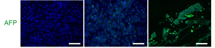
Application: IF/ICC    Species: Mouse    Sample:

Figure 4. Analysis of hepatic protein markers in mouse MSCs following 23 days of hepatic differentiation. Green immunofluorescence staining of ALB, AFP and CK19 in undifferentiated MSC control, 2D and 3D groups. Nuclei were stained blue with DAPI. Scale bars, 100 µm. MSCs, mesenchymal stem cells; ALB, albumin; AFP, α-fetoprotein; CK19, cytokeratin-19.

6). Clinicopathologic Features and Prognostic Factors in Alpha-Fetoprotein-Producing Colorectal Cancer: Analysis of 78 Cases. CELLULAR PHYSIOLOGY AND BIOCHEMISTRY (PubMed: 30522102)

Restrictive clause


Affinity Biosciences tests all products strictly. Citations are provided as a resource for additional applications that have not been validated by Affinity Biosciences. Please choose the appropriate format for each application and consult Materials and Methods sections for additional details about the use of any product in these publications.

For Research Use Only.
Not for use in diagnostic or therapeutic procedures. Not for resale. Not for distribution without written consent. Affinity Biosciences will not be held responsible for patent infringement or other violations that may occur with the use of our products. Affinity Biosciences, Affinity Biosciences Logo and all other trademarks are the property of Affinity Biosciences LTD.