Lamin B1 Antibody - #AF5161

| Product: | Lamin B1 Antibody |
| Catalog: | AF5161 |
| Description: | Rabbit polyclonal antibody to Lamin B1 |
| Application: | WB IHC IF/ICC |
| Reactivity: | Human, Mouse, Rat, Monkey, Fish |
| Prediction: | Pig, Bovine, Sheep, Rabbit, Xenopus |
| Mol.Wt.: | 66 kDa; 66kD(Calculated). |
| Uniprot: | P20700 |
| RRID: | AB_2837647 |
Related Downloads
Protocols
Product Info
*The optimal dilutions should be determined by the end user.
*Tips:
WB: For western blot detection of denatured protein samples. IHC: For immunohistochemical detection of paraffin sections (IHC-p) or frozen sections (IHC-f) of tissue samples. IF/ICC: For immunofluorescence detection of cell samples. ELISA(peptide): For ELISA detection of antigenic peptide.
Cite Format: Affinity Biosciences Cat# AF5161, RRID:AB_2837647.
Fold/Unfold
ADLD; lamin B1; Lamin-B1; LMN; LMN2; LMNB; Lmnb1; LMNB1_HUMAN; MGC111419; OTTHUMP00000159218;
Immunogens
- P20700 LMNB1_HUMAN:
- Protein BLAST With
- NCBI/
- ExPASy/
- Uniprot
MATATPVPPRMGSRAGGPTTPLSPTRLSRLQEKEELRELNDRLAVYIDKVRSLETENSALQLQVTEREEVRGRELTGLKALYETELADARRALDDTARERAKLQIELGKCKAEHDQLLLNYAKKESDLNGAQIKLREYEAALNSKDAALATALGDKKSLEGDLEDLKDQIAQLEASLAAAKKQLADETLLKVDLENRCQSLTEDLEFRKSMYEEEINETRRKHETRLVEVDSGRQIEYEYKLAQALHEMREQHDAQVRLYKEELEQTYHAKLENARLSSEMNTSTVNSAREELMESRMRIESLSSQLSNLQKESRACLERIQELEDLLAKEKDNSRRMLTDKEREMAEIRDQMQQQLNDYEQLLDVKLALDMEISAYRKLLEGEEERLKLSPSPSSRVTVSRASSSRSVRTTRGKRKRVDVEESEASSSVSISHSASATGNVCIEEIDVDGKFIRLKNTSEQDQPMGGWEMIRKIGDTSVSYKYTSRYVLKAGQTVTIWAANAGVTASPPTDLIWKNQNSWGTGEDVKVILKNSQGEEVAQRSTVFKTTIPEEEEEEEEAAGVVVEEELFHQQGTPRASNRSCAIM
Predictions
Score>80(red) has high confidence and is suggested to be used for WB detection. *The prediction model is mainly based on the alignment of immunogen sequences, the results are for reference only, not as the basis of quality assurance.
High(score>80) Medium(80>score>50) Low(score<50) No confidence
PTMs - P20700 As Substrate
| Site | PTM Type | Enzyme | Source |
|---|---|---|---|
| A2 | Acetylation | Uniprot | |
| T3 | Phosphorylation | Uniprot | |
| T5 | Phosphorylation | Uniprot | |
| R10 | Methylation | Uniprot | |
| S13 | Phosphorylation | Uniprot | |
| R14 | Methylation | Uniprot | |
| T19 | Phosphorylation | Uniprot | |
| T20 | Phosphorylation | Uniprot | |
| S23 | Phosphorylation | P06493 (CDK1) | Uniprot |
| T25 | Phosphorylation | Uniprot | |
| S28 | Phosphorylation | Uniprot | |
| K33 | Acetylation | Uniprot | |
| K33 | Ubiquitination | Uniprot | |
| R42 | Methylation | Uniprot | |
| Y46 | Phosphorylation | Uniprot | |
| K49 | Acetylation | Uniprot | |
| K49 | Ubiquitination | Uniprot | |
| S58 | Phosphorylation | Uniprot | |
| K79 | Acetylation | Uniprot | |
| K79 | Ubiquitination | Uniprot | |
| Y82 | Phosphorylation | Uniprot | |
| T84 | Phosphorylation | Uniprot | |
| K102 | Acetylation | Uniprot | |
| K102 | Sumoylation | Uniprot | |
| K102 | Ubiquitination | Uniprot | |
| K109 | Ubiquitination | Uniprot | |
| K111 | Acetylation | Uniprot | |
| K111 | Ubiquitination | Uniprot | |
| K123 | Acetylation | Uniprot | |
| K123 | Ubiquitination | Uniprot | |
| K124 | Ubiquitination | Uniprot | |
| S126 | Phosphorylation | Uniprot | |
| K134 | Acetylation | Uniprot | |
| K134 | Ubiquitination | Uniprot | |
| Y138 | Phosphorylation | Uniprot | |
| S144 | Phosphorylation | Uniprot | |
| K145 | Ubiquitination | Uniprot | |
| K156 | Acetylation | Uniprot | |
| K156 | Ubiquitination | Uniprot | |
| K157 | Acetylation | Uniprot | |
| K157 | Ubiquitination | Uniprot | |
| S158 | Phosphorylation | Uniprot | |
| K167 | Ubiquitination | Uniprot | |
| K181 | Acetylation | Uniprot | |
| K181 | Ubiquitination | Uniprot | |
| K182 | Ubiquitination | Uniprot | |
| R197 | Methylation | Uniprot | |
| C198 | S-Nitrosylation | Uniprot | |
| S200 | Phosphorylation | Uniprot | |
| K209 | Acetylation | Uniprot | |
| K209 | Ubiquitination | Uniprot | |
| S210 | Phosphorylation | Uniprot | |
| Y212 | Phosphorylation | Uniprot | |
| K222 | Ubiquitination | Uniprot | |
| S232 | Phosphorylation | Uniprot | |
| Y238 | Phosphorylation | Uniprot | |
| K241 | Sumoylation | Uniprot | |
| K241 | Ubiquitination | Uniprot | |
| R250 | Methylation | Uniprot | |
| K261 | Sumoylation | Uniprot | |
| K261 | Ubiquitination | Uniprot | |
| T267 | Phosphorylation | Uniprot | |
| Y268 | Phosphorylation | Uniprot | |
| K271 | Acetylation | Uniprot | |
| K271 | Ubiquitination | Uniprot | |
| S278 | Phosphorylation | Uniprot | |
| S279 | Phosphorylation | Uniprot | |
| T283 | Phosphorylation | Uniprot | |
| S284 | Phosphorylation | Uniprot | |
| T285 | Phosphorylation | Uniprot | |
| S288 | Phosphorylation | Uniprot | |
| R297 | Methylation | Uniprot | |
| R299 | Methylation | Uniprot | |
| S302 | Phosphorylation | Uniprot | |
| S304 | Phosphorylation | Uniprot | |
| S305 | Phosphorylation | Uniprot | |
| K312 | Acetylation | Uniprot | |
| K312 | Ubiquitination | Uniprot | |
| K330 | Ubiquitination | Uniprot | |
| T340 | Phosphorylation | Uniprot | |
| K342 | Ubiquitination | Uniprot | |
| Y360 | Phosphorylation | Uniprot | |
| S375 | Phosphorylation | Uniprot | |
| Y377 | Phosphorylation | Uniprot | |
| K379 | Acetylation | Uniprot | |
| K379 | Sumoylation | Uniprot | |
| K379 | Ubiquitination | Uniprot | |
| R387 | Methylation | Uniprot | |
| K389 | Sumoylation | Uniprot | |
| K389 | Ubiquitination | Uniprot | |
| S391 | Phosphorylation | Uniprot | |
| S393 | Phosphorylation | P06493 (CDK1) | Uniprot |
| S395 | Phosphorylation | P05771 (PRKCB) | Uniprot |
| S396 | Phosphorylation | Uniprot | |
| T399 | Phosphorylation | Uniprot | |
| S401 | Phosphorylation | Uniprot | |
| S404 | Phosphorylation | Uniprot | |
| S405 | Phosphorylation | P05771 (PRKCB) | Uniprot |
| S406 | Phosphorylation | Uniprot | |
| S408 | Phosphorylation | Uniprot | |
| T411 | Phosphorylation | Uniprot | |
| T412 | Phosphorylation | Uniprot | |
| K457 | Ubiquitination | Uniprot | |
| S460 | Phosphorylation | Uniprot | |
| K474 | Ubiquitination | Uniprot | |
| S479 | Phosphorylation | Uniprot | |
| S481 | Phosphorylation | Uniprot | |
| K483 | Acetylation | Uniprot | |
| K483 | Ubiquitination | Uniprot | |
| K528 | Ubiquitination | Uniprot | |
| K532 | Acetylation | Uniprot | |
| K532 | Ubiquitination | Uniprot | |
| S534 | Phosphorylation | Uniprot | |
| K547 | Ubiquitination | Uniprot | |
| T575 | Phosphorylation | Uniprot | |
| S579 | Phosphorylation | Uniprot |
Research Backgrounds
Lamins are components of the nuclear lamina, a fibrous layer on the nucleoplasmic side of the inner nuclear membrane, which is thought to provide a framework for the nuclear envelope and may also interact with chromatin.
B-type lamins undergo a series of modifications, such as farnesylation and phosphorylation. Increased phosphorylation of the lamins occurs before envelope disintegration and probably plays a role in regulating lamin associations.
Nucleus inner membrane>Lipid-anchor>Nucleoplasmic side.
Homodimer. Interacts with lamin-associated polypeptides IA, IB and 2. Interacts with SPAG4 and SEPT12.
Belongs to the intermediate filament family.
Research Fields
· Cellular Processes > Cell growth and death > Apoptosis. (View pathway)
References

Application: WB Species: Human Sample: HGC27 cells
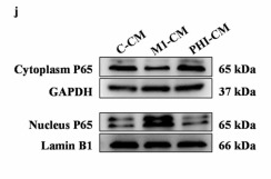
Application: WB Species: Mouse Sample: mHSCs
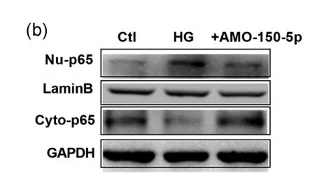
Application: WB Species: rat Sample: Neonatal cardiac fibroblasts (CFs)
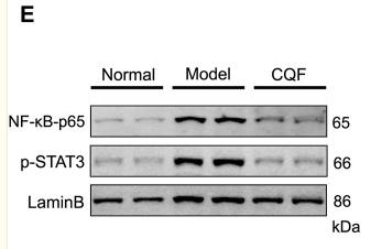
Application: WB Species: Mice Sample: colon tissue
Restrictive clause
Affinity Biosciences tests all products strictly. Citations are provided as a resource for additional applications that have not been validated by Affinity Biosciences. Please choose the appropriate format for each application and consult Materials and Methods sections for additional details about the use of any product in these publications.
For Research Use Only.
Not for use in diagnostic or therapeutic procedures. Not for resale. Not for distribution without written consent. Affinity Biosciences will not be held responsible for patent infringement or other violations that may occur with the use of our products. Affinity Biosciences, Affinity Biosciences Logo and all other trademarks are the property of Affinity Biosciences LTD.