Product: AQP1 Antibody
Catalog: AF5231
Description: Rabbit polyclonal antibody to AQP1
Application: WB IHC IF/ICC
Reactivity: Human, Mouse, Rat
Prediction: Horse, Dog
Mol.Wt.: 29 kDa.; 29kD(Calculated).
Uniprot: P29972
RRID: AB_2837717

View similar products>>

   Size Price Inventory
 100ul $280 In stock
 200ul $350 In stock

Lead Time: Same day delivery

For pricing and ordering contact:
Local distributors

Product Info

Source:
Rabbit
Application:
WB 1:500-1:2000, IHC 1:50-1:200, IF/ICC 1:100-1:500
*The optimal dilutions should be determined by the end user.
*Tips:

WB: For western blot detection of denatured protein samples. IHC: For immunohistochemical detection of paraffin sections (IHC-p) or frozen sections (IHC-f) of tissue samples. IF/ICC: For immunofluorescence detection of cell samples. ELISA(peptide): For ELISA detection of antigenic peptide.

Reactivity:
Human,Mouse,Rat
Prediction:
Horse(80%), Dog(80%)
Clonality:
Polyclonal
Specificity:
AQP1 Antibody detects endogenous levels of total AQP1.
RRID:
AB_2837717
Cite Format: Affinity Biosciences Cat# AF5231, RRID:AB_2837717.
Conjugate:
Unconjugated.
Purification:
The antiserum was purified by peptide affinity chromatography using SulfoLink™ Coupling Resin (Thermo Fisher Scientific).
Storage:
Rabbit IgG in phosphate buffered saline , pH 7.4, 150mM NaCl, 0.02% sodium azide and 50% glycerol. Store at -20 °C. Stable for 12 months from date of receipt.
Alias:

Fold/Unfold

AQP 1; AQP CHIP; AQP-1; AQP1; AQP1_HUMAN; aquaporin 1 (channel-forming integral protein, 28kDa, CO blood group); aquaporin 1 (Colton blood group); Aquaporin CHIP; Aquaporin-1; Aquaporin-CHIP; Aquaporin1; Channel forming integral protein 28kDa; Channel like integral membrane protein 28 kDa; CHIP 28; CHIP28; CO; Colton blood group; Growth factor induced delayed early response protein; MGC26324; Urine water channel; Water channel protein CHIP 29; Water channel protein CHIP29; Water channel protein for red blood cells and kidney proximal tubule;

Immunogens

Immunogen:
Uniprot:
Gene(ID):
Expression:
P29972 AQP1_HUMAN:

Detected in erythrocytes (at protein level). Expressed in a number of tissues including erythrocytes, renal tubules, retinal pigment epithelium, heart, lung, skeletal muscle, kidney and pancreas. Weakly expressed in brain, placenta and liver.

Description:
Forms a water-specific channel that provides the plasma membranes of red cells and kidney proximal tubules with high permeability to water, thereby permitting water to move in the direction of an osmotic gradient.
Sequence:
MASEFKKKLFWRAVVAEFLATTLFVFISIGSALGFKYPVGNNQTAVQDNVKVSLAFGLSIATLAQSVGHISGAHLNPAVTLGLLLSCQISIFRALMYIIAQCVGAIVATAILSGITSSLTGNSLGRNDLADGVNSGQGLGIEIIGTLQLVLCVLATTDRRRRDLGGSAPLAIGLSVALGHLLAIDYTGCGINPARSFGSAVITHNFSNHWIFWVGPFIGGALAVLIYDFILAPRSSDLTDRVKVWTSGQVEEYDLDADDINSRVEMKPK

Predictions

Predictions:

Score>80(red) has high confidence and is suggested to be used for WB detection. *The prediction model is mainly based on the alignment of immunogen sequences, the results are for reference only, not as the basis of quality assurance.

Species
Results
Score
Horse
80
Dog
80
Pig
0
Bovine
0
Sheep
0
Xenopus
0
Zebrafish
0
Chicken
0
Rabbit
0
Model Confidence:
High(score>80) Medium(80>score>50) Low(score<50) No confidence

PTMs - P29972 As Substrate

Site PTM Type Enzyme
T157 Phosphorylation P17252 (PRKCA)
S236 O-Glycosylation
T239 Phosphorylation P17252 (PRKCA)
Y253 Phosphorylation
S262 Phosphorylation

Research Backgrounds

Function:

Forms a water-specific channel that provides the plasma membranes of red cells and kidney proximal tubules with high permeability to water, thereby permitting water to move in the direction of an osmotic gradient.

Subcellular Location:

Cell membrane>Multi-pass membrane protein.

Extracellular region or secreted Cytosol Plasma membrane Cytoskeleton Lysosome Endosome Peroxisome ER Golgi apparatus Nucleus Mitochondrion Manual annotation Automatic computational assertionSubcellular location
Tissue Specificity:

Detected in erythrocytes (at protein level). Expressed in a number of tissues including erythrocytes, renal tubules, retinal pigment epithelium, heart, lung, skeletal muscle, kidney and pancreas. Weakly expressed in brain, placenta and liver.

Subunit Structure:

Homotetramer. Interacts with EPHB2; involved in endolymph production in the inner ear (By similarity). Identified in a complex with STOM.

Family&Domains:

Aquaporins contain two tandem repeats each containing three membrane-spanning domains and a pore-forming loop with the signature motif Asn-Pro-Ala (NPA).

Belongs to the MIP/aquaporin (TC 1.A.8) family.

Research Fields

· Organismal Systems > Endocrine system > Renin secretion.

· Organismal Systems > Excretory system > Proximal tubule bicarbonate reclamation.

References

1). Synergistic effect of constituent drugs of Baibutang on improving Yin-deficiency pulmonary fibrosis in rats. JOURNAL OF ETHNOPHARMACOLOGY (PubMed: 36535334) [IF=5.4]

2). MMP‐7 affects peritoneal ultrafiltration associated with elevated aquaporin‐1 expression via MAPK/ERK pathway in peritoneal mesothelial cells. Journal of Cellular and Molecular Medicine (PubMed: 34117704) [IF=5.3]

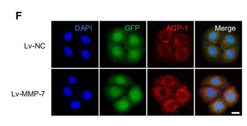
Application: IF/ICC    Species: Human    Sample: peritoneal mesothelial cells

FIGURE 4 Ectopic expression of MMP-7 enhanced AQP-1 expression and then altered cell volume in peritoneal mesothelial cells. HMrSV5 cells were infected with lentivirus expressing MMP-7. (a, b) The expression of MMP-7 in the lentivirus-transfected cells was determined by quantitative RT-PCR (a) and Western blotting analysis (b). (c) The MMP-7-overexpressing cells (Lv-MMP-7) and control cells (Lv-NC) were incubated in NS or dialysate with 4.25% glucose for 1 minute. The cell diameter of peritoneal mesothelial cells was evaluated by the Scepter 2.0 cell counter. The changes of cell diameter after glucose stimulation were calculated. (d-f) The mRNA level of AQP-1 was detected by quantitative RT-PCR (d), and the protein expression of AQP-1 was assessed by Western blotting assay (e). Besides, the cells were permeabilized by Triton-X 100 and immunofluorescence assay was used to detect the expression of AQP-1 (f). (g, h) The cells were treated with the indicated 50 ng/ml MMP-7 for 12 hours. The expression of AQP-1 in the HMrSV5 cell membrane was evaluated by Western blotting (g) and immunofluorescence assay (h), HMrSV5 cells were transfected with AQP-1-targeting siRNAs for 36 hours. The expression level of AQP-1 protein was assayed by immunoblotting. The #3 siRNA against AQP-1 was selected for the further analysis. (I) After transfecting with AQP-1-targeting siRNAs for 36 hours, HMrSV5 cells were incubated with 100 ng/ml MMP-7 for another 24 hours. Cell diameter of the peritoneal mesothelial cells was evaluated by the Scepter 2.0 cell counter (J). *P < 0.05, **P < 0.01, one of the three independent experiments is shown

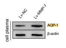
Application: WB    Species: Human    Sample: peritoneal mesothelial cells

FIGURE 4 Ectopic expression of MMP-7 enhanced AQP-1 expression and then altered cell volume in peritoneal mesothelial cells. HMrSV5 cells were infected with lentivirus expressing MMP-7. (a, b) The expression of MMP-7 in the lentivirus-transfected cells was determined by quantitative RT-PCR (a) and Western blotting analysis (b). (c) The MMP-7-overexpressing cells (Lv-MMP-7) and control cells (Lv-NC) were incubated in NS or dialysate with 4.25% glucose for 1 minute. The cell diameter of peritoneal mesothelial cells was evaluated by the Scepter 2.0 cell counter. The changes of cell diameter after glucose stimulation were calculated. (d-f) The mRNA level of AQP-1 was detected by quantitative RT-PCR (d), and the protein expression of AQP-1 was assessed by Western blotting assay (e). Besides, the cells were permeabilized by Triton-X 100 and immunofluorescence assay was used to detect the expression of AQP-1 (f). (g, h) The cells were treated with the indicated 50 ng/ml MMP-7 for 12 hours. The expression of AQP-1 in the HMrSV5 cell membrane was evaluated by Western blotting (g) and immunofluorescence assay (h), HMrSV5 cells were transfected with AQP-1-targeting siRNAs for 36 hours. The expression level of AQP-1 protein was assayed by immunoblotting. The #3 siRNA against AQP-1 was selected for the further analysis. (I) After transfecting with AQP-1-targeting siRNAs for 36 hours, HMrSV5 cells were incubated with 100 ng/ml MMP-7 for another 24 hours. Cell diameter of the peritoneal mesothelial cells was evaluated by the Scepter 2.0 cell counter (J). *P < 0.05, **P < 0.01, one of the three independent experiments is shown

3). Combination of pseudoephedrine and emodin ameliorates LPS-induced acute lung injury by regulating macrophage M1/M2 polarization through the VIP/cAMP/PKA pathway. Chinese Medicine (PubMed: 35123524) [IF=4.9]

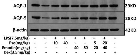
Application: WB    Species: Rat    Sample: lung tissues 

Fig. 1 Effect of Pseudoephedrine + emodin on pathological changes of lung tissues (×200), expression of AQP‐1, AQP‐5 in LPS‐induced ALI rats. A The chemical structure of Pseudoephedrine and Emodin. B Stained with hematoxylin and eosin. C The lung injury score was determined following a five-point scale from 0 to 4 as follows: 0, l, 2, 3, and 4 represent no damage, mild damage, moderate damage, severe damage, and very severe damage, respectively. Representative histological sections of the lungs were stained with hematoxylin and eosin (H & E staining, magnification ×200). D–F Western blot analysis was performed to detect AQP‐1 and AQP‐5 protein expression. K The dry/wet weight ratio and lung weight/body weight ratio were used to reflect the pulmonary edema. All data are expressed as mean ± S.D. (n = 5). ##p < 0.01, ###p < 0.001 vs. control group. *p < 0.05, **p < 0.01, ***p < 0.001 vs. LPS alone group. +p < 0.05, +++p < 0.001 vs. combined treatment group (5 + 20 mg/kg)

4). MiR-3194-3p Inhibits Breast Cancer Progression by Targeting Aquaporin1. Frontiers in Oncology (PubMed: 32903818) [IF=4.7]

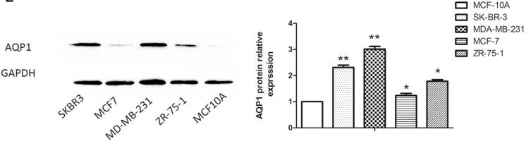
Application: WB    Species: human    Sample: BC tissues and cell lines

FIGURE 1 | The miR-3194-3p and AQP1 expression levels in BC tissues and cell lines. Comparing differences in the expression levels of miR-3194-3p between(A) BC and adjacent non-tumor tissues (n = 30); (B) BC cell lines and the control breast epithelial cells (MCF-10A); differences in AQP1 mRNA level between (C) BC and adjacent non-tumor tissue; (D) BC cell lines and MCF-10A; differences in AQP1 protein level between (E) BC cell lines and MCF-10A.

5). Hyperglycemia induces corneal endothelial dysfunction through attenuating mitophagy. EXPERIMENTAL EYE RESEARCH (PubMed: 34951999) [IF=3.4]

Restrictive clause


Affinity Biosciences tests all products strictly. Citations are provided as a resource for additional applications that have not been validated by Affinity Biosciences. Please choose the appropriate format for each application and consult Materials and Methods sections for additional details about the use of any product in these publications.

For Research Use Only.
Not for use in diagnostic or therapeutic procedures. Not for resale. Not for distribution without written consent. Affinity Biosciences will not be held responsible for patent infringement or other violations that may occur with the use of our products. Affinity Biosciences, Affinity Biosciences Logo and all other trademarks are the property of Affinity Biosciences LTD.