CD3 zeta Antibody - #AF5405
 The expression level of CAR (containing exogenous CD3ζ) in CAR-T cells was detected by western blotting using an anti-CD3ζ antibody, with blank T cells as negative control, and endogenous CD3ζ as a loading control. The experiments have been repeated for three times.CAR, chimeric antigen receptor; scFv, single chain variable fragment; VH, heavy chain variable region; VL, light chain variable region; MOI, multiplicity of infection;7-AAD, 7-aminoactinomycin D; Blank T, T cells not transduced; CAR-T, T cells transduced with CAR.")
| Product: | CD3 zeta Antibody |
| Catalog: | AF5405 |
| Description: | Rabbit polyclonal antibody to CD3 zeta |
| Application: | WB IF/ICC |
| Reactivity: | Human, Mouse |
| Prediction: | Pig, Bovine, Horse, Sheep, Rabbit |
| Mol.Wt.: | 19 kDa; 19kD(Calculated). |
| Uniprot: | P20963 |
| RRID: | AB_2837889 |
Related Downloads
Protocols
Product Info
*The optimal dilutions should be determined by the end user.
*Tips:
WB: For western blot detection of denatured protein samples. IHC: For immunohistochemical detection of paraffin sections (IHC-p) or frozen sections (IHC-f) of tissue samples. IF/ICC: For immunofluorescence detection of cell samples. ELISA(peptide): For ELISA detection of antigenic peptide.
Cite Format: Affinity Biosciences Cat# AF5405, RRID:AB_2837889.
Fold/Unfold
4930549J05Rik; A430104F18Rik; AW552088; CD247; CD247 antigen; Cd247 molecule; Cd3; CD3 antigen, zeta polypeptide, isoform CRA_b; CD3 antigen, zeta subunit; CD3-eta; CD3H; CD3Q; Cd3z; CD3Z antigen zeta polypeptide (TiT3 complex); CD3Z_HUMAN; CD3zeta; CD3zeta chain; MGC140430; T cell receptor T3 zeta chain; T cell surface glycoprotein CD3 zeta chain; T-cell antigen receptor complex, zeta subunit of CD3; T-cell receptor T3 zeta chain; T-cell surface glycoprotein CD3 zeta chain; T3z; TCR zeta chain; TCRk; TCRZ; TCRzeta;
Immunogens
CD3Z is expressed in normal lymphoid tissue and in peripheral blood mononuclear cells (PBMCs) (PubMed:11722641).
- P20963 CD3Z_HUMAN:
- Protein BLAST With
- NCBI/
- ExPASy/
- Uniprot
MKWKALFTAAILQAQLPITEAQSFGLLDPKLCYLLDGILFIYGVILTALFLRVKFSRSADAPAYQQGQNQLYNELNLGRREEYDVLDKRRGRDPEMGGKPQRRKNPQEGLYNELQKDKMAEAYSEIGMKGERRRGKGHDGLYQGLSTATKDTYDALHMQALPPR
Predictions
Score>80(red) has high confidence and is suggested to be used for WB detection. *The prediction model is mainly based on the alignment of immunogen sequences, the results are for reference only, not as the basis of quality assurance.
High(score>80) Medium(80>score>50) Low(score<50) No confidence
PTMs - P20963 As Substrate
| Site | PTM Type | Enzyme | Source |
|---|---|---|---|
| S58 | Phosphorylation | Uniprot | |
| Y64 | Phosphorylation | Uniprot | |
| Y72 | Phosphorylation | Uniprot | |
| Y83 | Phosphorylation | Uniprot | |
| K88 | Ubiquitination | Uniprot | |
| K99 | Ubiquitination | Uniprot | |
| K104 | Ubiquitination | Uniprot | |
| Y111 | Phosphorylation | P06241 (FYN) | Uniprot |
| K116 | Ubiquitination | Uniprot | |
| K118 | Ubiquitination | Uniprot | |
| Y123 | Phosphorylation | P06241 (FYN) | Uniprot |
| S124 | Phosphorylation | Uniprot | |
| K129 | Ubiquitination | Uniprot | |
| K136 | Ubiquitination | Uniprot | |
| Y142 | Phosphorylation | P06239 (LCK) | Uniprot |
| S146 | Phosphorylation | Uniprot | |
| T147 | Phosphorylation | Uniprot | |
| T149 | Phosphorylation | Uniprot | |
| K150 | Ubiquitination | Uniprot | |
| T152 | Phosphorylation | Uniprot | |
| Y153 | Phosphorylation | Uniprot |
Research Backgrounds
Part of the TCR-CD3 complex present on T-lymphocyte cell surface that plays an essential role in adaptive immune response. When antigen presenting cells (APCs) activate T-cell receptor (TCR), TCR-mediated signals are transmitted across the cell membrane by the CD3 chains CD3D, CD3E, CD3G and CD3Z. All CD3 chains contain immunoreceptor tyrosine-based activation motifs (ITAMs) in their cytoplasmic domain. Upon TCR engagement, these motifs become phosphorylated by Src family protein tyrosine kinases LCK and FYN, resulting in the activation of downstream signaling pathways. CD3Z ITAMs phosphorylation creates multiple docking sites for the protein kinase ZAP70 leading to ZAP70 phosphorylation and its conversion into a catalytically active enzyme. Plays an important role in intrathymic T-cell differentiation. Additionally, participates in the activity-dependent synapse formation of retinal ganglion cells (RGCs) in both the retina and dorsal lateral geniculate nucleus (dLGN) (By similarity).
Phosphorylated on Tyr residues after T-cell receptor triggering by LCK in association with CD4/CD8.
Membrane>Single-pass type I membrane protein.
CD3Z is expressed in normal lymphoid tissue and in peripheral blood mononuclear cells (PBMCs).
The TCR-CD3 complex is composed of a CD3D/CD3E and a CD3G/CD3E heterodimers that preferentially associate with TCRalpha and TCRbeta, respectively, to form TCRalpha/CD3E/CD3G and TCRbeta/CD3G/CD3E trimers. In turn, the hexamer interacts with CD3Z homodimer to form the TCR-CD3 complex. Alternatively, TCRalpha and TCRbeta can be replaced by TCRgamma and TCRdelta. Interacts with SLA. Interacts with TRAT1. Interacts with DOCK2. Interacts with SLA2. Interacts with SHB. Interacts with ZAP70. Interacts (tyrosine phosphorylated) with SHC1 (via SH2 domain). Interacts with PTPRC. Interacts with CRK; this interaction regulates CD3Z phosphorylation. Interacts (on T cell side) with CD81, ICAM1 and CD9 at immunological synapses between antigen-presenting cells and T cells. Interacts with CD160.
(Microbial infection) Interacts with HIV-1 Nef; this interaction up-regulates the expression of the Fas ligand (FASLG) at the cell surface.
(Microbial infection) Interacts with HIV-2 Nef protein; this interaction induces down-regulation of cell surface TCR/CD3 complexes.
The ITAM domains mediate interaction with SHB.
Belongs to the CD3Z/FCER1G family.
Research Fields
· Human Diseases > Infectious diseases: Parasitic > Chagas disease (American trypanosomiasis).
· Organismal Systems > Immune system > Natural killer cell mediated cytotoxicity. (View pathway)
· Organismal Systems > Immune system > Th1 and Th2 cell differentiation. (View pathway)
· Organismal Systems > Immune system > Th17 cell differentiation. (View pathway)
· Organismal Systems > Immune system > T cell receptor signaling pathway. (View pathway)
References
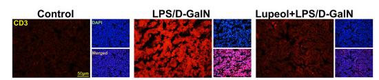
Application: IF/ICC Species: Mice Sample: liver tissue
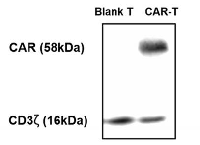
Application: WB Species: mouse Sample: CAR-T cells
Restrictive clause
Affinity Biosciences tests all products strictly. Citations are provided as a resource for additional applications that have not been validated by Affinity Biosciences. Please choose the appropriate format for each application and consult Materials and Methods sections for additional details about the use of any product in these publications.
For Research Use Only.
Not for use in diagnostic or therapeutic procedures. Not for resale. Not for distribution without written consent. Affinity Biosciences will not be held responsible for patent infringement or other violations that may occur with the use of our products. Affinity Biosciences, Affinity Biosciences Logo and all other trademarks are the property of Affinity Biosciences LTD.