Product: VCAM1 Antibody
Catalog: DF6082
Description: Rabbit polyclonal antibody to VCAM1
Application: WB IHC IF/ICC
Reactivity: Human, Mouse, Rat
Prediction: Pig, Bovine, Horse, Rabbit, Dog
Mol.Wt.: 80~120kDa; 81kD(Calculated).
Uniprot: P19320
RRID: AB_2838050

View similar products>>

   Size Price Inventory
 100ul $280 In stock
 200ul $350 In stock

Lead Time: Same day delivery

For pricing and ordering contact:
Local distributors

Product Info

Source:
Rabbit
Application:
WB 1:500-1:2000, IHC 1:50-1:200, IF/ICC 1:100-1:500
*The optimal dilutions should be determined by the end user.
*Tips:

WB: For western blot detection of denatured protein samples. IHC: For immunohistochemical detection of paraffin sections (IHC-p) or frozen sections (IHC-f) of tissue samples. IF/ICC: For immunofluorescence detection of cell samples. ELISA(peptide): For ELISA detection of antigenic peptide.

Reactivity:
Human,Mouse,Rat
Prediction:
Pig(82%), Bovine(82%), Horse(100%), Rabbit(91%), Dog(82%)
Clonality:
Polyclonal
Specificity:
VCAM1 Antibody detects endogenous levels of total VCAM1.
RRID:
AB_2838050
Cite Format: Affinity Biosciences Cat# DF6082, RRID:AB_2838050.
Conjugate:
Unconjugated.
Purification:
The antiserum was purified by peptide affinity chromatography using SulfoLink™ Coupling Resin (Thermo Fisher Scientific).
Storage:
Rabbit IgG in phosphate buffered saline , pH 7.4, 150mM NaCl, 0.02% sodium azide and 50% glycerol. Store at -20 °C. Stable for 12 months from date of receipt.
Alias:

Fold/Unfold

CD106; CD106 Antigen; INCAM 100; INCAM-100; L1CAM; MGC99561; V-CAM 1; Vascular Cell Adhesion Molecule 1; Vascular cell adhesion protein 1; VCAM 1; VCAM-1; VCAM1; VCAM1_HUMAN;

Immunogens

Immunogen:
Uniprot:
Gene(ID):
Expression:
P19320 VCAM1_HUMAN:

Expressed on inflamed vascular endothelium, as well as on macrophage-like and dendritic cell types in both normal and inflamed tissue.

Description:
Vascular cell adhesion protein 1 (VCAM1) is important in cell-cell recognition. It has been reported to function in leukocyte-endothelial cell adhesion, interact with the beta-1 integrin VLA4 on leukocytes, and mediate both adhesion and signal transduction. There are two isoforms; Isoform 1, a single-pass type I membrane protein, and isoform 2, a lipid-anchor (GPI-anchor). VCAM1 is expressed on inflamed vascular endothelium, as well as on macrophage-like and dendritic cell types in both normal and inflamed tissue. It is also expressed in the bone marrow (1).
Sequence:
MPGKMVVILGASNILWIMFAASQAFKIETTPESRYLAQIGDSVSLTCSTTGCESPFFSWRTQIDSPLNGKVTNEGTTSTLTMNPVSFGNEHSYLCTATCESRKLEKGIQVEIYSFPKDPEIHLSGPLEAGKPITVKCSVADVYPFDRLEIDLLKGDHLMKSQEFLEDADRKSLETKSLEVTFTPVIEDIGKVLVCRAKLHIDEMDSVPTVRQAVKELQVYISPKNTVISVNPSTKLQEGGSVTMTCSSEGLPAPEIFWSKKLDNGNLQHLSGNATLTLIAMRMEDSGIYVCEGVNLIGKNRKEVELIVQEKPFTVEISPGPRIAAQIGDSVMLTCSVMGCESPSFSWRTQIDSPLSGKVRSEGTNSTLTLSPVSFENEHSYLCTVTCGHKKLEKGIQVELYSFPRDPEIEMSGGLVNGSSVTVSCKVPSVYPLDRLEIELLKGETILENIEFLEDTDMKSLENKSLEMTFIPTIEDTGKALVCQAKLHIDDMEFEPKQRQSTQTLYVNVAPRDTTVLVSPSSILEEGSSVNMTCLSQGFPAPKILWSRQLPNGELQPLSENATLTLISTKMEDSGVYLCEGINQAGRSRKEVELIIQVTPKDIKLTAFPSESVKEGDTVIISCTCGNVPETWIILKKKAETGDTVLKSIDGAYTIRKAQLKDAGVYECESKNKVGSQLRSLTLDVQGRENNKDYFSPELLVLYFASSLIIPAIGMIIYFARKANMKGSYSLVEAQKSKV

Predictions

Predictions:

Score>80(red) has high confidence and is suggested to be used for WB detection. *The prediction model is mainly based on the alignment of immunogen sequences, the results are for reference only, not as the basis of quality assurance.

Species
Results
Score
Horse
100
Rabbit
91
Pig
82
Bovine
82
Dog
82
Chicken
70
Sheep
0
Xenopus
0
Zebrafish
0
Model Confidence:
High(score>80) Medium(80>score>50) Low(score<50) No confidence

PTMs - P19320 As Substrate

Site PTM Type Enzyme
S465 Phosphorylation
N561 N-Glycosylation
Y653 Phosphorylation
Y703 Phosphorylation
S706 Phosphorylation
S707 Phosphorylation
Y718 Phosphorylation

Research Backgrounds

Function:

Important in cell-cell recognition. Appears to function in leukocyte-endothelial cell adhesion. Interacts with integrin alpha-4/beta-1 (ITGA4/ITGB1) on leukocytes, and mediates both adhesion and signal transduction. The VCAM1/ITGA4/ITGB1 interaction may play a pathophysiologic role both in immune responses and in leukocyte emigration to sites of inflammation.

PTMs:

Sialoglycoprotein.

Subcellular Location:

Membrane>Single-pass type I membrane protein.

Extracellular region or secreted Cytosol Plasma membrane Cytoskeleton Lysosome Endosome Peroxisome ER Golgi apparatus Nucleus Mitochondrion Manual annotation Automatic computational assertionSubcellular location
Tissue Specificity:

Expressed on inflamed vascular endothelium, as well as on macrophage-like and dendritic cell types in both normal and inflamed tissue.

Family&Domains:

Either the first or the fourth Ig-like C2-type domain is required for VLA4-dependent cell adhesion.

Research Fields

· Environmental Information Processing > Signal transduction > NF-kappa B signaling pathway.   (View pathway)

· Environmental Information Processing > Signaling molecules and interaction > Cell adhesion molecules (CAMs).   (View pathway)

· Environmental Information Processing > Signal transduction > TNF signaling pathway.   (View pathway)

· Human Diseases > Infectious diseases: Parasitic > African trypanosomiasis.

· Human Diseases > Infectious diseases: Parasitic > Malaria.

· Human Diseases > Infectious diseases: Viral > HTLV-I infection.

· Organismal Systems > Immune system > Leukocyte transendothelial migration.   (View pathway)

References

1). A novel UV-curable extravascular stent to prevent restenosis of venous grafts. Composites Part B: Engineering [IF=13.1]

2). Novel Strategy for Isolation of Mice Bone Marrow–Derived Endothelial Cells (BMECs). Stem Cell Research & Therapy (PubMed: 33941266) [IF=7.5]

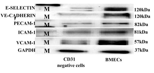
Application: WB    Species: mouse    Sample: primary bone marrow endothelial cells

Fig. 4 |Characterization of primary bone marrow endothelial cells by RT-QPCR and immunoblottinge .The immunoblotting analysis of primary bone marrow endothelial after 7 days of cultured further substantiates the purity of endothelial cells with protein ladder (M) (N= 3 indicates independent experiments for RT-qPCR and N=2 for immunoblotting with six mice/group(12 femora and 12 tibias). *p<0.05 **p<0.01, and ***p<0.001

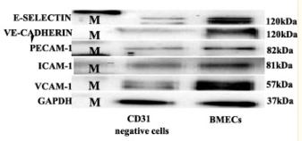
Application: WB    Species: Mice    Sample: bMECs

Fig. 4 Characterization of primary bone marrow endothelial cells by RT-QPCR and immunoblotting: a–d the relative mRNA expression of primary bone marrow endothelial cells compared to CD31 microbead-negative selected cells. The bar chart indicated the fold of the gene expression of primary BMECs compared to the CD31 microbead-negative selected cells. The fold change is quantified by the Pitfall method, followed by a Student’s test comparison between the CD31 microbead-negative selected cells and endothelial cells. P value ≤0.05 considered being statistically significant. e The immunoblotting analysis of primary bone marrow endothelial after 7 days of cultured further substantiates the purity of endothelial cells with protein ladder (M) (N= 3 indicates independent experiments for RT-qPCR and N=2 for immunoblotting with six mice/group (12 femora and 12 tibias). *p<0.05 **p<0.01, and ***p<0.001

3). NAG-1/GDF15 inhibits diabetic nephropathy via inhibiting AGE/RAGE-mediated inflammation signaling pathways in C57BL/6 mice and HK-2 cells. Life Sciences (PubMed: 36367498) [IF=6.1]

4). Galectin-3 exacerbates ox-LDL-mediated endothelial injury by inducing inflammation via integrin β1-RhoA-JNK signaling activation. JOURNAL OF CELLULAR PHYSIOLOGY (PubMed: 30536538) [IF=5.6]

Application: WB    Species: human    Sample: HUVECs cells

FIGURE 6| Gal‐3 induced expression of inflammatory factors promoted HUVECs injury via integrin β1‐RhoA‐JNK pathway. HUVECs,exposing to the ox‐LDL alone or in combination with Gal‐3 were further treated with integrin β1‐siRNA or JNK inhibitor (SP600125). The total and phosphorylated expression of p65,IKKα and IKKβ after the above treatment was examined by WB and demonstrated by histogram. (a and b).The levels of inflammatory cytokines and chemokines were then measured by ELISA. (c and d) The expression of adhesion molecules (ICAM‐1 and VCAM‐1) was detected by WB (e). *p < 0.05, **p < 0.01

5). Nepeta angustifolia attenuates responses to vascular inflammation in high glucose-induced human umbilical vein endothelial cells through heme oxygenase-1 induction. JOURNAL OF ETHNOPHARMACOLOGY (PubMed: 30419276) [IF=5.4]

6). Sanhuang Xiexin decoction ameliorates DSS-induced colitis in mice by regulating intestinal inflammation, intestinal barrier, and intestinal flora. JOURNAL OF ETHNOPHARMACOLOGY (PubMed: 35843414) [IF=5.4]

7). Salidroside ameliorates endothelial inflammation and oxidative stress by regulating the AMPK/NF-κB/NLRP3 signaling pathway in AGEs-induced HUVECs. European Journal of Pharmacology (PubMed: 31747547) [IF=5.0]

Application: WB    Species: human    Sample: HUVECs

Fig. 8. |An AMPK inhibitor abrogated the protective effect of salidroside against inflammation in AGEs-induced HUVECs.(A) Representative western blot results for AMPK/NF-κB p65/NLRP3 signaling pathway proteins in AGEs/salidroside treated-HUVECs with or without compound C.GAPDH was used as a loading control. (B) Histograms analysis of the western blot results. The data are representative of one experiment performed in triplicate and are expressed as the mean ± S.D.

8). Wenyang Huazhuo Tongluo formula alleviates pulmonary vascular injury and downregulates HIF-1α in bleomycin-induced systemic sclerosis mouse model. BMC Complementary Medicine and Therapies (PubMed: 35733188) [IF=3.9]

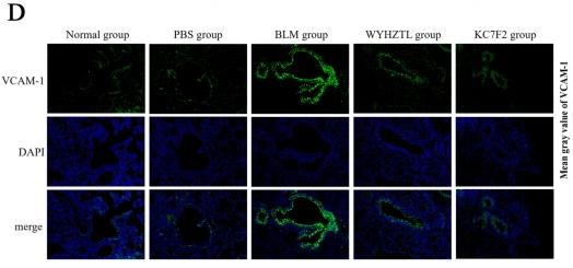
Application: IF/ICC    Species: Mouse    Sample:

Fig. 5Wenyang Huazhuo Tongluo Formula and KC7F2 can significantly inhibit the expression of vWF, SELE, ICAM-1 and VCAM-1 in bleomycin-induced SSc mouse model. The effect of Wenyang Huazhuo Tongluo Formula and KC7F2 on the expression of vWF (A), SELE (B), ICAM-1 (C) and VCAM-1 (D) was observed by immunofluorescence staining. compared with the PBS group, bleomycin can significantly upregulate the expression levels of vWF, SELE, ICAM-1 and VCAM-1 in endothelial cells, while Wenyang Huazhuo Tongluo Formula and KC7F2 can significantly reverse the bleomycin-induced upregulation of vWF, SELE, ICAM-1 and VCAM-1. The mean values ± SD was shown for each bar. * (P < 0.05) or ** (P < 0.01) or *** (P < 0.001) represents significance, ns represents no significance. Original magnification: × 20. BLM: Bleomycin, WYHZTL: Wenyang Huazhuo Tongluo formula

9). Evaluating the Effect of Circ-Sirt1 on the Expression of SIRT1 and Its Role in Pathology of Pulmonary Hypertension. CELL TRANSPLANTATION (PubMed: 35225027) [IF=3.3]

10). Therapeutic effect and mechanism of 4‑phenyl butyric acid on renal ischemia‑reperfusion injury in mice. Experimental and Therapeutic Medicine (PubMed: 35069825) [IF=2.7]

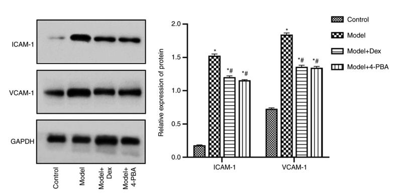
Application: WB    Species: mouse    Sample: kidneys

Figure 4. |Western blotting analysis of the expression levels of ICAM‑1 and VCAM‑1 in mouse kidneys following ischemia‑reperfusion, DEX and 4‑PBA treat‑ment. The right panel presents representative western blotting, and the left panel shows the relative expression levels. *P<0.05 vs. control; #P<0.05 vs. model.DEX, dexmedetomidine; 4‑PBA, 4‑phenylbutyric acid; ICAM‑1, intercellular adhesion molecule‑1; VCAM‑1, vascular adhesion molecule‑1.

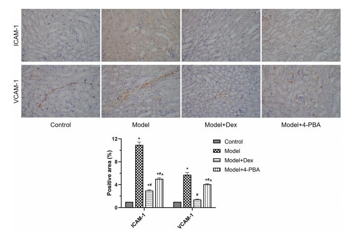
Application: IHC    Species: mouse    Sample: kidneys

Figure 5.| Immunohistochemistry assessment of the expression levels of ICAM‑1 and VCAM‑1 in mice following ischemia‑reperfusion and DEX and 4‑PBA treatment (magnification, x400). The upper panel presents immunohistochemistry results, and the lower panel shows the quantified relative expression levels.*P<0.05 vs. control; #P<0.05 vs. model; ^P<0.05 vs. model + DEX. DEX, dexmedetomidine; 4‑PBA, 4‑phenylbutyric acid; ICAM‑1, intercellular adhesion molecule‑1; VCAM‑1, vascular adhesion molecule‑1.

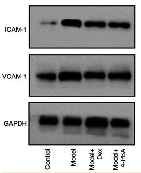
Application: WB    Species: Mouse    Sample: kidneys

Figure 4 Western blotting analysis of the expression levels of ICAM-1 and VCAM-1 in mouse kidneys following ischemia-reperfusion, DEX and 4-PBA treatment. The right panel presents representative western blotting, and the left panel shows the relative expression levels. *P<0.05 vs. control; #P<0.05 vs. model. DEX, dexmedetomidine; 4-PBA, 4-phenylbutyric acid; ICAM-1, intercellular adhesion molecule-1; VCAM-1, vascular adhesion molecule-1.

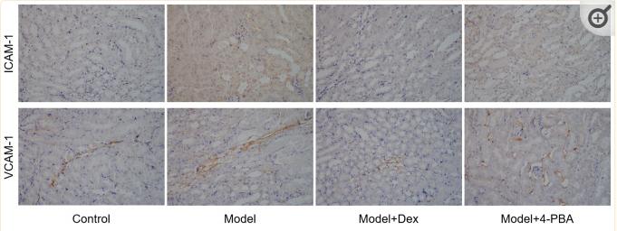
Application: IHC    Species: Mouse    Sample: kidneys

Figure 5 Immunohistochemistry assessment of the expression levels of ICAM-1 and VCAM-1 in mice following ischemia-reperfusion and DEX and 4-PBA treatment (magnification, x400). The upper panel presents immunohistochemistry results, and the lower panel shows the quantified relative expression levels. *P<0.05 vs. control; #P<0.05 vs. model; ^P<0.05 vs. model + DEX. DEX, dexmedetomidine; 4-PBA, 4-phenylbutyric acid; ICAM-1, intercellular adhesion molecule-1; VCAM-1, vascular adhesion molecule-1.

Load more

Restrictive clause


Affinity Biosciences tests all products strictly. Citations are provided as a resource for additional applications that have not been validated by Affinity Biosciences. Please choose the appropriate format for each application and consult Materials and Methods sections for additional details about the use of any product in these publications.

For Research Use Only.
Not for use in diagnostic or therapeutic procedures. Not for resale. Not for distribution without written consent. Affinity Biosciences will not be held responsible for patent infringement or other violations that may occur with the use of our products. Affinity Biosciences, Affinity Biosciences Logo and all other trademarks are the property of Affinity Biosciences LTD.