Product: Bim Antibody
Catalog: DF6093
Description: Rabbit polyclonal antibody to Bim
Application: WB
Reactivity: Human, Mouse, Rat
Prediction: Pig, Horse, Sheep, Rabbit, Dog
Mol.Wt.: 22kDa; 22kD(Calculated).
Uniprot: O43521
RRID: AB_2838061

View similar products>>

   Size Price Inventory
 100ul $280 In stock
 200ul $350 In stock

Lead Time: Same day delivery

For pricing and ordering contact:
Local distributors

Product Info

Source:
Rabbit
Application:
WB 1:500-1:2000
*The optimal dilutions should be determined by the end user.
*Tips:

WB: For western blot detection of denatured protein samples. IHC: For immunohistochemical detection of paraffin sections (IHC-p) or frozen sections (IHC-f) of tissue samples. IF/ICC: For immunofluorescence detection of cell samples. ELISA(peptide): For ELISA detection of antigenic peptide.

Reactivity:
Human,Mouse,Rat
Prediction:
Pig(100%), Horse(86%), Sheep(100%), Rabbit(100%), Dog(100%)
Clonality:
Polyclonal
Specificity:
Bim Antibody detects endogenous levels of total Bim.
RRID:
AB_2838061
Cite Format: Affinity Biosciences Cat# DF6093, RRID:AB_2838061.
Conjugate:
Unconjugated.
Purification:
The antiserum was purified by peptide affinity chromatography using SulfoLink™ Coupling Resin (Thermo Fisher Scientific).
Storage:
Rabbit IgG in phosphate buffered saline , pH 7.4, 150mM NaCl, 0.02% sodium azide and 50% glycerol. Store at -20 °C. Stable for 12 months from date of receipt.
Alias:

Fold/Unfold

BCL2 like 11; B2L11_HUMAN; BAM; Bcl 2 interacting protein Bim; Bcl 2 related ovarian death agonist; Bcl-2-like protein 11; BCL2 interacting mediator of cell death; BCL2 like 11 (apoptosis facilitator); BCL2 like protein 11; Bcl2-interacting mediator of cell death; Bcl2-L-11; Bcl2l11; BIM alpha6; BIM; BIM beta6; BIM beta7; BimEL; BimL; BOD;

Immunogens

Immunogen:
Uniprot:
Gene(ID):
Expression:
O43521 B2L11_HUMAN:

Isoform BimEL, isoform BimL and isoform BimS are the predominant isoforms and are widely expressed with tissue-specific variation. Isoform Bim-gamma is most abundantly expressed in small intestine and colon, and in lower levels in spleen, prostate, testis, heart, liver and kidney.

Description:
Bim/Bod is a pro-apoptotic protein belonging to the BH3-only group of Bcl-2 family members including Bad, Bid, Bik, Hrk and Noxa that contain a BH3 domain but lack other conserved BH1 or BH2 domains (1,2). Bim induces apoptosis by binding to and antagonizing anti-apoptotic members of the Bcl-2 family. Interactions have been observed with Bcl-2, Bcl-xL, Mcl-1, Bcl-w, Bfl-1 and BHRF-1 (1,2). Bim functions in regulating apoptosis associated with thymocyte negative selection and following growth factor withdrawal, during which Bim expression is elevated (3-6). Three major isoforms of Bim are generated by alternative splicing: BimEL, BimL and BimS (1). The shortest form, BimS, is the most cytotoxic and is generally only transiently expressed during apoptosis. The BimEL and BimL isoforms may be sequestered to the dynein motor complex through an interaction with the dynein light chain and released from this complex during apoptosis (7). Apoptotic activity of these longer isoforms may be regulated by phosphorylation (8,9). Environmental stress triggers Bim phosphorylation by JNK and results in its dissociation from the dynein complex and increased apoptotic activity.
Sequence:
MAKQPSDVSSECDREGRQLQPAERPPQLRPGAPTSLQTEPQGNPEGNHGGEGDSCPHGSPQGPLAPPASPGPFATRSPLFIFMRRSSLLSRSSSGYFSFDTDRSPAPMSCDKSTQTPSPPCQAFNHYLSAMASMRQAEPADMRPEIWIAQELRRIGDEFNAYYARRVFLNNYQAAEDHPRMVILRLLRYIVRLVWRMH

Predictions

Predictions:

Score>80(red) has high confidence and is suggested to be used for WB detection. *The prediction model is mainly based on the alignment of immunogen sequences, the results are for reference only, not as the basis of quality assurance.

Species
Results
Score
Pig
100
Sheep
100
Dog
100
Rabbit
100
Horse
86
Bovine
0
Xenopus
0
Zebrafish
0
Chicken
0
Model Confidence:
High(score>80) Medium(80>score>50) Low(score<50) No confidence

PTMs - O43521 As Substrate

Site PTM Type Enzyme
Phosphorylation
C12 S-Nitrosylation
S59 Phosphorylation
S65 Phosphorylation
S69 Phosphorylation Q16539 (MAPK14) , P45983 (MAPK8) , P45984 (MAPK9) , P53779 (MAPK10) , P28482 (MAPK1) , P27361 (MAPK3)
S77 Phosphorylation
S87 Phosphorylation P31749 (AKT1)
S93 Phosphorylation O14965 (AURKA)
S94 Phosphorylation O14965 (AURKA)
Y96 Phosphorylation
S98 Phosphorylation O14965 (AURKA)
S104 Phosphorylation P45983 (MAPK8)
S113 Phosphorylation
T116 Phosphorylation P53779 (MAPK10) , P45983 (MAPK8)
S118 Phosphorylation P45983 (MAPK8)

Research Backgrounds

Function:

Induces apoptosis and anoikis. Isoform BimL is more potent than isoform BimEL. Isoform Bim-alpha1, isoform Bim-alpha2 and isoform Bim-alpha3 induce apoptosis, although less potent than isoform BimEL, isoform BimL and isoform BimS. Isoform Bim-gamma induces apoptosis. Isoform Bim-alpha3 induces apoptosis possibly through a caspase-mediated pathway. Isoform BimAC and isoform BimABC lack the ability to induce apoptosis.

PTMs:

Phosphorylation at Ser-69 by MAPK1/MAPK3 leads to interaction with TRIM2 and polyubiquitination, followed by proteasomal degradation. Deubiquitination catalyzed by USP27X stabilizes the protein (By similarity).

Ubiquitination by TRIM2 following phosphorylation by MAPK1/MAPK3 leads to proteasomal degradation. Conversely, deubiquitination catalyzed by USP27X stabilizes the protein.

Subcellular Location:

Endomembrane system>Peripheral membrane protein.
Note: Associated with intracytoplasmic membranes.

Mitochondrion.
Note: Translocates from microtubules to mitochondria on loss of cell adherence.

Mitochondrion.

Mitochondrion.

Mitochondrion.

Extracellular region or secreted Cytosol Plasma membrane Cytoskeleton Lysosome Endosome Peroxisome ER Golgi apparatus Nucleus Mitochondrion Manual annotation Automatic computational assertionSubcellular location
Tissue Specificity:

Isoform BimEL, isoform BimL and isoform BimS are the predominant isoforms and are widely expressed with tissue-specific variation. Isoform Bim-gamma is most abundantly expressed in small intestine and colon, and in lower levels in spleen, prostate, testis, heart, liver and kidney.

Subunit Structure:

Forms heterodimers with a number of antiapoptotic Bcl-2 proteins, including MCL1, BCL2, BCL2L1 isoform Bcl-X(L), BCL2A1/BFL-1, BHRF1, and BCL2L2/BCLW. Isoform BimS and isoform Bim-alpha3 interact with BAX; this interaction may lead to BAX activation through conformational change. Does not heterodimerize with proapoptotic proteins such as BAD, BOK or BAK. Identified in a complex containing BCL2L11, DYNLL1 and BCL2L1 isoform Bcl-X(L); BH3 integrity is required for BCL2L1-binding. Interacts with YWHAZ. When phosphorylated, interacts with TRIM2; this interaction is associated with ubiquitination and degradation. Interacts with MCL1; may sequester BCL2L11 to prevent its pro-apoptotic activity. When phosphorylated, isoform BimEL interacts with USP27X; this interaction leads to BCL2L11 deubiquitination and stabilization. Interacts with GIMAP5.

Family&Domains:

The BH3 motif is required for interaction with Bcl-2 proteins and cytotoxicity.

Belongs to the Bcl-2 family.

References

1). Derlin1 functions as an oncogene in cervical cancer via AKT/mTOR signaling pathway. Biological Research (PubMed: 30808417) [IF=6.7]

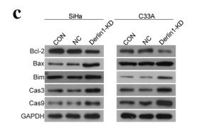
Application: WB    Species: human    Sample: CC cells

Fig.?4|Knockdown of Derlin1 induces the apoptosis of CC cells. a Apoptosis was detected by fow cytometry. b Quantitative results of apoptotic cell percentage. c The proteins expression of apoptosis-related proteins were detected by western blot. d Quantitative results of protein expression levels. N?=?3, *p?

2). Overexpression of CD59 inhibits apoptosis of T-acute lymphoblastic leukemia via AKT/Notch1 signaling pathway. Cancer Cell International (PubMed: 30636930) [IF=5.8]

Application: WB    Species: human    Sample: Jurkat cells

Fig.?4?|The activation of AKT, STAT5 and Notch1 signaling pathway are involve in the regulation of apoptosis by CD59 in Jurkat cells. The expression of CD59 (a), apoptosis-related proteins (c) and key proteins in signaling pathways (e) detected by western blot. Quantitative analysis of the protein expression of CD59 (b), apoptosis-related proteins (d) and key proteins in signaling pathways (f, g). All experiments were performed three times. *P?

3). Suppression of CHOP Reduces Neuronal Apoptosis and Rescues Cognitive Impairment Induced by Intermittent Hypoxia by Inhibiting Bax and Bak Activation. Neural Plasticity (PubMed: 34471408) [IF=3.1]

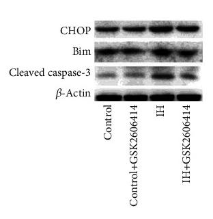
Application: WB    Species: Mouse    Sample: hippocampal tissues

Figure 3 GSK2606414 reduced the activation of caspase-3 via mitochondria-dependent apoptosis. (a) Representative western blots showed that GSK2606414 reduced the expression of CHOP, Bim, and cleaved caspase-3 in hippocampal tissue. The pooled data from three mice for each group are summarized in (b). (c) The western blot results showed that the expression of Bax and Bak in the mitochondria was obviously increased and that this effect was accompanied by a reduction in cytochrome c. These effects were prevented by the administration of GSK2606414. (d) Statistical data from three animals for each group are summarized. (e) Representative western blots showing that the cytoplasmic expression of Bax and Bak was obviously reduced and that of cytochrome C was significantly increased. GSK2606414 increased the expression of Bax and Bak and reduced cytochrome C. (f) Statistical data from three animals for each group are summarized. (g) The morphology of mitochondria in the hippocampal CA1 region in the (i) control group, (ii) GSK2606414 group, (iii) IH group, and (iv) IH + GSK2606414 group. In the control and GSK2606414 groups, intact mitochondria (indicated by arrowheads) with clear cristae were found. Much fewer cristae were found in the mitochondria from the IH group. GSK2606414 treatment preserved more intact mitochondria with discernable cristae. Scale bar: 0.25 μm. (h) The mitochondrial membrane potential (JC-1 fluorescence intensity ratio) was impaired by IH treatment and rescued by GSK2606414 treatment. ∗P < 0.05; ∗∗P < 0.01; ns: not significant.

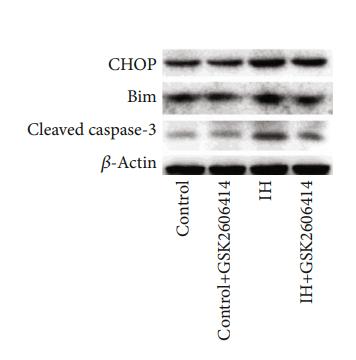
Application: WB    Species: mouse    Sample: hippocampal

Figure 3:| GSK2606414 reduced the activation of caspase-3 via mitochondria-dependent apoptosis. (a) Representative western blots showed that GSK2606414 reduced the expression of CHOP, Bim, and cleaved caspase-3 in hippocampal tissue.

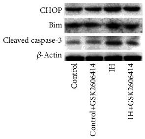
Application: WB    Species: Mice    Sample: hippocampal tissue

Figure 3 GSK2606414 reduced the activation of caspase-3 via mitochondria-dependent apoptosis. (a) Representative western blots showed that GSK2606414 reduced the expression of CHOP, Bim, and cleaved caspase-3 in hippocampal tissue. The pooled data from three mice for each group are summarized in (b). (c) The western blot results showed that the expression of Bax and Bak in the mitochondria was obviously increased and that this effect was accompanied by a reduction in cytochrome c. These effects were prevented by the administration of GSK2606414. (d) Statistical data from three animals for each group are summarized. (e) Representative western blots showing that the cytoplasmic expression of Bax and Bak was obviously reduced and that of cytochrome C was significantly increased. GSK2606414 increased the expression of Bax and Bak and reduced cytochrome C. (f) Statistical data from three animals for each group are summarized. (g) The morphology of mitochondria in the hippocampal CA1 region in the (i) control group, (ii) GSK2606414 group, (iii) IH group, and (iv) IH + GSK2606414 group. In the control and GSK2606414 groups, intact mitochondria (indicated by arrowheads) with clear cristae were found. Much fewer cristae were found in the mitochondria from the IH group. GSK2606414 treatment preserved more intact mitochondria with discernable cristae. Scale bar: 0.25 μm. (h) The mitochondrial membrane potential (JC-1 fluorescence intensity ratio) was impaired by IH treatment and rescued by GSK2606414 treatment. ∗P < 0.05; ∗∗P < 0.01; ns: not significant.

4). Quxie Capsule Inhibits Colon Tumor Growth Partially Through Foxo1-Mediated Apoptosis and Immune Modulation. INTEGRATIVE CANCER THERAPIES (PubMed: 31030593) [IF=2.9]

Application: WB    Species: mouse    Sample: tumor

Figure 2.| Quxie capsule (QX) inhibited cell proliferation and induced apoptosis in tumor tissues. (A) Ki-67 staining of tumor sections obtained from mice treated with (a) vehicle control or (b) QX. Quantification of Ki-67-positive cells in the tumor sections (c). (B)TUNEL staining of tumor sections obtained from mice treated with (a) vehicle control or (b) QX. Quantification of TUNEL-positive cells in the tumor sections (c). (C) Western blotting of proapoptotic proteins Bim, FasL, and cleaved caspase-3 expression in tumor tissues of QX-treated mice or vehicle control-treated mice. Data are presented as mean ± SD. *P < .05, **P < .01 versus vehicle control.

Restrictive clause


Affinity Biosciences tests all products strictly. Citations are provided as a resource for additional applications that have not been validated by Affinity Biosciences. Please choose the appropriate format for each application and consult Materials and Methods sections for additional details about the use of any product in these publications.

For Research Use Only.
Not for use in diagnostic or therapeutic procedures. Not for resale. Not for distribution without written consent. Affinity Biosciences will not be held responsible for patent infringement or other violations that may occur with the use of our products. Affinity Biosciences, Affinity Biosciences Logo and all other trademarks are the property of Affinity Biosciences LTD.