NeuN Antibody - #DF6145

| Product: | NeuN Antibody |
| Catalog: | DF6145 |
| Description: | Rabbit polyclonal antibody to NeuN |
| Application: | WB IHC IF/ICC |
| Reactivity: | Human, Mouse, Rat |
| Prediction: | Pig, Bovine, Horse, Sheep, Rabbit, Dog, Chicken |
| Mol.Wt.: | 34kDa; 34kD(Calculated). |
| Uniprot: | A6NFN3 |
| RRID: | AB_2838112 |
Related Downloads
Protocols
Product Info
*The optimal dilutions should be determined by the end user.
*Tips:
WB: For western blot detection of denatured protein samples. IHC: For immunohistochemical detection of paraffin sections (IHC-p) or frozen sections (IHC-f) of tissue samples. IF/ICC: For immunofluorescence detection of cell samples. ELISA(peptide): For ELISA detection of antigenic peptide.
Cite Format: Affinity Biosciences Cat# DF6145, RRID:AB_2838112.
Fold/Unfold
FLJ56884; FLJ58356; Fox-1 homolog C; fox1 homolog C; Fox3; FOX3NeuN; hexaribonucleotide binding protein 3; HRNBP3; NEUN; neuronal nuclei; Rbfox3; RFOX3_HUMAN; RNA binding protein fox-1 homolog 3; RNA binding protein, fox 1 homolog (C. elegans) 3;
Immunogens
- A6NFN3 RFOX3_HUMAN:
- Protein BLAST With
- NCBI/
- ExPASy/
- Uniprot
MAQPYPPAQYPPPPQNGIPAEYAPPPPHPTQDYSGQTPVPTEHGMTLYTPAQTHPEQPGSEASTQPIAGTQTVPQTDEAAQTDSQPLHPSDPTEKQQPKRLHVSNIPFRFRDPDLRQMFGQFGKILDVEIIFNERGSKGFGFVTFETSSDADRAREKLNGTIVEGRKIEVNNATARVMTNKKTGNPYTNGWKLNPVVGAVYGPEFYAVTGFPYPTTGTAVAYRGAHLRGRGRAVYNTFRAAPPPPPIPTYGAVVYQDGFYGAEIYGGYAAYRYAQPAAAAAAYSDSYGRVYAAADPYHHTIGPAATYSIGTM
Predictions
Score>80(red) has high confidence and is suggested to be used for WB detection. *The prediction model is mainly based on the alignment of immunogen sequences, the results are for reference only, not as the basis of quality assurance.
High(score>80) Medium(80>score>50) Low(score<50) No confidence
PTMs - A6NFN3 As Substrate
| Site | PTM Type | Enzyme | Source |
|---|---|---|---|
| S63 | Phosphorylation | Uniprot | |
| R116 | Methylation | Uniprot | |
| K167 | Ubiquitination | Uniprot | |
| Y187 | Phosphorylation | Uniprot | |
| T188 | Phosphorylation | Uniprot |
Research Backgrounds
Pre-mRNA alternative splicing regulator. Regulates alternative splicing of RBFOX2 to enhance the production of mRNA species that are targeted for nonsense-mediated decay (NMD).
Nucleus. Cytoplasm.
Note: Largely restricted to neuronal nuclei. However, significant cytoplasmic localization in neurons from brains from HIV-infected individuals with cognitive impairment.
References

Application: IF/ICC Species: rat Sample: dorsal column
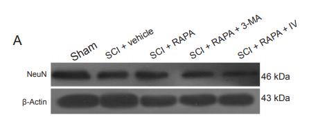
Application: WB Species: rat Sample: dorsal column

Application: IF/ICC Species: Rat Sample: PC12 cells
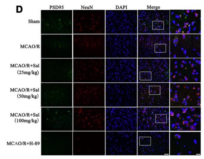
Application: IF/ICC Species: rat Sample:
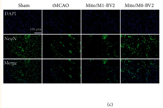
Application: IF/ICC Species: Rat Sample:
Restrictive clause
Affinity Biosciences tests all products strictly. Citations are provided as a resource for additional applications that have not been validated by Affinity Biosciences. Please choose the appropriate format for each application and consult Materials and Methods sections for additional details about the use of any product in these publications.
For Research Use Only.
Not for use in diagnostic or therapeutic procedures. Not for resale. Not for distribution without written consent. Affinity Biosciences will not be held responsible for patent infringement or other violations that may occur with the use of our products. Affinity Biosciences, Affinity Biosciences Logo and all other trademarks are the property of Affinity Biosciences LTD.