MyD88 Antibody - #DF6162
, blocked with antigen-specific peptides.
Lane 2: Hepg2 cells(serum starvation treatment).
Lane 3: Hela cells(heat-shock treatment).")
Related Downloads
Protocols
Product Info
*The optimal dilutions should be determined by the end user.
*Tips:
WB: For western blot detection of denatured protein samples. IHC: For immunohistochemical detection of paraffin sections (IHC-p) or frozen sections (IHC-f) of tissue samples. IF/ICC: For immunofluorescence detection of cell samples. ELISA(peptide): For ELISA detection of antigenic peptide.
Cite Format: Affinity Biosciences Cat# DF6162, RRID:AB_2838129.
Fold/Unfold
Mutant myeloid differentiation primary response 88; MYD 88; Myd88; MYD88_HUMAN; MYD88D; Myeloid differentiation marker 88; Myeloid differentiation primary response 88; Myeloid differentiation primary response gene (88); Myeloid differentiation primary response gene 88; Myeloid differentiation primary response gene; Myeloid differentiation primary response protein MyD88; OTTHUMP00000161718; OTTHUMP00000208595; OTTHUMP00000209058; OTTHUMP00000209059; OTTHUMP00000209060;
Immunogens
- Q99836 MYD88_HUMAN:
- Protein BLAST With
- NCBI/
- ExPASy/
- Uniprot
MAAGGPGAGSAAPVSSTSSLPLAALNMRVRRRLSLFLNVRTQVAADWTALAEEMDFEYLEIRQLETQADPTGRLLDAWQGRPGASVGRLLELLTKLGRDDVLLELGPSIEEDCQKYILKQQQEEAEKPLQVAAVDSSVPRTAELAGITTLDDPLGHMPERFDAFICYCPSDIQFVQEMIRQLEQTNYRLKLCVSDRDVLPGTCVWSIASELIEKRCRRMVVVVSDDYLQSKECDFQTKFALSLSPGAHQKRLIPIKYKAMKKEFPSILRFITVCDYTNPCTKSWFWTRLAKALSLP
Predictions
Score>80(red) has high confidence and is suggested to be used for WB detection. *The prediction model is mainly based on the alignment of immunogen sequences, the results are for reference only, not as the basis of quality assurance.
High(score>80) Medium(80>score>50) Low(score<50) No confidence
PTMs - Q99836 As Substrate
| Site | PTM Type | Enzyme | Source |
|---|---|---|---|
| K95 | Ubiquitination | Uniprot | |
| C113 | S-Nitrosylation | Uniprot | |
| K115 | Ubiquitination | Uniprot | |
| K119 | Ubiquitination | Uniprot | |
| K127 | Ubiquitination | Uniprot | |
| K190 | Ubiquitination | Uniprot | |
| K214 | Ubiquitination | Uniprot | |
| C216 | S-Nitrosylation | Uniprot | |
| K231 | Ubiquitination | Uniprot | |
| K238 | Ubiquitination | Uniprot | |
| S244 | Phosphorylation | Uniprot | |
| K250 | Ubiquitination | Uniprot | |
| K256 | Ubiquitination | Uniprot | |
| Y257 | Phosphorylation | Uniprot | |
| K262 | Ubiquitination | Uniprot | |
| Y276 | Phosphorylation | Uniprot | |
| K282 | Ubiquitination | Uniprot | |
| K291 | Ubiquitination | Uniprot |
Research Backgrounds
Adapter protein involved in the Toll-like receptor and IL-1 receptor signaling pathway in the innate immune response. Acts via IRAK1, IRAK2, IRF7 and TRAF6, leading to NF-kappa-B activation, cytokine secretion and the inflammatory response. Increases IL-8 transcription. Involved in IL-18-mediated signaling pathway. Activates IRF1 resulting in its rapid migration into the nucleus to mediate an efficient induction of IFN-beta, NOS2/INOS, and IL12A genes. MyD88-mediated signaling in intestinal epithelial cells is crucial for maintenance of gut homeostasis and controls the expression of the antimicrobial lectin REG3G in the small intestine (By similarity).
Ubiquitinated; undergoes 'Lys-63'-linked polyubiquitination. OTUD4 specifically hydrolyzes 'Lys-63'-linked polyubiquitinated MYD88.
Cytoplasm. Nucleus.
Ubiquitous.
Homodimer. Also forms heterodimers with TIRAP. Binds to TLR2, TLR4, TLR5, IRAK1, IRAK2 and IRAK4 via their respective TIR domains. Interacts with IL18R1. Interacts with BMX, IL1RL1, IKBKE and IRF7. Interacts with LRRFIP1 and LRRFIP2; this interaction positively regulates Toll-like receptor (TLR) signaling in response to agonist. Interacts with FLII. LRRFIP1 and LRRFIP2 compete with FLII for MYD88-binding. Interacts with IRF1. Upon IL1B treatment, forms a complex with PELI1, IRAK1, IRAK4 and TRAF6; this complex recruits MAP3K7/TAK1, TAB1 and TAB2 to mediate NF-kappa-B activation. Direct binding of SMAD6 to PELI1 prevents the complex formation and hence negatively regulates IL1R-TLR signaling and eventually NF-kappa-B-mediated gene expression. May interact with PIK3AP1. Interacts (via TIR domain) with DHX9 (via H2A and OB-fold regions); this interaction is direct. Interacts with OTUD4 deubiquitinase; the interaction is direct.
(Microbial infection) In case of infection, interacts with uropathogenic E.coli protein TcpC; suppressing Toll-like receptor (TLR)-mediated cytokine production.
(Microbial infection) In case of infection, interacts with uropathogenic E.faecalis protein TcpF; suppressing Toll-like receptor (TLR)-mediated cytokine production.
The intermediate domain (ID) is required for the phosphorylation and activation of IRAK.
Research Fields
· Environmental Information Processing > Signal transduction > MAPK signaling pathway. (View pathway)
· Environmental Information Processing > Signal transduction > NF-kappa B signaling pathway. (View pathway)
· Human Diseases > Infectious diseases: Bacterial > Salmonella infection.
· Human Diseases > Infectious diseases: Bacterial > Pertussis.
· Human Diseases > Infectious diseases: Bacterial > Legionellosis.
· Human Diseases > Infectious diseases: Parasitic > Leishmaniasis.
· Human Diseases > Infectious diseases: Parasitic > Chagas disease (American trypanosomiasis).
· Human Diseases > Infectious diseases: Parasitic > African trypanosomiasis.
· Human Diseases > Infectious diseases: Parasitic > Malaria.
· Human Diseases > Infectious diseases: Parasitic > Toxoplasmosis.
· Human Diseases > Infectious diseases: Bacterial > Tuberculosis.
· Human Diseases > Infectious diseases: Viral > Hepatitis B.
· Human Diseases > Infectious diseases: Viral > Measles.
· Human Diseases > Infectious diseases: Viral > Influenza A.
· Human Diseases > Infectious diseases: Viral > Herpes simplex infection.
· Organismal Systems > Immune system > Toll-like receptor signaling pathway. (View pathway)
· Organismal Systems > Immune system > NOD-like receptor signaling pathway. (View pathway)
References
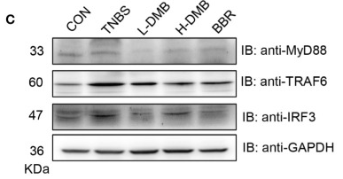
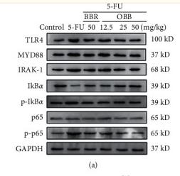
Application: WB Species: Mouse Sample: colonic tissue
Application: WB Species: mouse Sample: macrophages
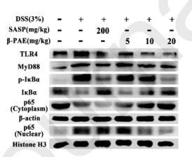
Application: WB Species: mouse Sample: colon

Application: WB Species: mouse Sample: Colons
Application: WB Species: Mice Sample:
Application: WB Species: Rat Sample: colonic tissue
Restrictive clause
Affinity Biosciences tests all products strictly. Citations are provided as a resource for additional applications that have not been validated by Affinity Biosciences. Please choose the appropriate format for each application and consult Materials and Methods sections for additional details about the use of any product in these publications.
For Research Use Only.
Not for use in diagnostic or therapeutic procedures. Not for resale. Not for distribution without written consent. Affinity Biosciences will not be held responsible for patent infringement or other violations that may occur with the use of our products. Affinity Biosciences, Affinity Biosciences Logo and all other trademarks are the property of Affinity Biosciences LTD.