TICAM1 Antibody - #DF6289

Related Downloads
Protocols
Product Info
*The optimal dilutions should be determined by the end user.
*Tips:
WB: For western blot detection of denatured protein samples. IHC: For immunohistochemical detection of paraffin sections (IHC-p) or frozen sections (IHC-f) of tissue samples. IF/ICC: For immunofluorescence detection of cell samples. ELISA(peptide): For ELISA detection of antigenic peptide.
Cite Format: Affinity Biosciences Cat# DF6289, RRID:AB_2838255.
Fold/Unfold
IIAE6; MGC35334; MyD88 3; Proline rich vinculin and TIR domain containing protein B; Proline-rich; PRVTIRB; Putative NF kappa B activating protein 502H; Putative NF-kappa-B-activating protein 502H; Putative NFkB activating protein; TCAM1_HUMAN; TICAM 1; TICAM-1; Ticam1; TIR domain containing adapter molecule 1; TIR domain containing adapter protein inducing IFN beta; TIR domain containing adaptor inducing interferon beta; TIR domain-containing adapter molecule 1; TIR domain-containing adapter protein inducing IFN-beta; Toll interleukin 1 receptor domain containing adapter protein inducing interferon beta; Toll like receptor adaptor molecule 1; Toll-interleukin-1 receptor domain-containing adapter protein inducing interferon beta; TRIF; TRIF protein; vinculin and TIR domain-containing protein B;
Immunogens
- Q8IUC6 TCAM1_HUMAN:
- Protein BLAST With
- NCBI/
- ExPASy/
- Uniprot
MACTGPSLPSAFDILGAAGQDKLLYLKHKLKTPRPGCQGQDLLHAMVLLKLGQETEARISLEALKADAVARLVARQWAGVDSTEDPEEPPDVSWAVARLYHLLAEEKLCPASLRDVAYQEAVRTLSSRDDHRLGELQDEARNRCGWDIAGDPGSIRTLQSNLGCLPPSSALPSGTRSLPRPIDGVSDWSQGCSLRSTGSPASLASNLEISQSPTMPFLSLHRSPHGPSKLCDDPQASLVPEPVPGGCQEPEEMSWPPSGEIASPPELPSSPPPGLPEVAPDATSTGLPDTPAAPETSTNYPVECTEGSAGPQSLPLPILEPVKNPCSVKDQTPLQLSVEDTTSPNTKPCPPTPTTPETSPPPPPPPPSSTPCSAHLTPSSLFPSSLESSSEQKFYNFVILHARADEHIALRVREKLEALGVPDGATFCEDFQVPGRGELSCLQDAIDHSAFIILLLTSNFDCRLSLHQVNQAMMSNLTRQGSPDCVIPFLPLESSPAQLSSDTASLLSGLVRLDEHSQIFARKVANTFKPHRLQARKAMWRKEQDTRALREQSQHLDGERMQAAALNAAYSAYLQSYLSYQAQMEQLQVAFGSHMSFGTGAPYGARMPFGGQVPLGAPPPFPTWPGCPQPPPLHAWQAGTPPPPSPQPAAFPQSLPFPQSPAFPTASPAPPQSPGLQPLIIHHAQMVQLGLNNHMWNQRGSQAPEDKTQEAE
Predictions
Score>80(red) has high confidence and is suggested to be used for WB detection. *The prediction model is mainly based on the alignment of immunogen sequences, the results are for reference only, not as the basis of quality assurance.
High(score>80) Medium(80>score>50) Low(score<50) No confidence
PTMs - Q8IUC6 As Substrate
| Site | PTM Type | Enzyme | Source |
|---|---|---|---|
| K65 | Ubiquitination | Uniprot | |
| S196 | Phosphorylation | Uniprot | |
| S199 | Phosphorylation | Uniprot | |
| S205 | Phosphorylation | Uniprot | |
| S210 | Phosphorylation | Uniprot | |
| S212 | Phosphorylation | Uniprot | |
| S223 | Phosphorylation | Uniprot | |
| S388 | Phosphorylation | Uniprot | |
| S389 | Phosphorylation | Uniprot | |
| S390 | Phosphorylation | Uniprot | |
| S500 | Phosphorylation | Uniprot | |
| Y570 | Phosphorylation | Uniprot | |
| Y573 | Phosphorylation | Uniprot | |
| Y577 | Phosphorylation | Uniprot | |
| K707 | Ubiquitination | Uniprot |
Research Backgrounds
Involved in innate immunity against invading pathogens. Adapter used by TLR3, TLR4 (through TICAM2) and TLR5 to mediate NF-kappa-B and interferon-regulatory factor (IRF) activation, and to induce apoptosis. Ligand binding to these receptors results in TRIF recruitment through its TIR domain. Distinct protein-interaction motifs allow recruitment of the effector proteins TBK1, TRAF6 and RIPK1, which in turn, lead to the activation of transcription factors IRF3 and IRF7, NF-kappa-B and FADD respectively. Phosphorylation by TBK1 on the pLxIS motif leads to recruitment and subsequent activation of the transcription factor IRF3 to induce expression of type I interferon and exert a potent immunity against invading pathogens. Component of a multi-helicase-TICAM1 complex that acts as a cytoplasmic sensor of viral double-stranded RNA (dsRNA) and plays a role in the activation of a cascade of antiviral responses including the induction of proinflammatory cytokines (By similarity).
Phosphorylated by TBK1. Following activation, phosphorylated by TBK1 at Ser-210 in the pLxIS motif. The phosphorylated pLxIS motif constitutes an IRF3-binding motif, leading to recruitment of the transcription factor IRF3 to induce type-I interferons and other cytokines.
Polyubiquitinated by TRIM38 with 'Lys-48'-linked chains, leading to proteasomal degradation.
(Microbial infection) Cleaved and degraded by hepatitis A virus (HAV) protein 3CD allowing the virus to disrupt host TLR3 signaling.
(Microbial infection) Cleaved by CVB3 protease 3C allowing the virus to disrupt host TLR3 signaling.
(Microbial infection) Cleaved by Seneca Valley virus protease 3C allowing the virus to disrupt host TLR3 signaling.
(Microbial infection) Cleaved by protease 3C of human enterovirus D68 (EV68) allowing the virus to disrupt host TLR3 signaling.
Cytoplasmic vesicle>Autophagosome. Cytoplasm>Cytosol. Mitochondrion.
Note: Colocalizes with UBQLN1 in the autophagosome (PubMed:21695056). Colocalizes in the cytosol with DDX1, DDX21 and DHX36. Colocalizes in the mitochondria with DDX1 and poly(I:C) RNA ligand. The multi-helicase-TICAM1 complex may translocate to the mitochondria upon poly(I:C) RNA ligand stimulation (By similarity).
Ubiquitously expressed but with higher levels in liver.
Homodimer. Found in a multi-helicase-TICAM1 complex at least composed of DHX36, DDX1, DDX21 and TICAM1; this complex exists in resting cells with or without poly(I:C) RNA ligand stimulation. Interacts (via TIR domain) with DDX21 (via C-terminus). Interacts (via TIR domain) with DHX36 (via C-terminus) (By similarity). Interacts with AZI2 and IRF7. Interacts with TICAM2 in TLR4 recruitment. Interaction with PIAS4 inhibits the TICAM1-induced NF-kappa-B, IRF and IFNB1 activation. Interacts with IKBKB and IKBKE. Interaction with SARM1 blocks TICAM1-dependent transcription factor activation. Interacts with TRAF3 (By similarity). Interacts (when phosphorylated) with IRF3; following activation and phosphorylation on the pLxIS motif by TBK1, recruits IRF3. Interacts with TBK1, TRAF6 and RIPK1 and these interactions are enhanced in the presence of WDFY1. Interacts with TRAFD1 (By similarity). Interacts with UBQLN1 (via UBA domain). Interacts with TLR4 in response to LPS in a WDFY1-dependent manner (By similarity). Interacts with WDFY1 in response to poly(I:C) (By similarity). Interacts (via the TIR domain) with TLR3 in response to poly(I:C) and this interaction is enhanced in the presence of WDFY1. Interacts with TRIM56. Component of a multi-helicase-TICAM1 complex that acts as a cytoplasmic sensor of viral double-stranded RNA (dsRNA) and plays a role in the activation of a cascade of antiviral responses including the induction of proinflammatory cytokines (By similarity). Interacts (via the TIR domain) with TLR5.
(Microbial infection) Interacts with Seneca Valley virus protease 3C; this interaction allows the cleavage of TICAM1/TRIF and subsequent suppression of host innate immunity.
(Microbial infection) Interacts (via C-terminus) with coxsackievirus B3 (CVB3) protease 3C.
The pLxIS motif constitutes an IRF3-binding motif: following phosphorylation by TBK1, the phosphorylated pLxIS motif of TICAM1 recruits IRF3 (PubMed:25636800). IRF3 is then phosphorylated and activated by TBK1 to induce type-I interferons and other cytokines (PubMed:25636800).
The N-terminal region is essential for activation of the IFNB promoter activity.
The N-terminal domain (TRIF-NTD) is globular and consists of two alpha-helical subdomains connected by a 14-residue linker. It shares structural similarity with IFIT family members N-terminal regions.
Research Fields
· Cellular Processes > Cell growth and death > Necroptosis. (View pathway)
· Environmental Information Processing > Signal transduction > NF-kappa B signaling pathway. (View pathway)
· Human Diseases > Infectious diseases: Bacterial > Pertussis.
· Human Diseases > Infectious diseases: Parasitic > Chagas disease (American trypanosomiasis).
· Human Diseases > Infectious diseases: Viral > Hepatitis C.
· Human Diseases > Infectious diseases: Viral > Hepatitis B.
· Human Diseases > Infectious diseases: Viral > Influenza A.
· Human Diseases > Infectious diseases: Viral > Human papillomavirus infection.
· Human Diseases > Infectious diseases: Viral > Herpes simplex infection.
· Organismal Systems > Immune system > Toll-like receptor signaling pathway. (View pathway)
· Organismal Systems > Immune system > NOD-like receptor signaling pathway. (View pathway)
References

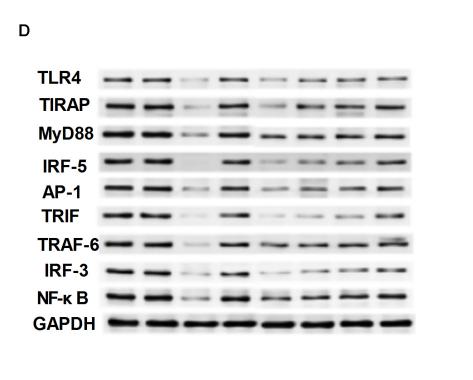
Application: WB Species: Mouse Sample: liver
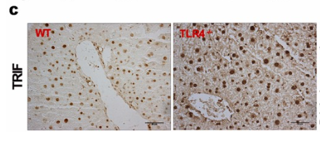
Application: IHC Species: Mouse Sample: liver
Application: WB Species: mouse Sample: RAW264.7cell
Application: WB Species: Mice Sample: cerebral cortex tissues

Application: WB Species: mouse Sample: BV-2 cells
Application: WB Species: Mice Sample: brain tissues
Restrictive clause
Affinity Biosciences tests all products strictly. Citations are provided as a resource for additional applications that have not been validated by Affinity Biosciences. Please choose the appropriate format for each application and consult Materials and Methods sections for additional details about the use of any product in these publications.
For Research Use Only.
Not for use in diagnostic or therapeutic procedures. Not for resale. Not for distribution without written consent. Affinity Biosciences will not be held responsible for patent infringement or other violations that may occur with the use of our products. Affinity Biosciences, Affinity Biosciences Logo and all other trademarks are the property of Affinity Biosciences LTD.