Product: PD-L1 Antibody
Catalog: DF6526
Description: Rabbit polyclonal antibody to PD-L1
Application: WB
Reactivity: Human, Mouse, Rat
Prediction: Bovine, Horse, Sheep, Rabbit, Dog
Mol.Wt.: 33kDa, 40~70kD(Glycosylated); 33kD(Calculated).
Uniprot: Q9NZQ7
RRID: AB_2838488

View similar products>>

   Size Price Inventory
 100ul $280 In stock
 200ul $350 In stock

Lead Time: Same day delivery

For pricing and ordering contact:
Local distributors

Product Info

Source:
Rabbit
Application:
WB 1:500-1:2000
*The optimal dilutions should be determined by the end user.
*Tips:

WB: For western blot detection of denatured protein samples. IHC: For immunohistochemical detection of paraffin sections (IHC-p) or frozen sections (IHC-f) of tissue samples. IF/ICC: For immunofluorescence detection of cell samples. ELISA(peptide): For ELISA detection of antigenic peptide.

Reactivity:
Human,Mouse,Rat
Prediction:
Bovine(100%), Horse(100%), Sheep(100%), Rabbit(100%), Dog(92%)
Clonality:
Polyclonal
Specificity:
CD274 Antibody detects endogenous levels of total CD274.
RRID:
AB_2838488
Cite Format: Affinity Biosciences Cat# DF6526, RRID:AB_2838488.
Conjugate:
Unconjugated.
Purification:
The antiserum was purified by peptide affinity chromatography using SulfoLink™ Coupling Resin (Thermo Fisher Scientific).
Storage:
Rabbit IgG in phosphate buffered saline , pH 7.4, 150mM NaCl, 0.02% sodium azide and 50% glycerol. Store at -20 °C. Stable for 12 months from date of receipt.
Alias:

Fold/Unfold

B7 H; B7 H1; B7 homolog 1; B7-H1; B7H; B7H1; CD 274; CD274; CD274 antigen; CD274 molecule; MGC142294; MGC142296; OTTHUMP00000021029; PD L1; PD-L1; PD1L1_HUMAN; PDCD1 ligand 1; PDCD1L1; PDCD1LG1; PDL 1; PDL1; Programmed cell death 1 ligand 1; Programmed death ligand 1; RGD1566211;

Immunogens

Immunogen:
Uniprot:
Gene(ID):
Expression:
Q9NZQ7 PD1L1_HUMAN:

Highly expressed in the heart, skeletal muscle, placenta and lung. Weakly expressed in the thymus, spleen, kidney and liver. Expressed on activated T- and B-cells, dendritic cells, keratinocytes and monocytes.

Description:
Programmed cell death ligand 1(CD274, or B7-H1, PD-L1), is the first member of B7 family to be discovered. B7 family molecules are type I transmembrane proteins belonging to the immunoglobulin superfamily. In concert with their CD28 family receptors, the B7s are key regulators of the adaptive immune response. CD274 is suggested a negative regulator of T and B cell, and play important role in mediating tolerance of lymphocytes to self-antigens. It also involved in the costimulatory signal, essential for T-cell proliferation and production of IL10 and IFNG, in an IL2-dependent and a PDCD1-independent manner.
Sequence:
MRIFAVFIFMTYWHLLNAFTVTVPKDLYVVEYGSNMTIECKFPVEKQLDLAALIVYWEMEDKNIIQFVHGEEDLKVQHSSYRQRARLLKDQLSLGNAALQITDVKLQDAGVYRCMISYGGADYKRITVKVNAPYNKINQRILVVDPVTSEHELTCQAEGYPKAEVIWTSSDHQVLSGKTTTTNSKREEKLFNVTSTLRINTTTNEIFYCTFRRLDPEENHTAELVIPELPLAHPPNERTHLVILGAILLCLGVALTFIFRLRKGRMMDVKKCGIQDTNSKKQSDTHLEET

Predictions

Predictions:

Score>80(red) has high confidence and is suggested to be used for WB detection. *The prediction model is mainly based on the alignment of immunogen sequences, the results are for reference only, not as the basis of quality assurance.

Species
Results
Score
Horse
100
Bovine
100
Sheep
100
Rabbit
100
Dog
92
Pig
0
Xenopus
0
Zebrafish
0
Chicken
0
Model Confidence:
High(score>80) Medium(80>score>50) Low(score<50) No confidence

PTMs - Q9NZQ7 As Substrate

Site PTM Type Enzyme
N192 N-Glycosylation
S195 Phosphorylation
K270 Acetylation
S283 Phosphorylation
T290 Phosphorylation

Research Backgrounds

Function:

Plays a critical role in induction and maintenance of immune tolerance to self. As a ligand for the inhibitory receptor PDCD1/PD-1, modulates the activation threshold of T-cells and limits T-cell effector response. Through a yet unknown activating receptor, may costimulate T-cell subsets that predominantly produce interleukin-10 (IL10).

The PDCD1-mediated inhibitory pathway is exploited by tumors to attenuate anti-tumor immunity and escape destruction by the immune system, thereby facilitating tumor survival. The interaction with PDCD1/PD-1 inhibits cytotoxic T lymphocytes (CTLs) effector function (By similarity). The blockage of the PDCD1-mediated pathway results in the reversal of the exhausted T-cell phenotype and the normalization of the anti-tumor response, providing a rationale for cancer immunotherapy (By similarity).

PTMs:

Ubiquitinated; STUB1 likely mediates polyubiquitination of PD-L1/CD274 triggering its degradation.

Subcellular Location:

Cell membrane>Single-pass type I membrane protein. Early endosome membrane>Single-pass type I membrane protein. Recycling endosome membrane>Single-pass type I membrane protein.
Note: Associates with CMTM6 at recycling endosomes, where it is protected from being targeted for lysosomal degradation.

Cell membrane>Single-pass type I membrane protein.

Endomembrane system>Single-pass type I membrane protein.

Extracellular region or secreted Cytosol Plasma membrane Cytoskeleton Lysosome Endosome Peroxisome ER Golgi apparatus Nucleus Mitochondrion Manual annotation Automatic computational assertionSubcellular location
Tissue Specificity:

Highly expressed in the heart, skeletal muscle, placenta and lung. Weakly expressed in the thymus, spleen, kidney and liver. Expressed on activated T- and B-cells, dendritic cells, keratinocytes and monocytes.

Subunit Structure:

Interacts with PDCD1. Interacts (via transmembrane domain) with CMTM4 and CMTM6.

Family&Domains:

Belongs to the immunoglobulin superfamily. BTN/MOG family.

Research Fields

· Environmental Information Processing > Signaling molecules and interaction > Cell adhesion molecules (CAMs).   (View pathway)

References

1). LncRNA MIAT correlates with immune infiltrates and drug reactions in hepatocellular carcinoma. International Immunopharmacology (PubMed: 33221703) [IF=5.6]

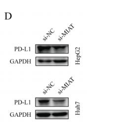
Application: WB    Species: Human    Sample: hepatocellular carcinoma tissue

Fig 7. The relationship between MIAT and PD-L1 and sorafenib resistance was verified in vitro. (A) There was a positive correlation between MIAT and PD-L1 expression in HCC tissue samples. (B) The expression of MIAT was positively correlated with CTLA4 in HCC tissue samples. (C) In HCC cells, the expression of PD-L1 mRNA decreased after the knockdown of MIAT. (D) Knockdown of MIAT down regulated the expression of PD-L1 protein in HCC cells. (E) The level of MIAT increased after sorafenib treatment. (F) The level of PD-L1 increased after sorafenib treatment. (G) MIAT affects the proliferation of HCC cells. *: P < 0.05; **: P < 0.01; ***: P < 0.001; ****: P < 0.0001.

2). miR‑576‑3p overexpression enhances cisplatin sensitivity of ovarian cancer cells by dysregulating PD‑L1 and cyclin D1. Molecular Medicine Reports (PubMed: 33236151) [IF=3.4]

3). Homeobox D9 drives the malignant phenotypes and enhances the Programmed death ligand-1 expression in non-small cell lung cancer cells via binding to Angiopoietin-2 promoter. World Journal of Surgical Oncology (PubMed: 36907878) [IF=3.2]

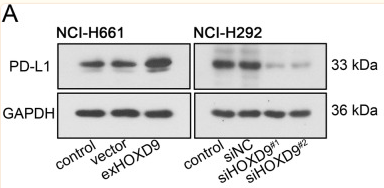
Application: WB    Species: Human    Sample: NSCLC cells

Fig. 7 HOXD9 has the ability to stimulate PD-L1 expression in NSCLC cells. NCI-H661 cells were transfected with exHOXD9 or empty vector, while NCI-H292 cells were transfected with siHOXD9#1, siHOXD9#2 or their negative control siNC. A After 48 h of transfection, total PD-L1 protein expression in two NSCLC cells (NCI-H661 and NCI-H292) was evaluated by western blot assay. B After 48 h of transfection, soluble PD-L1 protein expression in two NSCLC cells was evaluated by ELISA assay. C After 48 h of transfection, membrane PD-L1 protein expression in NCI-H292 cells was evaluated by flow cytometry analysis. MFI, relative mean fluorescence intensity. ** p 

4). lncRNA MIAT targets miR‐411‐5p/STAT3/PD‐L1 axis mediating hepatocellular carcinoma immune response. INTERNATIONAL JOURNAL OF EXPERIMENTAL PATHOLOGY (PubMed: 35429078) [IF=3.0]

5). PA-MSHA Regulates PD-L1 Expression in Hepatoma Cells. Immunological Investigations (PubMed: 36762677) [IF=2.8]

6). Screening of immunosuppressive factors for biomarkers of breast cancer malignancy phenotypes and subtype-specific targeted therapy. PeerJ (PubMed: 31293831) [IF=2.7]

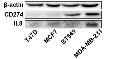
Application: WB    Species: human    Sample: T549 and MDA-MB-231 cell; MCF7 and T47D cell

Figure 8| qRT-PCR and western blot results. (A, B) qRT-PCR results showed that CD274 and IL8 were upregulated in the basal-like breast cancer cell lines BT549 and MDA-MB-231 (p < 0.0001). Similar to the qRT-PCR results, the western blot analysis (C–E) indicated that CD274 and IL8 protein were increased in the BT549 and MDA-MB-231 cell lines compared to the MCF7 and T47D cell lines. P < 0.001, p < 0.01, and p < 0.05

7). Depression promotes lung carcinoma progression by regulating the tumor microenvironment in tumor-bearing models of C57BL/6J mice. NEUROSCIENCE LETTERS (PubMed: 33781910) [IF=2.5]

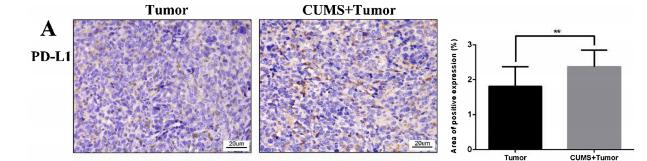
Application: IHC    Species: Mice    Sample: tumor tissue

Fig. 5. Relative protein levels of CD8, MMP2, MMP9, PD-L1 and VEGF in tumor tissue specimens. (A)- (E) The protein expression in the tumor tissues by immunohistochemical staining. Statistical analyses of single comparison were conducted through the Student’s t-test. All data are presented as mean ± SD, *p < 0.05, **p < 0.01, n = 3 per group. Bar: 50 μm (low magnification); 20 μm (high magnification).

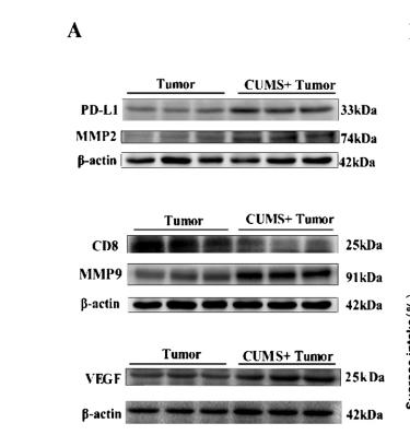
Application: WB    Species: Mice    Sample: tumor tissues

Fig. 6. WB of protein expression in tumor tissues and correlation analysis between the depression-like behaviors and protein expression. (A) The bands of protein expression. (B)-(F) Correlation analysis between the depression-like behaviors and protein expression. Statistical analyses of single comparison were conducted through the Student’s t-test. All data are presented as mean ± SD, *p < 0.05, **p < 0.01, n = 3 per group.

8). Screening of Immunosuppressive Factors for Identifying Breast Cancer Malignant Phenotypes Using mRNA Microarray Datasets.

Application: WB    Species: Human    Sample: BT549 and MDA- MB-231

Figure2 qRT-PCR and Western blot results (a) show that CD274 and IL8 were upregulated in the basal-like breast cancer cell lines, BT549 and MDA-MB-231 (p

9). Dying cell-released exosomal CXCL1 promotes breast cancer metastasis by activating TAM/PD-L1 signaling. bioRxiv

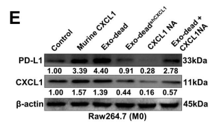
Application: WB    Species: Mouse    Sample: Raw264.7 cells

Figure 3. CXCL1exo-dead 404 induces macrophage M2 polarization by activating PD-L1 expression. (A) Chemokine array assay was conducted to characterize the differences in chemokine content between exo-dead and exo-alive. An ELISA was conducted to compare the relative CXCL1 content in exo-dead and exo-alive. (B) Changes in M2 phenotype polarization of Raw264.7 macrophages when treated with 10 ng/ml murine CXCL1, 50 μg/ml exo-dead, 50 μg/ml exo-deadshCXCL1 408 , 5 μg/ml CXCL1 neutralizing antibody (NA), or exo-dead and CXCL1-NA combination for 48 h. (C) Representative images of Transwell assay. Raw264.7 macrophages were treated as indicated for 48 h and then co-cultured with 4T1 cells. Scale bar: 200 μm. (D–F) Expression changes of CXCL1 and PD-L1 in Raw264.7 macrophages when treated as indicated for 48 h. Scale bar: 10 μm. (G) The results of flow cytometry assay suggested that 50 μg/ml exo-dead treatment for 48 h induced the M2 polarization of Raw264.7 macrophages by activating PD-L1 expression;

Restrictive clause


Affinity Biosciences tests all products strictly. Citations are provided as a resource for additional applications that have not been validated by Affinity Biosciences. Please choose the appropriate format for each application and consult Materials and Methods sections for additional details about the use of any product in these publications.

For Research Use Only.
Not for use in diagnostic or therapeutic procedures. Not for resale. Not for distribution without written consent. Affinity Biosciences will not be held responsible for patent infringement or other violations that may occur with the use of our products. Affinity Biosciences, Affinity Biosciences Logo and all other trademarks are the property of Affinity Biosciences LTD.