ODC1 Antibody - #DF6712
, using ODC1 Antibody. The lane on the left was treated with blocking peptide.")
| Product: | ODC1 Antibody |
| Catalog: | DF6712 |
| Description: | Rabbit polyclonal antibody to ODC1 |
| Application: | WB IHC |
| Reactivity: | Human, Mouse, Rat |
| Prediction: | Pig, Zebrafish, Bovine, Horse, Sheep, Rabbit, Dog, Chicken, Xenopus |
| Mol.Wt.: | 51kDa; 51kD(Calculated). |
| Uniprot: | P11926 |
| RRID: | AB_2838674 |
Related Downloads
Protocols
Product Info
*The optimal dilutions should be determined by the end user.
*Tips:
WB: For western blot detection of denatured protein samples. IHC: For immunohistochemical detection of paraffin sections (IHC-p) or frozen sections (IHC-f) of tissue samples. IF/ICC: For immunofluorescence detection of cell samples. ELISA(peptide): For ELISA detection of antigenic peptide.
Cite Format: Affinity Biosciences Cat# DF6712, RRID:AB_2838674.
Fold/Unfold
DCOR_HUMAN; Dodc1; Odc 1; ODC; Odc1; ODC2; Ornithine decarboxylase 1; Ornithine decarboxylase 2; Ornithine decarboxylase; Ornithine decarboxylase structural 1; RNODC;
Immunogens
- P11926 DCOR_HUMAN:
- Protein BLAST With
- NCBI/
- ExPASy/
- Uniprot
MNNFGNEEFDCHFLDEGFTAKDILDQKINEVSSSDDKDAFYVADLGDILKKHLRWLKALPRVTPFYAVKCNDSKAIVKTLAATGTGFDCASKTEIQLVQSLGVPPERIIYANPCKQVSQIKYAANNGVQMMTFDSEVELMKVARAHPKAKLVLRIATDDSKAVCRLSVKFGATLRTSRLLLERAKELNIDVVGVSFHVGSGCTDPETFVQAISDARCVFDMGAEVGFSMYLLDIGGGFPGSEDVKLKFEEITGVINPALDKYFPSDSGVRIIAEPGRYYVASAFTLAVNIIAKKIVLKEQTGSDDEDESSEQTFMYYVNDGVYGSFNCILYDHAHVKPLLQKRPKPDEKYYSSSIWGPTCDGLDRIVERCDLPEMHVGDWMLFENMGAYTVAAASTFNGFQRPTIYYVMSGPAWQLMQQFQNPDFPPEVEEQDASTLPVSCAWESGMKRHRAACASASINV
Predictions
Score>80(red) has high confidence and is suggested to be used for WB detection. *The prediction model is mainly based on the alignment of immunogen sequences, the results are for reference only, not as the basis of quality assurance.
High(score>80) Medium(80>score>50) Low(score<50) No confidence
PTMs - P11926 As Substrate
| Site | PTM Type | Enzyme | Source |
|---|---|---|---|
| K27 | Ubiquitination | Uniprot | |
| K37 | Ubiquitination | Uniprot | |
| K50 | Ubiquitination | Uniprot | |
| K57 | Ubiquitination | Uniprot | |
| K69 | Ubiquitination | Uniprot | |
| K78 | Ubiquitination | Uniprot | |
| K115 | Ubiquitination | Uniprot | |
| K141 | Ubiquitination | Uniprot | |
| K150 | Ubiquitination | Uniprot | |
| K161 | Ubiquitination | Uniprot | |
| K247 | Ubiquitination | Uniprot | |
| K261 | Ubiquitination | Uniprot | |
| S303 | Phosphorylation | Uniprot | |
| C360 | S-Nitrosylation | Uniprot |
Research Backgrounds
Catalyzes the first and rate-limiting step of polyamine biosynthesis that converts ornithine into putrescine, which is the precursor for the polyamines, spermidine and spermine. Polyamines are essential for cell proliferation and are implicated in cellular processes, ranging from DNA replication to apoptosis.
S-Nitrosylation inhibits the enzyme. S-Nitrosylated in vitro on 4 cysteine residues.
Homodimer. Only the dimer is catalytically active, as the active sites are constructed of residues from both monomers (Probable). Does not form a heterodimer with AZIN2 (By similarity).
Belongs to the Orn/Lys/Arg decarboxylase class-II family.
Research Fields
· Metabolism > Amino acid metabolism > Arginine and proline metabolism.
· Metabolism > Metabolism of other amino acids > Glutathione metabolism.
· Metabolism > Global and overview maps > Metabolic pathways.
References
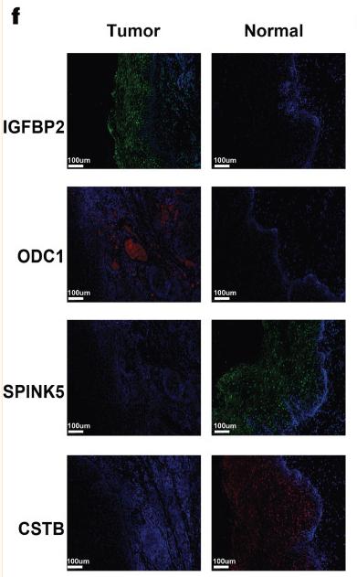
Application: IF/ICC Species: Human Sample: ESCC and non-malignant tissues
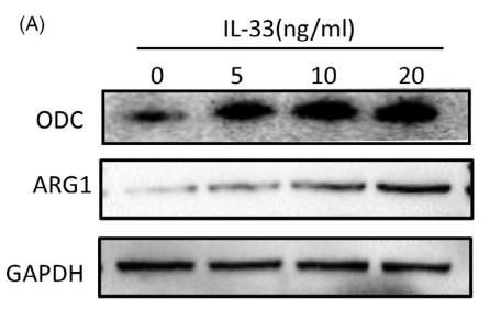
Application: WB Species: human Sample: M2 macrophages
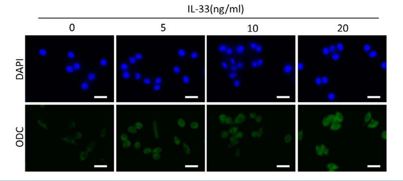
Application: IF/ICC Species: human Sample: M2 macrophages
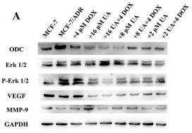
Application: WB Species: Human Sample: MCF-7/ADR Cells
Restrictive clause
Affinity Biosciences tests all products strictly. Citations are provided as a resource for additional applications that have not been validated by Affinity Biosciences. Please choose the appropriate format for each application and consult Materials and Methods sections for additional details about the use of any product in these publications.
For Research Use Only.
Not for use in diagnostic or therapeutic procedures. Not for resale. Not for distribution without written consent. Affinity Biosciences will not be held responsible for patent infringement or other violations that may occur with the use of our products. Affinity Biosciences, Affinity Biosciences Logo and all other trademarks are the property of Affinity Biosciences LTD.