TPSAB1 Antibody - #DF6758

| Product: | TPSAB1 Antibody |
| Catalog: | DF6758 |
| Description: | Rabbit polyclonal antibody to TPSAB1 |
| Application: | WB IHC IF/ICC |
| Reactivity: | Human, Mouse, Rat |
| Mol.Wt.: | 31kDa; 31kD(Calculated). |
| Uniprot: | Q15661 |
| RRID: | AB_2838720 |
Related Downloads
Protocols
Product Info
*The optimal dilutions should be determined by the end user.
*Tips:
WB: For western blot detection of denatured protein samples. IHC: For immunohistochemical detection of paraffin sections (IHC-p) or frozen sections (IHC-f) of tissue samples. IF/ICC: For immunofluorescence detection of cell samples. ELISA(peptide): For ELISA detection of antigenic peptide.
Cite Format: Affinity Biosciences Cat# DF6758, RRID:AB_2838720.
Fold/Unfold
alpha II; Lung tryptase; Mast cell alpha II tryptase; Mast cell beta I tryptase; Mast cell protease 7; Mast cell protease II; MCP 7; Pituitary tryptase; Skin tryptase; TPS 1; TPS1; TPS2; TPSAB1; TPSAB1 protein; TPSB1; Tryptase 1; Tryptase alpha 1; tryptase alpha I included; Tryptase alpha II; tryptase alpha II included; tryptase alpha included; tryptase alpha/beta 1; Tryptase beta 1; tryptase beta I included; Tryptase I; tryptase I included; Tryptase III; Tryptase skin;
Immunogens
Isoform 1 and isoform 2 are expressed in lung, stomach, spleen, heart and skin; in these tissues, isoform 1 is predominant. Isoform 2 is expressed in aorta, spleen, and breast tumor, with highest levels in the endothelial cells of some blood vessels surrounding the aorta, as well as those surrounding the tumor and low levels, if any, in mast cells (at protein level).
- Q15661 TRYB1_HUMAN:
- Protein BLAST With
- NCBI/
- ExPASy/
- Uniprot
MLNLLLLALPVLASRAYAAPAPGQALQRVGIVGGQEAPRSKWPWQVSLRVHGPYWMHFCGGSLIHPQWVLTAAHCVGPDVKDLAALRVQLREQHLYYQDQLLPVSRIIVHPQFYTAQIGADIALLELEEPVNVSSHVHTVTLPPASETFPPGMPCWVTGWGDVDNDERLPPPFPLKQVKVPIMENHICDAKYHLGAYTGDDVRIVRDDMLCAGNTRRDSCQGDSGGPLVCKVNGTWLQAGVVSWGEGCAQPNRPGIYTRVTYYLDWIHHYVPKKP
PTMs - Q15661 As Substrate
| Site | PTM Type | Enzyme | Source |
|---|---|---|---|
| Y97 | Phosphorylation | Uniprot | |
| C188 | S-Nitrosylation | Uniprot | |
| Y197 | Phosphorylation | Uniprot | |
| S219 | Phosphorylation | Uniprot | |
| S224 | Phosphorylation | Uniprot | |
| N233 | N-Glycosylation | Uniprot |
Research Backgrounds
Tryptase is the major neutral protease present in mast cells and is secreted upon the coupled activation-degranulation response of this cell type. May play a role in innate immunity. Isoform 2 cleaves large substrates, such as fibronectin, more efficiently than isoform 1, but seems less efficient toward small substrates.
Secreted.
Note: Released from the secretory granules upon mast cell activation.
Isoform 1 and isoform 2 are expressed in lung, stomach, spleen, heart and skin; in these tissues, isoform 1 is predominant. Isoform 2 is expressed in aorta, spleen, and breast tumor, with highest levels in the endothelial cells of some blood vessels surrounding the aorta, as well as those surrounding the tumor and low levels, if any, in mast cells (at protein level).
Homotetramer. The active tetramer is converted to inactive monomers at neutral and acidic pH in the absence of heparin. Low concentrations of inactive monomers become active monomers at pH 6.0 in the presence of heparin. When the concentration of active monomers is higher, they convert to active monomers and then to active tetramers. These monomers are active and functionally distinct from the tetrameric enzyme. In contrast to the hidden active sites in the tetrameric form, the active site of the monomeric form is accessible for macromolecular proteins and inhibitors eg: fibrinogen which is a substrate for the monomeric but not for the tetrameric form. The monomeric form forms a complex with SERPINB6.
Belongs to the peptidase S1 family. Tryptase subfamily.
References
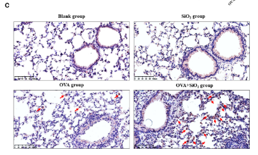
Application: IHC Species: Mouse Sample: Lung tissue
Restrictive clause
Affinity Biosciences tests all products strictly. Citations are provided as a resource for additional applications that have not been validated by Affinity Biosciences. Please choose the appropriate format for each application and consult Materials and Methods sections for additional details about the use of any product in these publications.
For Research Use Only.
Not for use in diagnostic or therapeutic procedures. Not for resale. Not for distribution without written consent. Affinity Biosciences will not be held responsible for patent infringement or other violations that may occur with the use of our products. Affinity Biosciences, Affinity Biosciences Logo and all other trademarks are the property of Affinity Biosciences LTD.