IL10 Antibody - #DF6894


 Representative figures of TNF-α, IL-1β and IL-10 protein. (D-F) Quantitative analyses of TNF-α, IL-1β and IL-10 protein. (mean ± S.D, n = 3).")

 14 days after OB, BI-1 overexpression in PFC-PL significantly blocked the reduction of the BI-1 expression in the PFC-PL of OB rats. (b) 14 days after OB, BI-1 overexpression in PFC-PL significantly blocked the induction of apoptotic and inflammatory signals by OB, including the decreased expressions of Bax, caspase-3, TNF-α, IL-6, and increased expressions of Bcl-2 and IL-10 in OB rats. Bars represent the mean ± SEM (n = 6, per group). *P < 0.05,**P < 0.01, ***P < 0.001, statistically significant. OB, olfactory bulbectomy; PFC-PL, prelimbic cortex.")
 Representative immunoblots of the nuclear NF-κB p65 in the hippocampus was shown, and Histone H3 was used as a loading control. (B) Representative immunoblots of TNF-α, IL-1β, IL-6 and IL-10 in the hippocampus, and β-actin protein was used here as an internal control.")
 western blotting and (B) reverse transcription-quantitative PCR. *P<0.05, **P<0.01. hAMSCs, human amniotic mesenchymal stem cells; TNF, tumor necrosis factor; IL, interleukin.")
 Western blots revealing that FOXO4-DRI decreases levels of the SASP factors IL-1β, IL-6 and TGF-β in testes of aged mice, but has no effect on testicular levels of IL-1α, IL-10 and TNF-α. Data depict the mean ± SD. n=6. *P<0.05. NS, nonsignificant.")
Representative Western blot picture, showing the expression levels of IL-6, IL-10,ZO1 and β-actin proteins in the four groups.")
, IL-6 (b) and IL-10 (c). Three samples per group were examined, and representative images are shown (Scale bar represents 50 µm).")


 Expression levels of NLRC3, TRAF6 and NF-κB p65 in LPS-stimulated RAW264.7 cells were detected using immunohistochemistry staining (magnification, ×400; scale bar, 200-µm). The mean optical density values of (B) NLRC3, (C) TRAF6 and (D) NF-κB p65 were quantified using ImageJ software. The protein expression levels of (E) NLRC3 and (F) TRAF6 were detected using western blotting and semi-quantified using Image Lab software. LPS represents protein from the 100 ng/ml LPS-treated group; Indo represents protein from the 8 µg/ml Indo and 100 ng/ml LPS-treated group; ARR represents protein from the 100 µg/ml ARR and 100 ng/ml LPS-treated group. Data are presented as the mean ± SD (n=3). ##P<0.01 vs. control group; *P<0.05, **P<0.01 vs. LPS group. ARR, α-rhamnrtin-3-α-rhamnoside; TRAF6, tumor necrosis factor-associated factor 6; NLRC3, NOD-like receptor family CARD domain containing 3; LPS, lipopolysaccharide; Indo, indomethacin.")
 Effects of ethanol on protein and mRNA levels of M1 macrophage markers (TNF-α, IL-1β) in liver tissue. (B) The protein and mRNA levels of M2 macrophage markers (IL-10, ARG-1) in liver tissue. (C) Immunofluorescence staining results of M1, M2 macrophage markers (CD86, CD206) in liver tissue of EtOH-fed mice. (D) The protein and mRNA levels of M1 macrophage (TNF-α, IL-1β) in ethanol-induced RAW264.7 cells. (E) The protein and mRNA levels of M2 macrophage markers (IL-10, ARG-1) in ethanol-induced RAW264.7 cells. The results are shown as relative expression against control expression without treatment. Data shown are the mean ± SD from three independent experiments. *p < 0.05, **p < 0.01 vs. CD-fed group or control group.")
 Relative mRNA level of IL10, Arg-1, and TGF-β1in the colon was measured through qPCR. Expression of IL-10, Arg-1, and TGF-β1 in colon tissue lysates were determined by Western blot from DSS-challenged mice. n = 3,.means ± SEM.*p < 0.05, **p < 0.01, ***p < 0.001.")
 Double immunofluorescent signals from CD80 and interleukin-1 in DSS-induced colitis tissue at different time points (original magnification, 200 ×). (B) Double immunofluorescent signals from CD206 and interleukin-10 in DSS-induced colitis tissue at different time points (original magnification, 200 ×).")
 Detection of relative protein expression levels of IL-10, phosphorylation levels of STAT3, STAT3 and SOCS3 by Western blot; (B–D), Statistics of the IL-10, phosphorylation levels of STAT3, STAT3 and SOCS3 Western blot results. Experiments were repeated three times and data were presented as the mean ± SEM (n = 4). * P < 0.05 vs. EpH4-Ev; # P < 0.05 vs. LPS; $ P < 0.05 vs. MSC; & P < 0.05 vs. MSC-GFP.")
 Representative images of western blot analysis of TGF-β1, IL-10, IFN-γ, IL-1β and α-tubulin in the hippocampus in three groups. (B–E) Quantification of the relative protein expression levels of TGF-β1, IL-10, IFN-γ, IL-1β. (*P < 0.05, vs. sham-operated group, #P < 0.05, vs. RHRSP group; n = 6/group).")
. Representative blots are shown. *P")
 western blot to determine FKN, IL-17, IL-6, Foxp3, IL-10, and GAPDH levels. (B) Total RNA was extracted from renal tissues, and mRNA were quantified by qRT-PCR. GAPDH protein and mRNA were used as internal controls. NC: C57BL/6 mice; LN: MRL/lpr mice; NC + IgG: NC group treated with an isotype antibody; LN + IgG: LN group treated with an isotype antibody; NC + rFKN: NC group treated with rFKN; LN + rFKN: LN group treated with rFKN; NC + aFKN: NC group treated with aFKN; and LN + aFKN: LN group treated with aFKN. *P < 0.05 vs. the control group; **P < 0.05 vs. the LN group. #P < 0.05, NC + rFKN vs. NC + aFKN; ##P < 0.05, LN + rFKN vs. LN + aFKN.")
 by using Western Blot. Results were normalized to β-actin. Values are expressed as mean ± SEM (n = 3) of three experiments compared with the control group (MCF-7 cells). Statistical analysis was performed by one way ANOVA followed by tukey’s multiple comparison test.")
 and F4/80 (green) immunofluorescence staining (scale bar, 50μm). B: CD206 (red) and F4/80 (green) immunofluorescence staining (scale bar, 50μm). C: Statistics of the percentage of CD86 positive macrophages in the lung tissues of mice in each group (n=3). D: Statistics of the percentage of CD206 positive macrophages in the lung tissues of mice in each group (n=3). E: mRNA levels of M1 macrophage-related markers (n=8). F: mRNA levels of M2 macrophage-related markers (n=8). G, H: Expression levels of IL-12 in lung cancer tissues (n=6). G, I: Expression levels of IL-10 in lung cancer tissues (n=6). J, K, L: Plasma levels of IL-12(J), IL-10(K) and IFN-γ(L) (n=8). Note: &, && respectively represent a significant difference compared with the Saline group (P")
| Product: | IL10 Antibody |
| Catalog: | DF6894 |
| Description: | Rabbit polyclonal antibody to IL10 |
| Application: | WB IHC IF/ICC |
| Cited expt.: | WB, IHC, IF/ICC |
| Reactivity: | Human, Mouse, Rat |
| Prediction: | Pig, Bovine, Horse, Sheep, Rabbit, Dog, Xenopus |
| Mol.Wt.: | 19kDa; 21kD(Calculated). |
| Uniprot: | P22301 |
| RRID: | AB_2838853 |
Related Downloads
Protocols
Product Info
*The optimal dilutions should be determined by the end user. For optimal experimental results, antibody reuse is not recommended.
*Tips:
WB: For western blot detection of denatured protein samples. IHC: For immunohistochemical detection of paraffin sections (IHC-p) or frozen sections (IHC-f) of tissue samples. IF/ICC: For immunofluorescence detection of cell samples. ELISA(peptide): For ELISA detection of antigenic peptide.
Cite Format: Affinity Biosciences Cat# DF6894, RRID:AB_2838853.
Fold/Unfold
CSIF; Cytokine synthesis inhibitory factor; GVHDS; IL 10; IL-10; IL10; IL10_HUMAN; IL10A; Interleukin 10; Interleukin-10; MGC126450; MGC126451; T-cell growth inhibitory factor; TGIF;
Immunogens
A synthesized peptide derived from human IL10, corresponding to a region within the internal amino acids.
Produced by a variety of cell lines, including T-cells, macrophages, mast cells and other cell types.
- P22301 IL10_HUMAN:
- Protein BLAST With
- NCBI/
- ExPASy/
- Uniprot
MHSSALLCCLVLLTGVRASPGQGTQSENSCTHFPGNLPNMLRDLRDAFSRVKTFFQMKDQLDNLLLKESLLEDFKGYLGCQALSEMIQFYLEEVMPQAENQDPDIKAHVNSLGENLKTLRLRLRRCHRFLPCENKSKAVEQVKNAFNKLQEKGIYKAMSEFDIFINYIEAYMTMKIRN
Predictions
Score>80(red) has high confidence and is suggested to be used for WB detection. *The prediction model is mainly based on the alignment of immunogen sequences, the results are for reference only, not as the basis of quality assurance.
High(score>80) Medium(80>score>50) Low(score<50) No confidence
Research Backgrounds
Major immune regulatory cytokine that acts on many cells of the immune system where it has profound anti-inflammatory functions, limiting excessive tissue disruption caused by inflammation. Mechanistically, IL10 binds to its heterotetrameric receptor comprising IL10RA and IL10RB leading to JAK1 and STAT2-mediated phosphorylation of STAT3. In turn, STAT3 translocates to the nucleus where it drives expression of anti-inflammatory mediators. Targets antigen-presenting cells (APCs) such as macrophages and monocytes and inhibits their release of pro-inflammatory cytokines including granulocyte-macrophage colony-stimulating factor /GM-CSF, granulocyte colony-stimulating factor/G-CSF, IL-1 alpha, IL-1 beta, IL-6, IL-8 and TNF-alpha. Interferes also with antigen presentation by reducing the expression of MHC-class II and co-stimulatory molecules, thereby inhibiting their ability to induce T cell activation. In addition, controls the inflammatory response of macrophages by reprogramming essential metabolic pathways including mTOR signaling (By similarity).
Secreted.
Produced by a variety of cell lines, including T-cells, macrophages, mast cells and other cell types.
Belongs to the IL-10 family.
Research Fields
· Environmental Information Processing > Signaling molecules and interaction > Cytokine-cytokine receptor interaction. (View pathway)
· Environmental Information Processing > Signal transduction > FoxO signaling pathway. (View pathway)
· Environmental Information Processing > Signal transduction > Jak-STAT signaling pathway. (View pathway)
· Human Diseases > Infectious diseases: Bacterial > Pertussis.
· Human Diseases > Infectious diseases: Parasitic > Leishmaniasis.
· Human Diseases > Infectious diseases: Parasitic > Chagas disease (American trypanosomiasis).
· Human Diseases > Infectious diseases: Parasitic > African trypanosomiasis.
· Human Diseases > Infectious diseases: Parasitic > Malaria.
· Human Diseases > Infectious diseases: Parasitic > Toxoplasmosis.
· Human Diseases > Infectious diseases: Parasitic > Amoebiasis.
· Human Diseases > Infectious diseases: Bacterial > Staphylococcus aureus infection.
· Human Diseases > Infectious diseases: Bacterial > Tuberculosis.
· Human Diseases > Infectious diseases: Viral > Epstein-Barr virus infection.
· Human Diseases > Immune diseases > Asthma.
· Human Diseases > Immune diseases > Autoimmune thyroid disease.
· Human Diseases > Immune diseases > Inflammatory bowel disease (IBD).
· Human Diseases > Immune diseases > Systemic lupus erythematosus.
· Human Diseases > Immune diseases > Allograft rejection.
· Organismal Systems > Immune system > T cell receptor signaling pathway. (View pathway)
· Organismal Systems > Immune system > Intestinal immune network for IgA production. (View pathway)
References
Application: WB Species: Mouse Sample: RAW264.7 cells
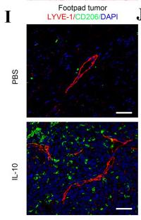
Application: IHC Species: Mice Sample: tumours
Restrictive clause
Affinity Biosciences tests all products strictly. Citations are provided as a resource for additional applications that have not been validated by Affinity Biosciences. Please choose the appropriate format for each application and consult Materials and Methods sections for additional details about the use of any product in these publications.
For Research Use Only.
Not for use in diagnostic or therapeutic procedures. Not for resale. Not for distribution without written consent. Affinity Biosciences will not be held responsible for patent infringement or other violations that may occur with the use of our products. Affinity Biosciences, Affinity Biosciences Logo and all other trademarks are the property of Affinity Biosciences LTD.