SIRT1 Antibody - #BF0189
, Jurkat (2), Hela (3), HEK293 (4) and A549 (5) cell lysate.")
Product Info
*The optimal dilutions should be determined by the end user.
*Tips:
WB: For western blot detection of denatured protein samples. IHC: For immunohistochemical detection of paraffin sections (IHC-p) or frozen sections (IHC-f) of tissue samples. IF/ICC: For immunofluorescence detection of cell samples. ELISA(peptide): For ELISA detection of antigenic peptide.
Cite Format: Affinity Biosciences Cat# BF0189, RRID:AB_2833834.
Fold/Unfold
75SirT1; hSIR2; hSIRT1; HST2, S. cerevisiae, homolog of; NAD dependent deacetylase sirtuin 1; NAD dependent protein deacetylase sirtuin 1; OTTHUMP00000198111; OTTHUMP00000198112; Regulatory protein SIR2 homolog 1; SIR1_HUMAN; SIR2; SIR2 like 1; SIR2 like protein 1; SIR2, S.cerevisiae, homolog-like 1; SIR2-like protein 1; SIR2ALPHA; SIR2L1; Sirt1; SirtT1 75 kDa fragment; Sirtuin (silent mating type information regulation 2 homolog) 1 (S. cerevisiae); Sirtuin 1; Sirtuin type 1;
Immunogens
Purified recombinant fragment of human SIRT1 expressed in E. Coli.
- Q96EB6 SIR1_HUMAN:
- Protein BLAST With
- NCBI/
- ExPASy/
- Uniprot
MADEAALALQPGGSPSAAGADREAASSPAGEPLRKRPRRDGPGLERSPGEPGGAAPEREVPAAARGCPGAAAAALWREAEAEAAAAGGEQEAQATAAAGEGDNGPGLQGPSREPPLADNLYDEDDDDEGEEEEEAAAAAIGYRDNLLFGDEIITNGFHSCESDEEDRASHASSSDWTPRPRIGPYTFVQQHLMIGTDPRTILKDLLPETIPPPELDDMTLWQIVINILSEPPKRKKRKDINTIEDAVKLLQECKKIIVLTGAGVSVSCGIPDFRSRDGIYARLAVDFPDLPDPQAMFDIEYFRKDPRPFFKFAKEIYPGQFQPSLCHKFIALSDKEGKLLRNYTQNIDTLEQVAGIQRIIQCHGSFATASCLICKYKVDCEAVRGDIFNQVVPRCPRCPADEPLAIMKPEIVFFGENLPEQFHRAMKYDKDEVDLLIVIGSSLKVRPVALIPSSIPHEVPQILINREPLPHLHFDVELLGDCDVIINELCHRLGGEYAKLCCNPVKLSEITEKPPRTQKELAYLSELPPTPLHVSEDSSSPERTSPPDSSVIVTLLDQAAKSNDDLDVSESKGCMEEKPQEVQTSRNVESIAEQMENPDLKNVGSSTGEKNERTSVAGTVRKCWPNRVAKEQISRRLDGNQYLFLPPNRYIFHGAEVYSDSEDDVLSSSSCGSNSDSGTCQSPSLEEPMEDESEIEEFYNGLEDEPDVPERAGGAGFGTDGDDQEAINEAISVKQEVTDMNYPSNKS
PTMs - Q96EB6 As Substrate
| Site | PTM Type | Enzyme | Source |
|---|---|---|---|
| A2 | Acetylation | Uniprot | |
| S14 | Phosphorylation | Uniprot | |
| S16 | Phosphorylation | Uniprot | |
| S26 | Phosphorylation | Uniprot | |
| S27 | Phosphorylation | P45983 (MAPK8) , Q96RR4 (CAMKK2) , Q9H2X6 (HIPK2) | Uniprot |
| S47 | Phosphorylation | P45983 (MAPK8) , Q96RR4 (CAMKK2) | Uniprot |
| S159 | Phosphorylation | Uniprot | |
| S162 | Phosphorylation | Uniprot | |
| S172 | Phosphorylation | Uniprot | |
| S173 | Phosphorylation | Uniprot | |
| K238 | Ubiquitination | Uniprot | |
| K248 | Ubiquitination | Uniprot | |
| Y280 | Phosphorylation | Uniprot | |
| Y301 | Phosphorylation | Uniprot | |
| K311 | Ubiquitination | Uniprot | |
| K314 | Ubiquitination | Uniprot | |
| K328 | Ubiquitination | Uniprot | |
| S333 | Phosphorylation | Uniprot | |
| K335 | Ubiquitination | Uniprot | |
| K338 | Ubiquitination | Uniprot | |
| T344 | Phosphorylation | P54646 (PRKAA2) | Uniprot |
| Y428 | Phosphorylation | Uniprot | |
| K430 | Acetylation | Uniprot | |
| S441 | Phosphorylation | Uniprot | |
| S442 | Phosphorylation | Uniprot | |
| K444 | Acetylation | Uniprot | |
| K499 | Ubiquitination | Uniprot | |
| K506 | Ubiquitination | Uniprot | |
| K513 | Ubiquitination | Uniprot | |
| T530 | Phosphorylation | P06493 (CDK1) , Q13627 (DYRK1A) , P45983 (MAPK8) | Uniprot |
| S535 | Phosphorylation | Uniprot | |
| S538 | Phosphorylation | Uniprot | |
| S539 | Phosphorylation | Uniprot | |
| S540 | Phosphorylation | P06493 (CDK1) | Uniprot |
| T544 | Phosphorylation | Uniprot | |
| S545 | Phosphorylation | Uniprot | |
| S549 | Phosphorylation | Uniprot | |
| K561 | Ubiquitination | Uniprot | |
| S562 | Phosphorylation | Uniprot | |
| S569 | Phosphorylation | Uniprot | |
| K572 | Ubiquitination | Uniprot | |
| C574 | S-Nitrosylation | Uniprot | |
| K578 | Acetylation | Uniprot | |
| K578 | Ubiquitination | Uniprot | |
| S590 | Phosphorylation | Uniprot | |
| K601 | Ubiquitination | Uniprot | |
| K610 | Ubiquitination | Uniprot | |
| T614 | Phosphorylation | Uniprot | |
| S615 | Phosphorylation | Uniprot | |
| T619 | Phosphorylation | Uniprot | |
| K622 | Methylation | Uniprot | |
| S659 | Phosphorylation | P68400 (CSNK2A1) | Uniprot |
| S661 | Phosphorylation | P68400 (CSNK2A1) | Uniprot |
| S682 | Phosphorylation | Q9H2X6 (HIPK2) | Uniprot |
| T719 | Phosphorylation | Uniprot | |
| K734 | Sumoylation | Uniprot | |
| S747 | Phosphorylation | Uniprot |
Research Backgrounds
NAD-dependent protein deacetylase that links transcriptional regulation directly to intracellular energetics and participates in the coordination of several separated cellular functions such as cell cycle, response to DNA damage, metabolism, apoptosis and autophagy. Can modulate chromatin function through deacetylation of histones and can promote alterations in the methylation of histones and DNA, leading to transcriptional repression. Deacetylates a broad range of transcription factors and coregulators, thereby regulating target gene expression positively and negatively. Serves as a sensor of the cytosolic ratio of NAD(+)/NADH which is altered by glucose deprivation and metabolic changes associated with caloric restriction. Is essential in skeletal muscle cell differentiation and in response to low nutrients mediates the inhibitory effect on skeletal myoblast differentiation which also involves 5'-AMP-activated protein kinase (AMPK) and nicotinamide phosphoribosyltransferase (NAMPT) (By similarity). Component of the eNoSC (energy-dependent nucleolar silencing) complex, a complex that mediates silencing of rDNA in response to intracellular energy status and acts by recruiting histone-modifying enzymes. The eNoSC complex is able to sense the energy status of cell: upon glucose starvation, elevation of NAD(+)/NADP(+) ratio activates SIRT1, leading to histone H3 deacetylation followed by dimethylation of H3 at 'Lys-9' (H3K9me2) by SUV39H1 and the formation of silent chromatin in the rDNA locus. Deacetylates 'Lys-266' of SUV39H1, leading to its activation. Inhibits skeletal muscle differentiation by deacetylating PCAF and MYOD1. Deacetylates H2A and 'Lys-26' of H1-4. Deacetylates 'Lys-16' of histone H4 (in vitro). Involved in NR0B2/SHP corepression function through chromatin remodeling: Recruited to LRH1 target gene promoters by NR0B2/SHP thereby stimulating histone H3 and H4 deacetylation leading to transcriptional repression. Proposed to contribute to genomic integrity via positive regulation of telomere length; however, reports on localization to pericentromeric heterochromatin are conflicting (By similarity). Proposed to play a role in constitutive heterochromatin (CH) formation and/or maintenance through regulation of the available pool of nuclear SUV39H1. Upon oxidative/metabolic stress decreases SUV39H1 degradation by inhibiting SUV39H1 polyubiquitination by MDM2. This increase in SUV39H1 levels enhances SUV39H1 turnover in CH, which in turn seems to accelerate renewal of the heterochromatin which correlates with greater genomic integrity during stress response. Deacetylates 'Lys-382' of p53/TP53 and impairs its ability to induce transcription-dependent proapoptotic program and modulate cell senescence. Deacetylates TAF1B and thereby represses rDNA transcription by the RNA polymerase I (By similarity). Deacetylates MYC, promotes the association of MYC with MAX and decreases MYC stability leading to compromised transformational capability. Deacetylates FOXO3 in response to oxidative stress thereby increasing its ability to induce cell cycle arrest and resistance to oxidative stress but inhibiting FOXO3-mediated induction of apoptosis transcriptional activity; also leading to FOXO3 ubiquitination and protesomal degradation. Appears to have a similar effect on MLLT7/FOXO4 in regulation of transcriptional activity and apoptosis. Deacetylates DNMT1; thereby impairs DNMT1 methyltransferase-independent transcription repressor activity, modulates DNMT1 cell cycle regulatory function and DNMT1-mediated gene silencing. Deacetylates RELA/NF-kappa-B p65 thereby inhibiting its transactivating potential and augments apoptosis in response to TNF-alpha. Deacetylates HIF1A, KAT5/TIP60, RB1 and HIC1. Deacetylates FOXO1 resulting in its nuclear retention and enhancement of its transcriptional activity leading to increased gluconeogenesis in liver. Inhibits E2F1 transcriptional activity and apoptotic function, possibly by deacetylation. Involved in HES1- and HEY2-mediated transcriptional repression. In cooperation with MYCN seems to be involved in transcriptional repression of DUSP6/MAPK3 leading to MYCN stabilization by phosphorylation at 'Ser-62'. Deacetylates MEF2D. Required for antagonist-mediated transcription suppression of AR-dependent genes which may be linked to local deacetylation of histone H3. Represses HNF1A-mediated transcription (By similarity). Required for the repression of ESRRG by CREBZF. Deacetylates NR1H3 and NR1H2 and deacetylation of NR1H3 at 'Lys-434' positively regulates transcription of NR1H3:RXR target genes, promotes NR1H3 proteosomal degradation and results in cholesterol efflux; a promoter clearing mechanism after reach round of transcription is proposed. Involved in lipid metabolism. Implicated in regulation of adipogenesis and fat mobilization in white adipocytes by repression of PPARG which probably involves association with NCOR1 and SMRT/NCOR2 (By similarity). Deacetylates p300/EP300 and PRMT1 (By similarity). Deacetylates ACSS2 leading to its activation, and HMGCS1 deacetylation. Involved in liver and muscle metabolism. Through deacetylation and activation of PPARGC1A is required to activate fatty acid oxidation in skeletal muscle under low-glucose conditions and is involved in glucose homeostasis. Involved in regulation of PPARA and fatty acid beta-oxidation in liver. Involved in positive regulation of insulin secretion in pancreatic beta cells in response to glucose; the function seems to imply transcriptional repression of UCP2. Proposed to deacetylate IRS2 thereby facilitating its insulin-induced tyrosine phosphorylation. Deacetylates SREBF1 isoform SREBP-1C thereby decreasing its stability and transactivation in lipogenic gene expression. Involved in DNA damage response by repressing genes which are involved in DNA repair, such as XPC and TP73, deacetylating XRCC6/Ku70, and facilitating recruitment of additional factors to sites of damaged DNA, such as SIRT1-deacetylated NBN can recruit ATM to initiate DNA repair and SIRT1-deacetylated XPA interacts with RPA2. Also involved in DNA repair of DNA double-strand breaks by homologous recombination and specifically single-strand annealing independently of XRCC6/Ku70 and NBN. Transcriptional suppression of XPC probably involves an E2F4:RBL2 suppressor complex and protein kinase B (AKT) signaling. Transcriptional suppression of TP73 probably involves E2F4 and PCAF. Deacetylates WRN thereby regulating its helicase and exonuclease activities and regulates WRN nuclear translocation in response to DNA damage. Deacetylates APEX1 at 'Lys-6' and 'Lys-7' and stimulates cellular AP endonuclease activity by promoting the association of APEX1 to XRCC1. Increases p53/TP53-mediated transcription-independent apoptosis by blocking nuclear translocation of cytoplasmic p53/TP53 and probably redirecting it to mitochondria. Deacetylates XRCC6/Ku70 at 'Lys-539' and 'Lys-542' causing it to sequester BAX away from mitochondria thereby inhibiting stress-induced apoptosis. Is involved in autophagy, presumably by deacetylating ATG5, ATG7 and MAP1LC3B/ATG8. Deacetylates AKT1 which leads to enhanced binding of AKT1 and PDK1 to PIP3 and promotes their activation. Proposed to play role in regulation of STK11/LBK1-dependent AMPK signaling pathways implicated in cellular senescence which seems to involve the regulation of the acetylation status of STK11/LBK1. Can deacetylate STK11/LBK1 and thereby increase its activity, cytoplasmic localization and association with STRAD; however, the relevance of such activity in normal cells is unclear. In endothelial cells is shown to inhibit STK11/LBK1 activity and to promote its degradation. Deacetylates SMAD7 at 'Lys-64' and 'Lys-70' thereby promoting its degradation. Deacetylates CIITA and augments its MHC class II transactivation and contributes to its stability. Deacetylates MECOM/EVI1. Deacetylates PML at 'Lys-487' and this deacetylation promotes PML control of PER2 nuclear localization. During the neurogenic transition, represses selective NOTCH1-target genes through histone deacetylation in a BCL6-dependent manner and leading to neuronal differentiation. Regulates the circadian expression of several core clock genes, including ARNTL/BMAL1, RORC, PER2 and CRY1 and plays a critical role in maintaining a controlled rhythmicity in histone acetylation, thereby contributing to circadian chromatin remodeling. Deacetylates ARNTL/BMAL1 and histones at the circadian gene promoters in order to facilitate repression by inhibitory components of the circadian oscillator (By similarity). Deacetylates PER2, facilitating its ubiquitination and degradation by the proteosome (By similarity). Protects cardiomyocytes against palmitate-induced apoptosis (By similarity). Deacetylates XBP1 isoform 2; deacetylation decreases protein stability of XBP1 isoform 2 and inhibits its transcriptional activity. Deacetylates PCK1 and directs its activity toward phosphoenolpyruvate production promoting gluconeogenesis. Involved in the CCAR2-mediated regulation of PCK1 and NR1D1. Deacetylates CTNB1 at 'Lys-49'. In POMC (pro-opiomelanocortin) neurons, required for leptin-induced activation of PI3K signaling (By similarity). In addition to protein deacetylase activity, also acts as protein-lysine deacylase: acts as a protein depropionylase by mediating depropionylation of Osterix (SP7) (By similarity).
Deacetylates 'Lys-382' of p53/TP53, however with lower activity than isoform 1. In combination, the two isoforms exert an additive effect. Isoform 2 regulates p53/TP53 expression and cellular stress response and is in turn repressed by p53/TP53 presenting a SIRT1 isoform-dependent auto-regulatory loop.
(Microbial infection) In case of HIV-1 infection, interacts with and deacetylates the viral Tat protein. The viral Tat protein inhibits SIRT1 deacetylation activity toward RELA/NF-kappa-B p65, thereby potentiates its transcriptional activity and SIRT1 is proposed to contribute to T-cell hyperactivation during infection.
Catalytically inactive 75SirT1 may be involved in regulation of apoptosis. May be involved in protecting chondrocytes from apoptotic death by associating with cytochrome C and interfering with apoptosome assembly.
Methylated on multiple lysine residues; methylation is enhanced after DNA damage and is dispensable for deacetylase activity toward p53/TP53.
Phosphorylated. Phosphorylated by STK4/MST1, resulting in inhibition of SIRT1-mediated p53/TP53 deacetylation. Phosphorylation by MAPK8/JNK1 at Ser-27, Ser-47, and Thr-530 leads to increased nuclear localization and enzymatic activity. Phosphorylation at Thr-530 by DYRK1A and DYRK3 activates deacetylase activity and promotes cell survival. Phosphorylation by mammalian target of rapamycin complex 1 (mTORC1) at Ser-47 inhibits deacetylation activity. Phosphorylated by CaMK2, leading to increased p53/TP53 and NF-kappa-B p65/RELA deacetylation activity (By similarity). Phosphorylation at Ser-27 implicating MAPK9 is linked to protein stability. There is some ambiguity for some phosphosites: Ser-159/Ser-162 and Thr-544/Ser-545.
Proteolytically cleaved by cathepsin B upon TNF-alpha treatment to yield catalytic inactive but stable SirtT1 75 kDa fragment (75SirT1).
S-nitrosylated by GAPDH, leading to inhibit the NAD-dependent protein deacetylase activity.
Acetylated at various Lys residues. Deacetylated via an autocatalytic mechanism. Autodeacetylation at Lys-238 promotes its protein deacetylase activity.
Nucleus>PML body. Cytoplasm. Nucleus.
Note: Recruited to the nuclear bodies via its interaction with PML (PubMed:12006491). Colocalized with APEX1 in the nucleus (PubMed:19934257). May be found in nucleolus, nuclear euchromatin, heterochromatin and inner membrane (PubMed:15469825). Shuttles between nucleus and cytoplasm (By similarity). Colocalizes in the nucleus with XBP1 isoform 2 (PubMed:20955178).
Cytoplasm. Mitochondrion.
Widely expressed.
Interacts with XBP1 isoform 2. Found in a complex with PCAF and MYOD1. Interacts with FOXO1; the interaction deacetylates FOXO1, resulting in its nuclear retention and promotion of its transcriptional activity Component of the eNoSC complex, composed of SIRT1, SUV39H1 and RRP8. Interacts with HES1, HEY2 and PML. Interacts with RPS19BP1/AROS. Interacts with CCAR2 (via N-terminus); the interaction disrupts the interaction between SIRT1 and p53/TP53. Interacts with SETD7; the interaction induces the dissociation of SIRT1 from p53/TP53 and increases p53/TP53 activity. Interacts with MYCN, NR1I2, CREBZF, TSC2, TLE1, FOS, JUN, NR0B2, PPARG, NCOR, IRS1, IRS2 and NMNAT1. Interacts with HNF1A; the interaction occurs under nutrient restriction. Interacts with SUZ12; the interaction mediates the association with the PRC4 histone methylation complex which is specific as an association with PCR2 and PCR3 complex variants is not found. Interacts with BCL6; leads to a epigenetic repression of specific target genes. Interacts with CLOCK, ARNTL/BMAL1 and PER2 (By similarity). Interacts with PPARA; the interaction seems to be modulated by NAD(+) levels. Interacts with NR1H3 and this interaction is inhibited in the presence of CCAR2. Interacts with CHEK2. Interacts with p53/TP53. Exhibits a preferential interaction with sumoylated CCAR2 over its unmodified form. Interacts with PACS2. Interacts with SIRT7 (By similarity).
(Microbial infection) Interacts with HIV-1 Tat.
Belongs to the sirtuin family. Class I subfamily.
Research Fields
· Cellular Processes > Cell growth and death > Cellular senescence. (View pathway)
· Environmental Information Processing > Signal transduction > FoxO signaling pathway. (View pathway)
· Environmental Information Processing > Signal transduction > AMPK signaling pathway. (View pathway)
· Human Diseases > Substance dependence > Amphetamine addiction.
· Human Diseases > Cancers: Overview > MicroRNAs in cancer.
· Organismal Systems > Aging > Longevity regulating pathway. (View pathway)
· Organismal Systems > Aging > Longevity regulating pathway - multiple species. (View pathway)
· Organismal Systems > Endocrine system > Glucagon signaling pathway.
References
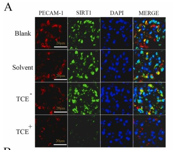
Application: IF/ICC Species: Mouse Sample: endothelial cells

Application: WB Species: Human Sample: HepG2 cells

Application: IF/ICC Species: Human Sample: HepG2 cells
Restrictive clause
Affinity Biosciences tests all products strictly. Citations are provided as a resource for additional applications that have not been validated by Affinity Biosciences. Please choose the appropriate format for each application and consult Materials and Methods sections for additional details about the use of any product in these publications.
For Research Use Only.
Not for use in diagnostic or therapeutic procedures. Not for resale. Not for distribution without written consent. Affinity Biosciences will not be held responsible for patent infringement or other violations that may occur with the use of our products. Affinity Biosciences, Affinity Biosciences Logo and all other trademarks are the property of Affinity Biosciences LTD.