GFAP Antibody - #BF0345
, rat brain(2), mouse brain(3) lysates.")
| Product: | GFAP Antibody |
| Catalog: | BF0345 |
| Description: | Mouse monoclonal antibody to GFAP |
| Application: | WB IHC-P |
| Reactivity: | Human, Mouse, Rat |
| Mol.Wt.: | 50kDa; 50kD(Calculated). |
| Uniprot: | P14136 |
| RRID: | AB_2833847 |
Product Info
*The optimal dilutions should be determined by the end user.
*Tips:
WB: For western blot detection of denatured protein samples. IHC: For immunohistochemical detection of paraffin sections (IHC-p) or frozen sections (IHC-f) of tissue samples. IF/ICC: For immunofluorescence detection of cell samples. ELISA(peptide): For ELISA detection of antigenic peptide.
Cite Format: Affinity Biosciences Cat# BF0345, RRID:AB_2833847.
Fold/Unfold
wu:fb34h11; ALXDRD; cb345; etID36982.3; FLJ42474; FLJ45472; GFAP; GFAP_HUMAN; gfapl; Glial fibrillary acidic protein; Intermediate filament protein; wu:fk42c12; xx:af506734; zgc:110485;
Immunogens
Purified recombinant fragment of human GFAP expressed in E. Coli.
- P14136 GFAP_HUMAN:
- Protein BLAST With
- NCBI/
- ExPASy/
- Uniprot
MERRRITSAARRSYVSSGEMMVGGLAPGRRLGPGTRLSLARMPPPLPTRVDFSLAGALNAGFKETRASERAEMMELNDRFASYIEKVRFLEQQNKALAAELNQLRAKEPTKLADVYQAELRELRLRLDQLTANSARLEVERDNLAQDLATVRQKLQDETNLRLEAENNLAAYRQEADEATLARLDLERKIESLEEEIRFLRKIHEEEVRELQEQLARQQVHVELDVAKPDLTAALKEIRTQYEAMASSNMHEAEEWYRSKFADLTDAAARNAELLRQAKHEANDYRRQLQSLTCDLESLRGTNESLERQMREQEERHVREAASYQEALARLEEEGQSLKDEMARHLQEYQDLLNVKLALDIEIATYRKLLEGEENRITIPVQTFSNLQIRETSLDTKSVSEGHLKRNIVVKTVEMRDGEVIKESKQEHKDVM
PTMs - P14136 As Substrate
| Site | PTM Type | Enzyme | Source |
|---|---|---|---|
| T7 | Phosphorylation | Q96GD4 (AURKB) , P17612 (PRKACA) , Q13464 (ROCK1) | Uniprot |
| S8 | Phosphorylation | P17612 (PRKACA) | Uniprot |
| R11 | Methylation | Uniprot | |
| R12 | Methylation | Uniprot | |
| S13 | Phosphorylation | Q13464 (ROCK1) , P17612 (PRKACA) , Q96GD4 (AURKB) | Uniprot |
| Y14 | Phosphorylation | Uniprot | |
| S17 | Phosphorylation | Uniprot | |
| S38 | Phosphorylation | Q96GD4 (AURKB) , Q13464 (ROCK1) , P17612 (PRKACA) | Uniprot |
| K95 | Acetylation | Uniprot | |
| Y116 | Phosphorylation | Uniprot | |
| T131 | Phosphorylation | Uniprot | |
| T150 | Phosphorylation | Uniprot | |
| K154 | Acetylation | Uniprot | |
| Y172 | Phosphorylation | Uniprot | |
| K189 | Acetylation | Uniprot | |
| K228 | Acetylation | Uniprot | |
| T240 | Phosphorylation | Uniprot | |
| Y242 | Phosphorylation | Uniprot | |
| K260 | Acetylation | Uniprot | |
| S305 | Phosphorylation | Uniprot | |
| Y324 | Phosphorylation | Uniprot | |
| K339 | Acetylation | Uniprot | |
| Y349 | Phosphorylation | Uniprot |
Research Backgrounds
GFAP, a class-III intermediate filament, is a cell-specific marker that, during the development of the central nervous system, distinguishes astrocytes from other glial cells.
Phosphorylated by PKN1.
Cytoplasm.
Note: Associated with intermediate filaments.
Expressed in cells lacking fibronectin.
Interacts with SYNM (By similarity). Isoform 3 interacts with PSEN1 (via N-terminus).
Belongs to the intermediate filament family.
Research Fields
· Environmental Information Processing > Signal transduction > Jak-STAT signaling pathway. (View pathway)
References
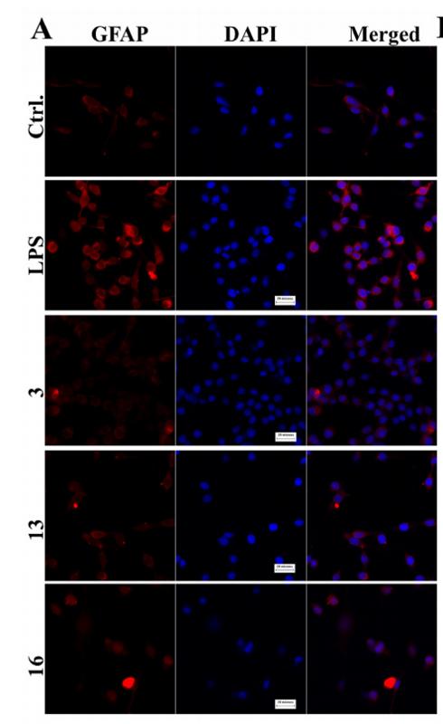
Application: IF/ICC Species: Mouse Sample: BV2 and C6 cells
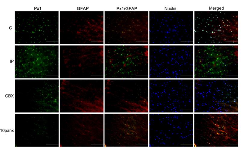
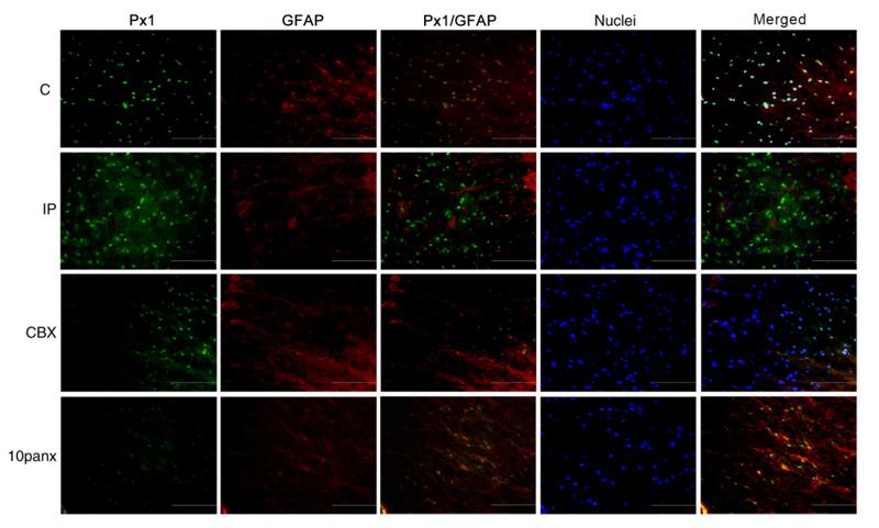
Application: IF/ICC Species: rat Sample:
Application: IF/ICC Species: Sample:
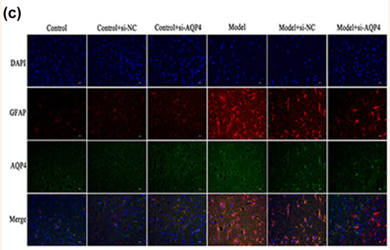
Application: IF/ICC Species: Mouse Sample:
Restrictive clause
Affinity Biosciences tests all products strictly. Citations are provided as a resource for additional applications that have not been validated by Affinity Biosciences. Please choose the appropriate format for each application and consult Materials and Methods sections for additional details about the use of any product in these publications.
For Research Use Only.
Not for use in diagnostic or therapeutic procedures. Not for resale. Not for distribution without written consent. Affinity Biosciences will not be held responsible for patent infringement or other violations that may occur with the use of our products. Affinity Biosciences, Affinity Biosciences Logo and all other trademarks are the property of Affinity Biosciences LTD.