beta-Actin Antibody - #T0022

| Product: | beta-Actin Antibody |
| Catalog: | T0022 |
| Description: | Mouse monoclonal antibody to beta-Actin |
| Application: | WB IHC IF/ICC ELISA |
| Reactivity: | Human, Mouse, Rat, Pig, Zebrafish, Bovine, Sheep, Rabbit, Goat, Guinea pig, Dog, Monkey, Hamster, Duck, Chicken, Fish |
| Mol.Wt.: | 43KD; 42kD(Calculated). |
| Uniprot: | P60709 |
| RRID: | AB_2839417 |
Product Info
*The optimal dilutions should be determined by the end user.
*Tips:
WB: For western blot detection of denatured protein samples. IHC: For immunohistochemical detection of paraffin sections (IHC-p) or frozen sections (IHC-f) of tissue samples. IF/ICC: For immunofluorescence detection of cell samples. ELISA(peptide): For ELISA detection of antigenic peptide.
Cite Format: Affinity Biosciences Cat# T0022, RRID:AB_2839417.
Fold/Unfold
ACTB;Actin; cytoplasmic 1; Beta-actin; Beta actin; BRWS1; Actin; beta; Beta cytoskeletal actin; PS1TP5-binding protein 1; PS1TP5BP1;
Immunogens
Full-length beta-Actin protein of human.
- P60709 ACTB_HUMAN:
- Protein BLAST With
- NCBI/
- ExPASy/
- Uniprot
MDDDIAALVVDNGSGMCKAGFAGDDAPRAVFPSIVGRPRHQGVMVGMGQKDSYVGDEAQSKRGILTLKYPIEHGIVTNWDDMEKIWHHTFYNELRVAPEEHPVLLTEAPLNPKANREKMTQIMFETFNTPAMYVAIQAVLSLYASGRTTGIVMDSGDGVTHTVPIYEGYALPHAILRLDLAGRDLTDYLMKILTERGYSFTTTAEREIVRDIKEKLCYVALDFEQEMATAASSSSLEKSYELPDGQVITIGNERFRCPEALFQPSFLGMESCGIHETTFNSIMKCDVDIRKDLYANTVLSGGTTMYPGIADRMQKEITALAPSTMKIKIIAPPERKYSVWIGGSILASLSTFQQMWISKQEYDESGPSIVHRKCF
PTMs - P60709 As Substrate
| Site | PTM Type | Enzyme | Source |
|---|---|---|---|
| M1 | Acetylation | Uniprot | |
| D2 | Acetylation | Uniprot | |
| S14 | Phosphorylation | Uniprot | |
| C17 | S-Nitrosylation | Uniprot | |
| K18 | Methylation | Uniprot | |
| K18 | Ubiquitination | Uniprot | |
| S33 | Phosphorylation | Uniprot | |
| K50 | Acetylation | Uniprot | |
| K50 | Methylation | Uniprot | |
| K50 | Ubiquitination | Uniprot | |
| S52 | Phosphorylation | Uniprot | |
| Y53 | Phosphorylation | Uniprot | |
| S60 | Phosphorylation | Uniprot | |
| K61 | Acetylation | Uniprot | |
| K61 | Sumoylation | Uniprot | |
| K61 | Ubiquitination | Uniprot | |
| T66 | Phosphorylation | Uniprot | |
| K68 | Methylation | Uniprot | |
| K68 | Sumoylation | Uniprot | |
| Y69 | Phosphorylation | Uniprot | |
| H73 | Methylation | Uniprot | |
| T77 | Phosphorylation | Uniprot | |
| K84 | Methylation | Uniprot | |
| K84 | Sumoylation | Uniprot | |
| K84 | Ubiquitination | Uniprot | |
| T89 | Phosphorylation | Uniprot | |
| Y91 | Phosphorylation | Uniprot | |
| T106 | Phosphorylation | Uniprot | |
| K113 | Acetylation | Uniprot | |
| K113 | Sumoylation | Uniprot | |
| K113 | Ubiquitination | Uniprot | |
| T120 | Phosphorylation | Uniprot | |
| Y143 | Phosphorylation | Uniprot | |
| S155 | Phosphorylation | Uniprot | |
| T160 | Phosphorylation | Uniprot | |
| T162 | Phosphorylation | Uniprot | |
| Y166 | Phosphorylation | Uniprot | |
| Y169 | Phosphorylation | Uniprot | |
| T186 | Phosphorylation | Uniprot | |
| Y188 | Phosphorylation | Uniprot | |
| K191 | Acetylation | Uniprot | |
| K191 | Methylation | Uniprot | |
| K191 | Ubiquitination | Uniprot | |
| T194 | Phosphorylation | Uniprot | |
| Y198 | Phosphorylation | Uniprot | |
| S199 | Phosphorylation | Uniprot | |
| T201 | Phosphorylation | Uniprot | |
| T202 | Phosphorylation | Uniprot | |
| T203 | Phosphorylation | Uniprot | |
| K213 | Acetylation | Uniprot | |
| K213 | Ubiquitination | Uniprot | |
| K215 | Ubiquitination | Uniprot | |
| C217 | S-Nitrosylation | Uniprot | |
| Y218 | Phosphorylation | Uniprot | |
| T229 | Phosphorylation | Uniprot | |
| S233 | Phosphorylation | Uniprot | |
| S235 | Phosphorylation | Uniprot | |
| K238 | Ubiquitination | Uniprot | |
| S239 | Phosphorylation | Uniprot | |
| Y240 | Phosphorylation | Uniprot | |
| T249 | Phosphorylation | Uniprot | |
| C257 | S-Nitrosylation | Uniprot | |
| S265 | Phosphorylation | Uniprot | |
| S271 | Phosphorylation | Uniprot | |
| C272 | S-Nitrosylation | Uniprot | |
| K284 | Sumoylation | Uniprot | |
| K284 | Ubiquitination | Uniprot | |
| C285 | S-Nitrosylation | Uniprot | |
| K291 | Sumoylation | Uniprot | |
| K291 | Ubiquitination | Uniprot | |
| Y294 | Phosphorylation | Uniprot | |
| T297 | Phosphorylation | Uniprot | |
| S300 | Phosphorylation | Uniprot | |
| T303 | Phosphorylation | Uniprot | |
| T304 | Phosphorylation | Uniprot | |
| Y306 | Phosphorylation | Uniprot | |
| K315 | Acetylation | Uniprot | |
| K315 | Sumoylation | Uniprot | |
| K315 | Ubiquitination | Uniprot | |
| T318 | Phosphorylation | Uniprot | |
| S323 | Phosphorylation | Uniprot | |
| T324 | Phosphorylation | Uniprot | |
| K326 | Acetylation | Uniprot | |
| K326 | Methylation | Uniprot | |
| K326 | Sumoylation | Uniprot | |
| K326 | Ubiquitination | Uniprot | |
| K328 | Acetylation | Uniprot | |
| K328 | Sumoylation | Uniprot | |
| K328 | Ubiquitination | Uniprot | |
| K336 | Sumoylation | Uniprot | |
| S348 | Phosphorylation | Uniprot | |
| K359 | Ubiquitination | Uniprot | |
| Y362 | Phosphorylation | Uniprot | |
| S365 | Phosphorylation | Uniprot | |
| S368 | Phosphorylation | Uniprot | |
| K373 | Ubiquitination | Uniprot | |
| C374 | S-Nitrosylation | Uniprot |
Research Backgrounds
Actin is a highly conserved protein that polymerizes to produce filaments that form cross-linked networks in the cytoplasm of cells. Actin exists in both monomeric (G-actin) and polymeric (F-actin) forms, both forms playing key functions, such as cell motility and contraction. In addition to their role in the cytoplasmic cytoskeleton, G- and F-actin also localize in the nucleus, and regulate gene transcription and motility and repair of damaged DNA.
ISGylated.
Oxidation of Met-44 and Met-47 by MICALs (MICAL1, MICAL2 or MICAL3) to form methionine sulfoxide promotes actin filament depolymerization. MICAL1 and MICAL2 produce the (R)-S-oxide form. The (R)-S-oxide form is reverted by MSRB1 and MSRB2, which promote actin repolymerization.
Monomethylation at Lys-84 (K84me1) regulates actin-myosin interaction and actomyosin-dependent processes. Demethylation by ALKBH4 is required for maintaining actomyosin dynamics supporting normal cleavage furrow ingression during cytokinesis and cell migration.
Methylated at His-73 by SETD3. Methylation at His-73 is required for smooth muscle contraction of the laboring uterus during delivery (By similarity).
N-terminal acetylation by NAA80 affects actin filament depolymerization and elongation, including elongation driven by formins. In contrast, filament nucleation by the Arp2/3 complex is not affected.
(Microbial infection) Monomeric actin is cross-linked by V.cholerae toxins RtxA and VgrG1 in case of infection: bacterial toxins mediate the cross-link between Lys-50 of one monomer and Glu-270 of another actin monomer, resulting in formation of highly toxic actin oligomers that cause cell rounding. The toxin can be highly efficient at very low concentrations by acting on formin homology family proteins: toxic actin oligomers bind with high affinity to formins and adversely affect both nucleation and elongation abilities of formins, causing their potent inhibition in both profilin-dependent and independent manners.
Cytoplasm>Cytoskeleton. Nucleus.
Note: Localized in cytoplasmic mRNP granules containing untranslated mRNAs.
Polymerization of globular actin (G-actin) leads to a structural filament (F-actin) in the form of a two-stranded helix. Each actin can bind to 4 others. Identified in a IGF2BP1-dependent mRNP granule complex containing untranslated mRNAs. Component of the BAF complex, which includes at least actin (ACTB), ARID1A, ARID1B/BAF250, SMARCA2, SMARCA4/BRG1, ACTL6A/BAF53, ACTL6B/BAF53B, SMARCE1/BAF57 SMARCC1/BAF155, SMARCC2/BAF170, SMARCB1/SNF5/INI1, and one or more of SMARCD1/BAF60A, SMARCD2/BAF60B, or SMARCD3/BAF60C. In muscle cells, the BAF complex also contains DPF3. Found in a complex with XPO6, Ran, ACTB and PFN1. Interacts with XPO6 and EMD. Interacts with ERBB2. Interacts with GCSAM. Interacts with TBC1D21 (By similarity). Interacts with CPNE1 (via VWFA domain) and CPNE4 (via VWFA domain) (By similarity). Interacts with DHX9 (via C-terminus); this interaction is direct and mediates the attachment to nuclear ribonucleoprotein complexes. Interacts with FAM107A.
Belongs to the actin family.
Research Fields
· Cellular Processes > Transport and catabolism > Phagosome. (View pathway)
· Cellular Processes > Cell growth and death > Apoptosis. (View pathway)
· Cellular Processes > Cellular community - eukaryotes > Focal adhesion. (View pathway)
· Cellular Processes > Cellular community - eukaryotes > Adherens junction. (View pathway)
· Cellular Processes > Cellular community - eukaryotes > Tight junction. (View pathway)
· Cellular Processes > Cell motility > Regulation of actin cytoskeleton. (View pathway)
· Environmental Information Processing > Signal transduction > Rap1 signaling pathway. (View pathway)
· Environmental Information Processing > Signal transduction > Hippo signaling pathway. (View pathway)
· Human Diseases > Infectious diseases: Bacterial > Bacterial invasion of epithelial cells.
· Human Diseases > Infectious diseases: Bacterial > Vibrio cholerae infection.
· Human Diseases > Infectious diseases: Bacterial > Pathogenic Escherichia coli infection.
· Human Diseases > Infectious diseases: Bacterial > Shigellosis.
· Human Diseases > Infectious diseases: Bacterial > Salmonella infection.
· Human Diseases > Infectious diseases: Viral > Influenza A.
· Human Diseases > Cancers: Overview > Proteoglycans in cancer.
· Human Diseases > Cancers: Specific types > Hepatocellular carcinoma. (View pathway)
· Human Diseases > Cardiovascular diseases > Hypertrophic cardiomyopathy (HCM).
· Human Diseases > Cardiovascular diseases > Arrhythmogenic right ventricular cardiomyopathy (ARVC).
· Human Diseases > Cardiovascular diseases > Dilated cardiomyopathy (DCM).
· Human Diseases > Cardiovascular diseases > Viral myocarditis.
· Organismal Systems > Immune system > Platelet activation. (View pathway)
· Organismal Systems > Immune system > Leukocyte transendothelial migration. (View pathway)
· Organismal Systems > Endocrine system > Thyroid hormone signaling pathway. (View pathway)
· Organismal Systems > Endocrine system > Oxytocin signaling pathway.
· Organismal Systems > Digestive system > Gastric acid secretion.
References
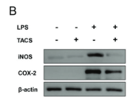
Application: WB Species: Mouse Sample: RAW264.7 cells
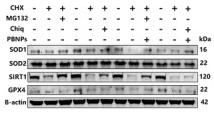
Application: WB Species: Rat Sample: NP cells

Application: WB Species: human Sample: hepatoma cell lines
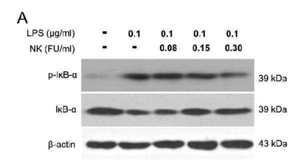
Application: WB Species: Mice Sample: RAW264.7 cells
Application: WB Species: Mouse Sample:
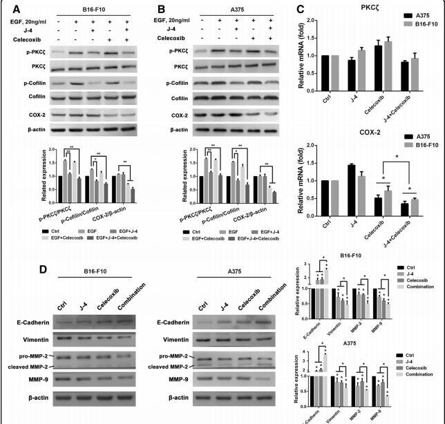
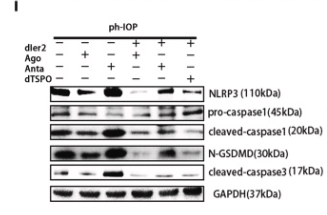
Application: WB Species: mouse Sample: B16F10
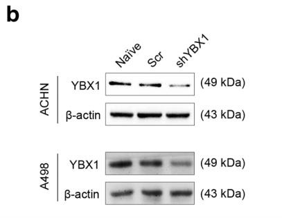
Application: WB Species: human Sample: A498
Restrictive clause
Affinity Biosciences tests all products strictly. Citations are provided as a resource for additional applications that have not been validated by Affinity Biosciences. Please choose the appropriate format for each application and consult Materials and Methods sections for additional details about the use of any product in these publications.
For Research Use Only.
Not for use in diagnostic or therapeutic procedures. Not for resale. Not for distribution without written consent. Affinity Biosciences will not be held responsible for patent infringement or other violations that may occur with the use of our products. Affinity Biosciences, Affinity Biosciences Logo and all other trademarks are the property of Affinity Biosciences LTD.