Tubulin alpha Antibody - #AF7010

| Product: | Tubulin alpha Antibody |
| Catalog: | AF7010 |
| Description: | Rabbit polyclonal antibody to Tubulin alpha |
| Application: | WB IHC IF/ICC |
| Reactivity: | Human, Mouse, Rat, Pig, Bovine, Rabbit, Chicken, Plants, Fish |
| Prediction: | Pig, Bovine, Horse, Sheep, Dog |
| Mol.Wt.: | 50kDa; 50kD(Calculated). |
| Uniprot: | P68363 | Q71U36 |
| RRID: | AB_2839418 |
Product Info
*The optimal dilutions should be determined by the end user.
*Tips:
WB: For western blot detection of denatured protein samples. IHC: For immunohistochemical detection of paraffin sections (IHC-p) or frozen sections (IHC-f) of tissue samples. IF/ICC: For immunofluorescence detection of cell samples. ELISA(peptide): For ELISA detection of antigenic peptide.
Cite Format: Affinity Biosciences Cat# AF7010, RRID:AB_2839418.
Fold/Unfold
TUBA1A; Alpha-tubulin 3; B-ALPHA-1; Hum-a-tub1; TUBA3; Tubulin alpha-3 chain; Tubulin B-alpha-1; Tubulin alpha 1a; Tubulin alpha; Tubulin Alpha 1a; Tubulin alpha-1A chain;Tubulin alpha-1B chain;
Immunogens
- P68363 TBA1B_HUMAN:
- Protein BLAST With
- NCBI/
- ExPASy/
- Uniprot
MRECISIHVGQAGVQIGNACWELYCLEHGIQPDGQMPSDKTIGGGDDSFNTFFSETGAGKHVPRAVFVDLEPTVIDEVRTGTYRQLFHPEQLITGKEDAANNYARGHYTIGKEIIDLVLDRIRKLADQCTGLQGFLVFHSFGGGTGSGFTSLLMERLSVDYGKKSKLEFSIYPAPQVSTAVVEPYNSILTTHTTLEHSDCAFMVDNEAIYDICRRNLDIERPTYTNLNRLISQIVSSITASLRFDGALNVDLTEFQTNLVPYPRIHFPLATYAPVISAEKAYHEQLSVAEITNACFEPANQMVKCDPRHGKYMACCLLYRGDVVPKDVNAAIATIKTKRSIQFVDWCPTGFKVGINYQPPTVVPGGDLAKVQRAVCMLSNTTAIAEAWARLDHKFDLMYAKRAFVHWYVGEGMEEGEFSEAREDMAALEKDYEEVGVDSVEGEGEEEGEEY
- Q71U36 TBA1A_HUMAN:
- Protein BLAST With
- NCBI/
- ExPASy/
- Uniprot
MRECISIHVGQAGVQIGNACWELYCLEHGIQPDGQMPSDKTIGGGDDSFNTFFSETGAGKHVPRAVFVDLEPTVIDEVRTGTYRQLFHPEQLITGKEDAANNYARGHYTIGKEIIDLVLDRIRKLADQCTGLQGFLVFHSFGGGTGSGFTSLLMERLSVDYGKKSKLEFSIYPAPQVSTAVVEPYNSILTTHTTLEHSDCAFMVDNEAIYDICRRNLDIERPTYTNLNRLIGQIVSSITASLRFDGALNVDLTEFQTNLVPYPRIHFPLATYAPVISAEKAYHEQLSVAEITNACFEPANQMVKCDPRHGKYMACCLLYRGDVVPKDVNAAIATIKTKRTIQFVDWCPTGFKVGINYQPPTVVPGGDLAKVQRAVCMLSNTTAIAEAWARLDHKFDLMYAKRAFVHWYVGEGMEEGEFSEAREDMAALEKDYEEVGVDSVEGEGEEEGEEY
Predictions
Score>80(red) has high confidence and is suggested to be used for WB detection. *The prediction model is mainly based on the alignment of immunogen sequences, the results are for reference only, not as the basis of quality assurance.
High(score>80) Medium(80>score>50) Low(score<50) No confidence
PTMs - P68363/Q71U36 As Substrate
| Site | PTM Type | Enzyme | Source |
|---|---|---|---|
| S6 | Phosphorylation | Uniprot | |
| Y24 | Phosphorylation | Uniprot | |
| S38 | Phosphorylation | Uniprot | |
| K40 | Acetylation | Uniprot | |
| K40 | Methylation | Uniprot | |
| K40 | Ubiquitination | Uniprot | |
| T41 | Phosphorylation | Uniprot | |
| S48 | Phosphorylation | Uniprot | |
| T51 | Phosphorylation | Uniprot | |
| S54 | Phosphorylation | Uniprot | |
| T56 | Phosphorylation | Uniprot | |
| K60 | Acetylation | Uniprot | |
| K60 | Sumoylation | Uniprot | |
| K60 | Ubiquitination | Uniprot | |
| T73 | Phosphorylation | Uniprot | |
| T80 | Phosphorylation | Uniprot | |
| T82 | Phosphorylation | Uniprot | |
| Y83 | Phosphorylation | Uniprot | |
| T94 | Phosphorylation | Uniprot | |
| K96 | Acetylation | Uniprot | |
| K96 | Sumoylation | Uniprot | |
| K96 | Ubiquitination | Uniprot | |
| Y103 | Phosphorylation | Uniprot | |
| Y108 | Phosphorylation | Uniprot | |
| T109 | Phosphorylation | Uniprot | |
| K112 | Acetylation | Uniprot | |
| K112 | Sumoylation | Uniprot | |
| K112 | Ubiquitination | Uniprot | |
| K124 | Acetylation | Uniprot | |
| K124 | Ubiquitination | Uniprot | |
| S151 | Phosphorylation | Uniprot | |
| S158 | Phosphorylation | Uniprot | |
| Y161 | Phosphorylation | Uniprot | |
| K163 | Acetylation | Uniprot | |
| K163 | Methylation | Uniprot | |
| K163 | Ubiquitination | Uniprot | |
| K164 | Ubiquitination | Uniprot | |
| S165 | Phosphorylation | Uniprot | |
| K166 | Ubiquitination | Uniprot | |
| Y172 | Phosphorylation | Uniprot | |
| Y185 | Phosphorylation | Uniprot | |
| S187 | Phosphorylation | Uniprot | |
| T190 | Phosphorylation | Uniprot | |
| T191 | Phosphorylation | Uniprot | |
| T193 | Phosphorylation | Uniprot | |
| T194 | Phosphorylation | Uniprot | |
| S198 | Phosphorylation | Uniprot | |
| Y210 | Phosphorylation | Uniprot | |
| T223 | Phosphorylation | Uniprot | |
| Y224 | Phosphorylation | Uniprot | |
| T225 | Phosphorylation | Uniprot | |
| S236 | Phosphorylation | Uniprot | |
| S237 | Phosphorylation | Uniprot | |
| T253 | Phosphorylation | Uniprot | |
| T257 | Phosphorylation | Uniprot | |
| Y262 | Phosphorylation | Uniprot | |
| T271 | Phosphorylation | Uniprot | |
| Y272 | Phosphorylation | Uniprot | |
| S277 | Phosphorylation | Uniprot | |
| K280 | Ubiquitination | Uniprot | |
| Y282 | Phosphorylation | Uniprot | |
| S287 | Phosphorylation | Uniprot | |
| T292 | Phosphorylation | Uniprot | |
| K304 | Ubiquitination | Uniprot | |
| K311 | Acetylation | Uniprot | |
| K311 | Ubiquitination | Uniprot | |
| Y312 | Phosphorylation | Uniprot | |
| Y319 | Phosphorylation | Uniprot | |
| K326 | Acetylation | Uniprot | |
| K326 | Sumoylation | Uniprot | |
| K326 | Ubiquitination | Uniprot | |
| T334 | Phosphorylation | Uniprot | |
| K336 | Acetylation | Uniprot | |
| K336 | Sumoylation | Uniprot | |
| K336 | Ubiquitination | Uniprot | |
| T337 | Phosphorylation | Uniprot | |
| K338 | Methylation | Uniprot | |
| K338 | Ubiquitination | Uniprot | |
| T340 | Phosphorylation | Uniprot | |
| T349 | Phosphorylation | Uniprot | |
| K352 | Acetylation | Uniprot | |
| K352 | Sumoylation | Uniprot | |
| K352 | Ubiquitination | Uniprot | |
| Y357 | Phosphorylation | Uniprot | |
| T361 | Phosphorylation | Uniprot | |
| K370 | Acetylation | Uniprot | |
| K370 | Sumoylation | Uniprot | |
| K370 | Ubiquitination | Uniprot | |
| S379 | Phosphorylation | Uniprot | |
| K394 | Acetylation | Uniprot | |
| K394 | Sumoylation | Uniprot | |
| K394 | Ubiquitination | Uniprot | |
| Y399 | Phosphorylation | Uniprot | |
| K401 | Acetylation | Uniprot | |
| K401 | Sumoylation | Uniprot | |
| K401 | Ubiquitination | Uniprot | |
| Y408 | Phosphorylation | Uniprot | |
| S419 | Phosphorylation | Uniprot | |
| K430 | Ubiquitination | Uniprot | |
| Y432 | Phosphorylation | Uniprot | |
| S439 | Phosphorylation | Uniprot | |
| Y451 | Phosphorylation | Uniprot |
| Site | PTM Type | Enzyme | Source |
|---|---|---|---|
| S6 | Phosphorylation | Uniprot | |
| Y24 | Phosphorylation | Uniprot | |
| S38 | Phosphorylation | Uniprot | |
| K40 | Acetylation | Uniprot | |
| K40 | Methylation | Uniprot | |
| K40 | Ubiquitination | Uniprot | |
| T41 | Phosphorylation | Uniprot | |
| S48 | Phosphorylation | Uniprot | |
| T51 | Phosphorylation | Uniprot | |
| S54 | Phosphorylation | Uniprot | |
| T56 | Phosphorylation | Uniprot | |
| K60 | Acetylation | Uniprot | |
| K60 | Sumoylation | Uniprot | |
| K60 | Ubiquitination | Uniprot | |
| T73 | Phosphorylation | Uniprot | |
| T80 | Phosphorylation | Uniprot | |
| T82 | Phosphorylation | Uniprot | |
| Y83 | Phosphorylation | Uniprot | |
| T94 | Phosphorylation | Uniprot | |
| K96 | Acetylation | Uniprot | |
| K96 | Sumoylation | Uniprot | |
| K96 | Ubiquitination | Uniprot | |
| Y103 | Phosphorylation | Uniprot | |
| Y108 | Phosphorylation | Uniprot | |
| T109 | Phosphorylation | Uniprot | |
| K112 | Acetylation | Uniprot | |
| K112 | Sumoylation | Uniprot | |
| K112 | Ubiquitination | Uniprot | |
| K124 | Acetylation | Uniprot | |
| K124 | Ubiquitination | Uniprot | |
| S151 | Phosphorylation | Uniprot | |
| S158 | Phosphorylation | Uniprot | |
| Y161 | Phosphorylation | Uniprot | |
| K163 | Acetylation | Uniprot | |
| K163 | Methylation | Uniprot | |
| K163 | Ubiquitination | Uniprot | |
| K164 | Ubiquitination | Uniprot | |
| K166 | Ubiquitination | Uniprot | |
| Y172 | Phosphorylation | Uniprot | |
| Y185 | Phosphorylation | Uniprot | |
| S187 | Phosphorylation | Uniprot | |
| T190 | Phosphorylation | Uniprot | |
| T191 | Phosphorylation | Uniprot | |
| T193 | Phosphorylation | Uniprot | |
| T194 | Phosphorylation | Uniprot | |
| S198 | Phosphorylation | Uniprot | |
| Y210 | Phosphorylation | Uniprot | |
| T223 | Phosphorylation | Uniprot | |
| Y224 | Phosphorylation | Uniprot | |
| T225 | Phosphorylation | Uniprot | |
| S232 | Phosphorylation | Uniprot | |
| S236 | Phosphorylation | Uniprot | |
| S237 | Phosphorylation | Uniprot | |
| T239 | Phosphorylation | Uniprot | |
| S241 | Phosphorylation | Uniprot | |
| T253 | Phosphorylation | Uniprot | |
| T257 | Phosphorylation | Uniprot | |
| Y262 | Phosphorylation | Uniprot | |
| T271 | Phosphorylation | Uniprot | |
| Y272 | Phosphorylation | Uniprot | |
| S277 | Phosphorylation | Uniprot | |
| K280 | Acetylation | Uniprot | |
| K280 | Ubiquitination | Uniprot | |
| Y282 | Phosphorylation | Uniprot | |
| S287 | Phosphorylation | Uniprot | |
| T292 | Phosphorylation | Uniprot | |
| C295 | S-Nitrosylation | Uniprot | |
| K304 | Ubiquitination | Uniprot | |
| K311 | Acetylation | Uniprot | |
| K311 | Ubiquitination | Uniprot | |
| Y312 | Phosphorylation | Uniprot | |
| C315 | S-Nitrosylation | Uniprot | |
| C316 | S-Nitrosylation | Uniprot | |
| Y319 | Phosphorylation | Uniprot | |
| K326 | Acetylation | Uniprot | |
| K326 | Sumoylation | Uniprot | |
| K326 | Ubiquitination | Uniprot | |
| T334 | Phosphorylation | Uniprot | |
| K336 | Acetylation | Uniprot | |
| K336 | Sumoylation | Uniprot | |
| K336 | Ubiquitination | Uniprot | |
| T337 | Phosphorylation | Uniprot | |
| K338 | Methylation | Uniprot | |
| K338 | Ubiquitination | Uniprot | |
| R339 | Methylation | Uniprot | |
| S340 | Phosphorylation | Uniprot | |
| C347 | S-Nitrosylation | Uniprot | |
| T349 | Phosphorylation | Uniprot | |
| K352 | Acetylation | Uniprot | |
| K352 | Sumoylation | Uniprot | |
| K352 | Ubiquitination | Uniprot | |
| Y357 | Phosphorylation | Uniprot | |
| T361 | Phosphorylation | Uniprot | |
| K370 | Acetylation | Uniprot | |
| K370 | Sumoylation | Uniprot | |
| K370 | Ubiquitination | Uniprot | |
| C376 | S-Nitrosylation | Uniprot | |
| S379 | Phosphorylation | Uniprot | |
| K394 | Acetylation | Uniprot | |
| K394 | Methylation | Uniprot | |
| K394 | Sumoylation | Uniprot | |
| K394 | Ubiquitination | Uniprot | |
| Y399 | Phosphorylation | Uniprot | |
| K401 | Acetylation | Uniprot | |
| K401 | Sumoylation | Uniprot | |
| K401 | Ubiquitination | Uniprot | |
| Y408 | Phosphorylation | Uniprot | |
| S419 | Phosphorylation | Uniprot | |
| K430 | Ubiquitination | Uniprot | |
| Y432 | Phosphorylation | Uniprot | |
| S439 | Phosphorylation | Uniprot | |
| Y451 | Phosphorylation | Uniprot |
Research Backgrounds
Tubulin is the major constituent of microtubules. It binds two moles of GTP, one at an exchangeable site on the beta chain and one at a non-exchangeable site on the alpha chain.
Some glutamate residues at the C-terminus are polyglutamylated, resulting in polyglutamate chains on the gamma-carboxyl group. Polyglutamylation plays a key role in microtubule severing by spastin (SPAST). SPAST preferentially recognizes and acts on microtubules decorated with short polyglutamate tails: severing activity by SPAST increases as the number of glutamates per tubulin rises from one to eight, but decreases beyond this glutamylation threshold.
Some glutamate residues at the C-terminus are monoglycylated but not polyglycylated due to the absence of functional TTLL10 in human. Monoglycylation is mainly limited to tubulin incorporated into axonemes (cilia and flagella). Both polyglutamylation and monoglycylation can coexist on the same protein on adjacent residues, and lowering glycylation levels increases polyglutamylation, and reciprocally. The precise function of monoglycylation is still unclear (Probable).
Acetylation of alpha chains at Lys-40 is located inside the microtubule lumen. This modification has been correlated with increased microtubule stability, intracellular transport and ciliary assembly.
Methylation of alpha chains at Lys-40 is found in mitotic microtubules and is required for normal mitosis and cytokinesis contributing to genomic stability.
Nitration of Tyr-451 is irreversible and interferes with normal dynein intracellular distribution.
Undergoes a tyrosination/detyrosination cycle, the cyclic removal and re-addition of a C-terminal tyrosine residue by the enzymes tubulin tyrosine carboxypeptidase (VASH1 or VASH2) and tubulin tyrosine ligase (TTL), respectively.
Tyrosination promotes microtubule interaction with CAP-Gly domain-containing proteins such as CLIP1, CLIP2 and DCTN1 (By similarity). Tyrosination regulates the initiation of dynein-dynactin motility via interaction with DCTN1, which brings the dynein-dynactin complex into contact with microtubules. In neurons, tyrosinated tubulins mediate the initiation of retrograde vesicle transport (By similarity).
Detyrosination is involved in metaphase plate congression by guiding chromosomes during mitosis: detyrosination promotes interaction with CENPE, promoting pole-proximal transport of chromosomes toward the equator. Detyrosination increases microtubules-dependent mechanotransduction in dystrophic cardiac and skeletal muscle. In cardiomyocytes, detyrosinated microtubules are required to resist to contractile compression during contraction: detyrosination promotes association with desmin (DES) at force-generating sarcomeres, leading to buckled microtubules and mechanical resistance to contraction (By similarity).
Cytoplasm>Cytoskeleton.
Dimer of alpha and beta chains. A typical microtubule is a hollow water-filled tube with an outer diameter of 25 nm and an inner diameter of 15 nM. Alpha-beta heterodimers associate head-to-tail to form protofilaments running lengthwise along the microtubule wall with the beta-tubulin subunit facing the microtubule plus end conferring a structural polarity. Microtubules usually have 13 protofilaments but different protofilament numbers can be found in some organisms and specialized cells.
Belongs to the tubulin family.
Tubulin is the major constituent of microtubules. It binds two moles of GTP, one at an exchangeable site on the beta chain and one at a non-exchangeable site on the alpha chain.
Some glutamate residues at the C-terminus are polyglutamylated, resulting in polyglutamate chains on the gamma-carboxyl group. Polyglutamylation plays a key role in microtubule severing by spastin (SPAST). SPAST preferentially recognizes and acts on microtubules decorated with short polyglutamate tails: severing activity by SPAST increases as the number of glutamates per tubulin rises from one to eight, but decreases beyond this glutamylation threshold.
Some glutamate residues at the C-terminus are monoglycylated but not polyglycylated due to the absence of functional TTLL10 in human. Monoglycylation is mainly limited to tubulin incorporated into axonemes (cilia and flagella). Both polyglutamylation and monoglycylation can coexist on the same protein on adjacent residues, and lowering glycylation levels increases polyglutamylation, and reciprocally. The precise function of monoglycylation is still unclear (Probable).
Acetylation of alpha chains at Lys-40 is located inside the microtubule lumen. This modification has been correlated with increased microtubule stability, intracellular transport and ciliary assembly.
Methylation of alpha chains at Lys-40 is found in mitotic microtubules and is required for normal mitosis and cytokinesis contributing to genomic stability.
Nitration of Tyr-451 is irreversible and interferes with normal dynein intracellular distribution.
Undergoes a tyrosination/detyrosination cycle, the cyclic removal and re-addition of a C-terminal tyrosine residue by the enzymes tubulin tyrosine carboxypeptidase (VASH1 or VASH2) and tubulin tyrosine ligase (TTL), respectively.
Tyrosination promotes microtubule interaction with CAP-Gly domain-containing proteins such as CLIP1, CLIP2 and DCTN1. Tyrosination regulates the initiation of dynein-dynactin motility via interaction with DCTN1, which brings the dynein-dynactin complex into contact with microtubules. In neurons, tyrosinated tubulins mediate the initiation of retrograde vesicle transport.
Detyrosination is involved in metaphase plate congression by guiding chromosomes during mitosis: detyrosination promotes interaction with CENPE, promoting pole-proximal transport of chromosomes toward the equator. Detyrosination increases microtubules-dependent mechanotransduction in dystrophic cardiac and skeletal muscle. In cardiomyocytes, detyrosinated microtubules are required to resist to contractile compression during contraction: detyrosination promotes association with desmin (DES) at force-generating sarcomeres, leading to buckled microtubules and mechanical resistance to contraction (By similarity).
Cytoplasm>Cytoskeleton.
Expressed at a high level in fetal brain.
Dimer of alpha and beta chains. A typical microtubule is a hollow water-filled tube with an outer diameter of 25 nm and an inner diameter of 15 nM. Alpha-beta heterodimers associate head-to-tail to form protofilaments running lengthwise along the microtubule wall with the beta-tubulin subunit facing the microtubule plus end conferring a structural polarity. Microtubules usually have 13 protofilaments but different protofilament numbers can be found in some organisms and specialized cells. Interacts with SETD2; the interaction is independent on alpha-tubulin acetylation on Lys-40.
Belongs to the tubulin family.
Research Fields
· Cellular Processes > Transport and catabolism > Phagosome. (View pathway)
· Cellular Processes > Cell growth and death > Apoptosis. (View pathway)
· Cellular Processes > Cellular community - eukaryotes > Tight junction. (View pathway)
· Cellular Processes > Cellular community - eukaryotes > Gap junction. (View pathway)
· Human Diseases > Infectious diseases: Bacterial > Pathogenic Escherichia coli infection.
References

Application: WB Species: human Sample: MRC-5 fibroblasts
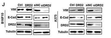
Application: WB Species: mouse Sample: tumor cells
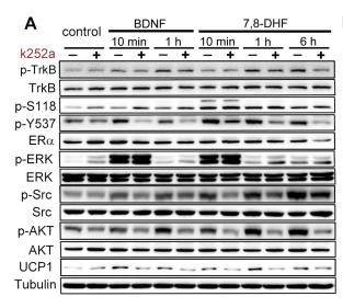
Application: WB Species: Sample:
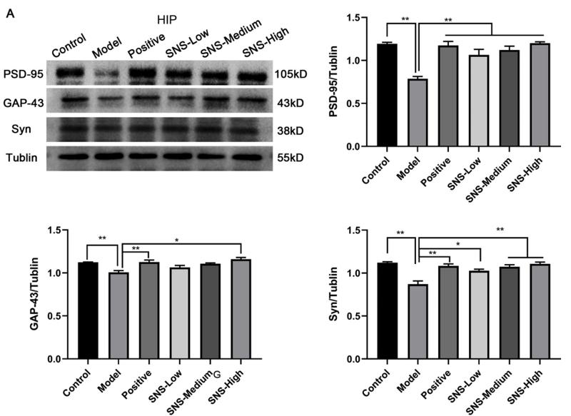
Application: WB Species: rat Sample: HIP
Restrictive clause
Affinity Biosciences tests all products strictly. Citations are provided as a resource for additional applications that have not been validated by Affinity Biosciences. Please choose the appropriate format for each application and consult Materials and Methods sections for additional details about the use of any product in these publications.
For Research Use Only.
Not for use in diagnostic or therapeutic procedures. Not for resale. Not for distribution without written consent. Affinity Biosciences will not be held responsible for patent infringement or other violations that may occur with the use of our products. Affinity Biosciences, Affinity Biosciences Logo and all other trademarks are the property of Affinity Biosciences LTD.