FGFR2 Antibody - #AF0159

| Product: | FGFR2 Antibody |
| Catalog: | AF0159 |
| Description: | Rabbit polyclonal antibody to FGFR2 |
| Application: | WB IHC |
| Reactivity: | Human, Mouse, Rat |
| Prediction: | Pig, Zebrafish, Bovine, Horse, Rabbit, Dog, Chicken, Xenopus |
| Mol.Wt.: | 90kD, 140kDa; 92kD(Calculated). |
| Uniprot: | P21802 |
| RRID: | AB_2833340 |
Product Info
*The optimal dilutions should be determined by the end user.
*Tips:
WB: For western blot detection of denatured protein samples. IHC: For immunohistochemical detection of paraffin sections (IHC-p) or frozen sections (IHC-f) of tissue samples. IF/ICC: For immunofluorescence detection of cell samples. ELISA(peptide): For ELISA detection of antigenic peptide.
Cite Format: Affinity Biosciences Cat# AF0159, RRID:AB_2833340.
Fold/Unfold
bacteria-expressed kinase; BBDS; BEK; BEK fibroblast growth factor receptor; BFR1; CD332; CD332 antigen; CEK3; CFD1; Craniofacial dysostosis 1; ECT1; FGF receptor; FGFR 2; FGFR-2; Fgfr2; FGFR2_HUMAN; Fibroblast growth factor receptor 2; Hydroxyaryl protein kinase; Jackson Weiss syndrome; JWS; K SAM; K-sam; Keratinocyte growth factor receptor 2; Keratinocyte growth factor receptor; KGFR; KSAM; protein tyrosine kinase, receptor like 14; soluble FGFR4 variant 4; TK14; TK25;
Immunogens
- P21802 FGFR2_HUMAN:
- Protein BLAST With
- NCBI/
- ExPASy/
- Uniprot
MVSWGRFICLVVVTMATLSLARPSFSLVEDTTLEPEEPPTKYQISQPEVYVAAPGESLEVRCLLKDAAVISWTKDGVHLGPNNRTVLIGEYLQIKGATPRDSGLYACTASRTVDSETWYFMVNVTDAISSGDDEDDTDGAEDFVSENSNNKRAPYWTNTEKMEKRLHAVPAANTVKFRCPAGGNPMPTMRWLKNGKEFKQEHRIGGYKVRNQHWSLIMESVVPSDKGNYTCVVENEYGSINHTYHLDVVERSPHRPILQAGLPANASTVVGGDVEFVCKVYSDAQPHIQWIKHVEKNGSKYGPDGLPYLKVLKAAGVNTTDKEIEVLYIRNVTFEDAGEYTCLAGNSIGISFHSAWLTVLPAPGREKEITASPDYLEIAIYCIGVFLIACMVVTVILCRMKNTTKKPDFSSQPAVHKLTKRIPLRRQVTVSAESSSSMNSNTPLVRITTRLSSTADTPMLAGVSEYELPEDPKWEFPRDKLTLGKPLGEGCFGQVVMAEAVGIDKDKPKEAVTVAVKMLKDDATEKDLSDLVSEMEMMKMIGKHKNIINLLGACTQDGPLYVIVEYASKGNLREYLRARRPPGMEYSYDINRVPEEQMTFKDLVSCTYQLARGMEYLASQKCIHRDLAARNVLVTENNVMKIADFGLARDINNIDYYKKTTNGRLPVKWMAPEALFDRVYTHQSDVWSFGVLMWEIFTLGGSPYPGIPVEELFKLLKEGHRMDKPANCTNELYMMMRDCWHAVPSQRPTFKQLVEDLDRILTLTTNEEYLDLSQPLEQYSPSYPDTRSSCSSGDDSVFSPDPMPYEPCLPQYPHINGSVKT
Predictions
Score>80(red) has high confidence and is suggested to be used for WB detection. *The prediction model is mainly based on the alignment of immunogen sequences, the results are for reference only, not as the basis of quality assurance.
High(score>80) Medium(80>score>50) Low(score<50) No confidence
PTMs - P21802 As Substrate
| Site | PTM Type | Enzyme | Source |
|---|---|---|---|
| Phosphorylation | Uniprot | ||
| S347 | Phosphorylation | Uniprot | |
| S437 | Phosphorylation | Uniprot | |
| S440 | Phosphorylation | Uniprot | |
| T442 | Phosphorylation | Uniprot | |
| T448 | Phosphorylation | Uniprot | |
| T449 | Phosphorylation | Uniprot | |
| S452 | Phosphorylation | Uniprot | |
| S453 | Phosphorylation | Uniprot | |
| T454 | Phosphorylation | Uniprot | |
| T457 | Phosphorylation | Uniprot | |
| Y466 | Phosphorylation | P21802 (FGFR2) | Uniprot |
| S533 | Phosphorylation | Uniprot | |
| K539 | Acetylation | Uniprot | |
| Y586 | Phosphorylation | P21802 (FGFR2) | Uniprot |
| S587 | Phosphorylation | Uniprot | |
| Y588 | Phosphorylation | P21802 (FGFR2) | Uniprot |
| Y608 | Phosphorylation | Uniprot | |
| Y616 | Phosphorylation | Uniprot | |
| Y656 | Phosphorylation | P21802 (FGFR2) | Uniprot |
| Y657 | Phosphorylation | P21802 (FGFR2) | Uniprot |
| Y733 | Phosphorylation | Uniprot | |
| K751 | Ubiquitination | Uniprot | |
| Y769 | Phosphorylation | P21802 (FGFR2) | Uniprot |
| S780 | Phosphorylation | Uniprot | |
| S782 | Phosphorylation | Q02156 (PRKCE) | Uniprot |
| S788 | Phosphorylation | Uniprot | |
| S789 | Phosphorylation | Uniprot | |
| S791 | Phosphorylation | Uniprot | |
| S792 | Phosphorylation | Uniprot | |
| Y805 | Phosphorylation | Uniprot | |
| Y812 | Phosphorylation | Uniprot |
PTMs - P21802 As Enzyme
Research Backgrounds
Tyrosine-protein kinase that acts as cell-surface receptor for fibroblast growth factors and plays an essential role in the regulation of cell proliferation, differentiation, migration and apoptosis, and in the regulation of embryonic development. Required for normal embryonic patterning, trophoblast function, limb bud development, lung morphogenesis, osteogenesis and skin development. Plays an essential role in the regulation of osteoblast differentiation, proliferation and apoptosis, and is required for normal skeleton development. Promotes cell proliferation in keratinocytes and immature osteoblasts, but promotes apoptosis in differentiated osteoblasts. Phosphorylates PLCG1, FRS2 and PAK4. Ligand binding leads to the activation of several signaling cascades. Activation of PLCG1 leads to the production of the cellular signaling molecules diacylglycerol and inositol 1,4,5-trisphosphate. Phosphorylation of FRS2 triggers recruitment of GRB2, GAB1, PIK3R1 and SOS1, and mediates activation of RAS, MAPK1/ERK2, MAPK3/ERK1 and the MAP kinase signaling pathway, as well as of the AKT1 signaling pathway. FGFR2 signaling is down-regulated by ubiquitination, internalization and degradation. Mutations that lead to constitutive kinase activation or impair normal FGFR2 maturation, internalization and degradation lead to aberrant signaling. Over-expressed FGFR2 promotes activation of STAT1.
Autophosphorylated. Binding of FGF family members together with heparan sulfate proteoglycan or heparin promotes receptor dimerization and autophosphorylation on several tyrosine residues. Autophosphorylation occurs in trans between the two FGFR molecules present in the dimer. Phosphorylation at Tyr-769 is essential for interaction with PLCG1.
N-glycosylated in the endoplasmic reticulum. The N-glycan chains undergo further maturation to an Endo H-resistant form in the Golgi apparatus.
Ubiquitinated. FGFR2 is rapidly ubiquitinated after autophosphorylation, leading to internalization and degradation. Subject to degradation both in lysosomes and by the proteasome.
Cell membrane>Single-pass type I membrane protein. Golgi apparatus. Cytoplasmic vesicle.
Note: Detected on osteoblast plasma membrane lipid rafts. After ligand binding, the activated receptor is rapidly internalized and degraded.
Cell membrane>Single-pass type I membrane protein.
Note: After ligand binding, the activated receptor is rapidly internalized and degraded.
Cell membrane>Single-pass type I membrane protein.
Note: After ligand binding, the activated receptor is rapidly internalized and degraded.
Secreted.
Secreted.
Monomer. Homodimer after ligand binding. Interacts predominantly with FGF1 and FGF2, but can also interact with FGF3, FGF4, FGF6, FGF7, FGF8, FGF9, FGF10, FGF17, FGF18 and FGF22 (in vitro). Ligand specificity is determined by tissue-specific expression of isoforms, and differences in the third Ig-like domain are crucial for ligand specificity. Isoform 1 has high affinity for FGF1 and FGF2, but low affinity for FGF7. Isoform 3 has high affinity for FGF1 and FGF7, and has much higher affinity for FGF7 than isoform 1 (in vitro). Affinity for fibroblast growth factors (FGFs) is increased by heparan sulfate glycosaminoglycans that function as coreceptors. Likewise, KLB increases the affinity for FGF19 and FGF21. Interacts with PLCG1, GRB2 and PAK4. Interacts with FLRT2 (By similarity).
The second and third Ig-like domains directly interact with fibroblast growth factors (FGF) and heparan sulfate proteoglycans. Alternative splicing events affecting the third Ig-like domain are crucial for ligand selectivity.
Belongs to the protein kinase superfamily. Tyr protein kinase family. Fibroblast growth factor receptor subfamily.
Research Fields
· Cellular Processes > Transport and catabolism > Endocytosis. (View pathway)
· Cellular Processes > Cellular community - eukaryotes > Signaling pathways regulating pluripotency of stem cells. (View pathway)
· Cellular Processes > Cell motility > Regulation of actin cytoskeleton. (View pathway)
· Environmental Information Processing > Signal transduction > MAPK signaling pathway. (View pathway)
· Environmental Information Processing > Signal transduction > Ras signaling pathway. (View pathway)
· Environmental Information Processing > Signal transduction > Rap1 signaling pathway. (View pathway)
· Environmental Information Processing > Signal transduction > PI3K-Akt signaling pathway. (View pathway)
· Human Diseases > Drug resistance: Antineoplastic > EGFR tyrosine kinase inhibitor resistance.
· Human Diseases > Cancers: Overview > Pathways in cancer. (View pathway)
· Human Diseases > Cancers: Specific types > Prostate cancer. (View pathway)
· Human Diseases > Cancers: Specific types > Gastric cancer. (View pathway)
· Human Diseases > Cancers: Overview > Central carbon metabolism in cancer. (View pathway)
References
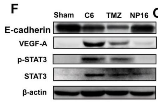
Application: WB Species: rat Sample: cardiomyocytes
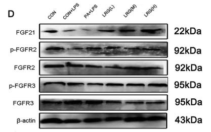
Application: WB Species: Mice Sample: diabetic mice

Application: WB Species: Goats Sample: epithelial cell
Restrictive clause
Affinity Biosciences tests all products strictly. Citations are provided as a resource for additional applications that have not been validated by Affinity Biosciences. Please choose the appropriate format for each application and consult Materials and Methods sections for additional details about the use of any product in these publications.
For Research Use Only.
Not for use in diagnostic or therapeutic procedures. Not for resale. Not for distribution without written consent. Affinity Biosciences will not be held responsible for patent infringement or other violations that may occur with the use of our products. Affinity Biosciences, Affinity Biosciences Logo and all other trademarks are the property of Affinity Biosciences LTD.