Collagen I Antibody - #AF0134

 Diabetes was mediated by STZ in rats. Subsequently, bilateral
OVX was carried out. The rats were fed for 8 weeks, and body weight was detected once a week. Then the animals were fasted
overnight, and fasting blood glucose and blood insulin contents were assessed using the commercial kits. Finally, all the animals
were sacrificed, and rat femur tissues were collected for following experiments. (d) Detection of BMD. (e) ALP staining was
performed in femur tissues. Scale bar = 100 μm. (f) Measurement of miR-340-5p expression by qRT-PCR. (g) Evaluation of OCN,
collagen-I, and RUNX2 levels with immunoblotting. β-actin was used as the internal reference. STZ, streptozotocin; OVX, ovariectomy;
BMD, bone mineral density; ALP, alkaline phosphatase; OCN, osteocalcin. Data were expressed as means ± SD (N = 6 per group). #
P < 0.05, ##P < 0.01, and ###P < 0.001 versus sham group; **P < 0.01 and ***P < 0.001 versus OVX group.")
, Protein levels of pro COLI, α-SMA, and RhoGDIα in MRC-5 cells following TGF-β1 stimulation for 1 h, as analysed by western blotting (n = 3).")
 Immunohistochemistry analysis of FSP-1 and α-SMA in sections of lung tissues. Scale bar: 50 μm. (b, c) Protein expressions of α-
SMA, COL-I, and TGF-β1 were detected by western blot. GAPDH was conducted as a loading control. (d, e) Protein expressions of
p-MEK1/2, MEK1/2, p-ERK1/2, ERK1/2 were examined by western blot. GAPDH was conducted as a loading control. One-way ANOVA,
*p < 0.05, ***p < 0.001, ****p < 0.0001")
 H&E stain-
ing with injury score of lung tissues. Red arrow:
edema and epithelial proliferation; Black arrow:
inflammatory infiltration. Scale bar =100 μ m.
(b) Masson trichrome staining with quantita-
tive histological fibrosis scoring (Ashcroft
score) of lung tissues. Blue: collagen fiber; Red:
muscle fiber. Scale bar =100 μ m. (c) The con-
tent of hydroxyproline in lung tissues. (d) The
protein levels of collagen-I. (e-g) The concen-
tration of TNF- α , IL-1β, and IL-6 in mice serum.
Data were expressed as mean ± SD. n = 6 in
each group. **p < 0.01 vs. control; #p < 0.05,
##p < 0.01 vs. BLM.")
 knockdown reduced myocardial fibrosis 4 weeks after myocardial infarction (MI). (A) Representative images of myocardial tissues with Masson staining (magnification ×200, scale bar=100 µm). Blue staining indicates the collagen. (B) Western blot analysis of collagen I, collagen III, and α-SMA in myocardial tissues. β-actin served as the loading control.")
 Representative (a1) longitudinal and (a2) transverse H&E staining of left ventricle. Scale bar, 50 μm. (b) Staining of Masson trichrome; dark blue indicates collagen fibers. Scale bar, 50 μm. (c) Sirus red staining; note the dense collagen staining. Scale bar, 50 μm. Immunohistochemical staining of (d) collagen I and (e) collagen III. Scale bar, 50 μm. (f) Oil Red O staining quantifies lipid accumulation. Scale bar, 50 μm. (g) The TUNEL assay was performed to identify DNA nicks; red plots indicate TUNEL-positive cells. Scale bar, 50 μm. (h) Representative transmission electron micrographs. Scale bar, 2 μm (h) Quantitative analysis of the cardiomyocyte size. (i) Semiquantification of collagen volume in total left ventricle. Data are presented as mean ± SD. ∗∗P < 0.01 CON vs. SHAM; ##P < 0.01 SG vs. SHAM. n = 6 in each group.")
 ALP activity in the femoral heads. (b) mRNA levels of OCN, COL I, OPN, and Runx2 in the femoral heads. (c) Expression levels of OCN, COL I, OPN, and Runx2 in the femoral heads. (d) Representative images of OCN staining and COL I staining (scale bar, 200 μm) in the femoral heads. ∗, p < 0.05; ∗∗, p < 0.01; ns, no significant. ALP, alkaline phosphatase; OCN, osteocalcin; COL I, type I collagen; OPN, osteopontin; Runx2, Runt-related transcription factor 2.")
 ALP activity in the femoral heads. (b) mRNA levels of OCN, COL I, OPN, and Runx2 in the femoral heads. (c) Expression levels of OCN, COL I, OPN, and Runx2 in the femoral heads. (d) Representative images of OCN staining and COL I staining (scale bar, 200 μm) in the femoral heads. ∗, p < 0.05; ∗∗, p < 0.01; ns, no significant. ALP, alkaline phosphatase; OCN, osteocalcin; COL I, type I collagen; OPN, osteopontin; Runx2, Runt-related transcription factor 2.")
Product Info
*The optimal dilutions should be determined by the end user. For optimal experimental results, antibody reuse is not recommended.
*Tips:
WB: For western blot detection of denatured protein samples. IHC: For immunohistochemical detection of paraffin sections (IHC-p) or frozen sections (IHC-f) of tissue samples. IF/ICC: For immunofluorescence detection of cell samples. ELISA(peptide): For ELISA detection of antigenic peptide.
Cite Format: Affinity Biosciences Cat# AF0134, RRID:AB_2813771.
Fold/Unfold
Alpha 1 type I collagen; Alpha 2 type I collagen; alpha 2 type I procollagen; alpha 2(I) procollagen; alpha 2(I)-collagen; Alpha-1 type I collagen; alpha1(I) procollagen; CO1A1_HUMAN; COL1A1; COL1A2; collagen alpha 1 chain type I; Collagen alpha-1(I) chain; collagen alpha-1(I) chain preproprotein; Collagen I alpha 1 polypeptide; Collagen I alpha 2 polypeptide; collagen of skin, tendon and bone, alpha-1 chain; collagen of skin, tendon and bone, alpha-2 chain; Collagen type I alpha 1; Collagen type I alpha 2; EDSC; OI1; OI2; OI3; OI4; pro-alpha-1 collagen type 1; type I proalpha 1; type I procollagen alpha 1 chain; Type I procollagen;
Immunogens
A synthesized peptide derived from human Collagen I, corresponding to a region within N-terminal amino acids.
Forms the fibrils of tendon, ligaments and bones. In bones the fibrils are mineralized with calcium hydroxyapatite.
P08123 CO1A2_HUMAN:Forms the fibrils of tendon, ligaments and bones. In bones the fibrils are mineralized with calcium hydroxyapatite.
- P02452 CO1A1_HUMAN:
- Protein BLAST With
- NCBI/
- ExPASy/
- Uniprot
MFSFVDLRLLLLLAATALLTHGQEEGQVEGQDEDIPPITCVQNGLRYHDRDVWKPEPCRICVCDNGKVLCDDVICDETKNCPGAEVPEGECCPVCPDGSESPTDQETTGVEGPKGDTGPRGPRGPAGPPGRDGIPGQPGLPGPPGPPGPPGPPGLGGNFAPQLSYGYDEKSTGGISVPGPMGPSGPRGLPGPPGAPGPQGFQGPPGEPGEPGASGPMGPRGPPGPPGKNGDDGEAGKPGRPGERGPPGPQGARGLPGTAGLPGMKGHRGFSGLDGAKGDAGPAGPKGEPGSPGENGAPGQMGPRGLPGERGRPGAPGPAGARGNDGATGAAGPPGPTGPAGPPGFPGAVGAKGEAGPQGPRGSEGPQGVRGEPGPPGPAGAAGPAGNPGADGQPGAKGANGAPGIAGAPGFPGARGPSGPQGPGGPPGPKGNSGEPGAPGSKGDTGAKGEPGPVGVQGPPGPAGEEGKRGARGEPGPTGLPGPPGERGGPGSRGFPGADGVAGPKGPAGERGSPGPAGPKGSPGEAGRPGEAGLPGAKGLTGSPGSPGPDGKTGPPGPAGQDGRPGPPGPPGARGQAGVMGFPGPKGAAGEPGKAGERGVPGPPGAVGPAGKDGEAGAQGPPGPAGPAGERGEQGPAGSPGFQGLPGPAGPPGEAGKPGEQGVPGDLGAPGPSGARGERGFPGERGVQGPPGPAGPRGANGAPGNDGAKGDAGAPGAPGSQGAPGLQGMPGERGAAGLPGPKGDRGDAGPKGADGSPGKDGVRGLTGPIGPPGPAGAPGDKGESGPSGPAGPTGARGAPGDRGEPGPPGPAGFAGPPGADGQPGAKGEPGDAGAKGDAGPPGPAGPAGPPGPIGNVGAPGAKGARGSAGPPGATGFPGAAGRVGPPGPSGNAGPPGPPGPAGKEGGKGPRGETGPAGRPGEVGPPGPPGPAGEKGSPGADGPAGAPGTPGPQGIAGQRGVVGLPGQRGERGFPGLPGPSGEPGKQGPSGASGERGPPGPMGPPGLAGPPGESGREGAPGAEGSPGRDGSPGAKGDRGETGPAGPPGAPGAPGAPGPVGPAGKSGDRGETGPAGPTGPVGPVGARGPAGPQGPRGDKGETGEQGDRGIKGHRGFSGLQGPPGPPGSPGEQGPSGASGPAGPRGPPGSAGAPGKDGLNGLPGPIGPPGPRGRTGDAGPVGPPGPPGPPGPPGPPSAGFDFSFLPQPPQEKAHDGGRYYRADDANVVRDRDLEVDTTLKSLSQQIENIRSPEGSRKNPARTCRDLKMCHSDWKSGEYWIDPNQGCNLDAIKVFCNMETGETCVYPTQPSVAQKNWYISKNPKDKRHVWFGESMTDGFQFEYGGQGSDPADVAIQLTFLRLMSTEASQNITYHCKNSVAYMDQQTGNLKKALLLQGSNEIEIRAEGNSRFTYSVTVDGCTSHTGAWGKTVIEYKTTKTSRLPIIDVAPLDVGAPDQEFGFDVGPVCFL
- P08123 CO1A2_HUMAN:
- Protein BLAST With
- NCBI/
- ExPASy/
- Uniprot
MLSFVDTRTLLLLAVTLCLATCQSLQEETVRKGPAGDRGPRGERGPPGPPGRDGEDGPTGPPGPPGPPGPPGLGGNFAAQYDGKGVGLGPGPMGLMGPRGPPGAAGAPGPQGFQGPAGEPGEPGQTGPAGARGPAGPPGKAGEDGHPGKPGRPGERGVVGPQGARGFPGTPGLPGFKGIRGHNGLDGLKGQPGAPGVKGEPGAPGENGTPGQTGARGLPGERGRVGAPGPAGARGSDGSVGPVGPAGPIGSAGPPGFPGAPGPKGEIGAVGNAGPAGPAGPRGEVGLPGLSGPVGPPGNPGANGLTGAKGAAGLPGVAGAPGLPGPRGIPGPVGAAGATGARGLVGEPGPAGSKGESGNKGEPGSAGPQGPPGPSGEEGKRGPNGEAGSAGPPGPPGLRGSPGSRGLPGADGRAGVMGPPGSRGASGPAGVRGPNGDAGRPGEPGLMGPRGLPGSPGNIGPAGKEGPVGLPGIDGRPGPIGPAGARGEPGNIGFPGPKGPTGDPGKNGDKGHAGLAGARGAPGPDGNNGAQGPPGPQGVQGGKGEQGPPGPPGFQGLPGPSGPAGEVGKPGERGLHGEFGLPGPAGPRGERGPPGESGAAGPTGPIGSRGPSGPPGPDGNKGEPGVVGAVGTAGPSGPSGLPGERGAAGIPGGKGEKGEPGLRGEIGNPGRDGARGAPGAVGAPGPAGATGDRGEAGAAGPAGPAGPRGSPGERGEVGPAGPNGFAGPAGAAGQPGAKGERGAKGPKGENGVVGPTGPVGAAGPAGPNGPPGPAGSRGDGGPPGMTGFPGAAGRTGPPGPSGISGPPGPPGPAGKEGLRGPRGDQGPVGRTGEVGAVGPPGFAGEKGPSGEAGTAGPPGTPGPQGLLGAPGILGLPGSRGERGLPGVAGAVGEPGPLGIAGPPGARGPPGAVGSPGVNGAPGEAGRDGNPGNDGPPGRDGQPGHKGERGYPGNIGPVGAAGAPGPHGPVGPAGKHGNRGETGPSGPVGPAGAVGPRGPSGPQGIRGDKGEPGEKGPRGLPGLKGHNGLQGLPGIAGHHGDQGAPGSVGPAGPRGPAGPSGPAGKDGRTGHPGTVGPAGIRGPQGHQGPAGPPGPPGPPGPPGVSGGGYDFGYDGDFYRADQPRSAPSLRPKDYEVDATLKSLNNQIETLLTPEGSRKNPARTCRDLRLSHPEWSSGYYWIDPNQGCTMDAIKVYCDFSTGETCIRAQPENIPAKNWYRSSKDKKHVWLGETINAGSQFEYNVEGVTSKEMATQLAFMRLLANYASQNITYHCKNSIAYMDEETGNLKKAVILQGSNDVELVAEGNSRFTYTVLVDGCSKKTNEWGKTIIEYKTNKPSRLPFLDIAPLDIGGADQEFFVDIGPVCFK
Predictions
Score>80(red) has high confidence and is suggested to be used for WB detection. *The prediction model is mainly based on the alignment of immunogen sequences, the results are for reference only, not as the basis of quality assurance.
High(score>80) Medium(80>score>50) Low(score<50) No confidence
Research Backgrounds
Type I collagen is a member of group I collagen (fibrillar forming collagen).
Contains mostly 4-hydroxyproline. Proline residues at the third position of the tripeptide repeating unit (G-X-Y) are hydroxylated in some or all of the chains.
Contains 3-hydroxyproline at a few sites. This modification occurs on the first proline residue in the sequence motif Gly-Pro-Hyp, where Hyp is 4-hydroxyproline.
Lysine residues at the third position of the tripeptide repeating unit (G-X-Y) are 5-hydroxylated in some or all of the chains.
O-glycosylated on hydroxylated lysine residues. The O-linked glycan consists of a Glc-Gal disaccharide.
Secreted>Extracellular space>Extracellular matrix.
Forms the fibrils of tendon, ligaments and bones. In bones the fibrils are mineralized with calcium hydroxyapatite.
The C-terminal propeptide, also known as COLFI domain, have crucial roles in tissue growth and repair by controlling both the intracellular assembly of procollagen molecules and the extracellular assembly of collagen fibrils. It binds a calcium ion which is essential for its function (By similarity).
Belongs to the fibrillar collagen family.
Type I collagen is a member of group I collagen (fibrillar forming collagen).
Prolines at the third position of the tripeptide repeating unit (G-X-Y) are hydroxylated in some or all of the chains.
Secreted>Extracellular space>Extracellular matrix.
Forms the fibrils of tendon, ligaments and bones. In bones the fibrils are mineralized with calcium hydroxyapatite.
The C-terminal propeptide, also known as COLFI domain, have crucial roles in tissue growth and repair by controlling both the intracellular assembly of procollagen molecules and the extracellular assembly of collagen fibrils. It binds a calcium ion which is essential for its function.
Belongs to the fibrillar collagen family.
Research Fields
· Cellular Processes > Cellular community - eukaryotes > Focal adhesion. (View pathway)
· Environmental Information Processing > Signal transduction > PI3K-Akt signaling pathway. (View pathway)
· Environmental Information Processing > Signaling molecules and interaction > ECM-receptor interaction. (View pathway)
· Human Diseases > Infectious diseases: Parasitic > Amoebiasis.
· Human Diseases > Infectious diseases: Viral > Human papillomavirus infection.
· Organismal Systems > Immune system > Platelet activation. (View pathway)
· Organismal Systems > Endocrine system > Relaxin signaling pathway.
· Organismal Systems > Digestive system > Protein digestion and absorption.
References
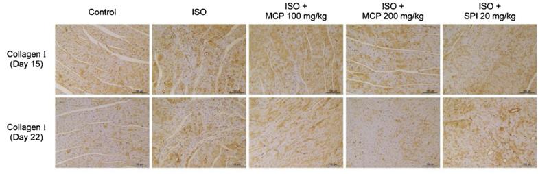
Application: IHC Species: rat Sample: heart
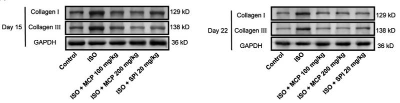
Application: WB Species: rat Sample: heart
Application: WB Species: mouse Sample: MLE‐12 cells

Application: IHC Species: Mouse Sample: lung tissue
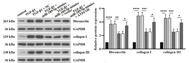
Application: WB Species: human Sample: HK2 cells

Application: WB Species: Rat Sample:

Application: WB Species: Human Sample:
Restrictive clause
Affinity Biosciences tests all products strictly. Citations are provided as a resource for additional applications that have not been validated by Affinity Biosciences. Please choose the appropriate format for each application and consult Materials and Methods sections for additional details about the use of any product in these publications.
For Research Use Only.
Not for use in diagnostic or therapeutic procedures. Not for resale. Not for distribution without written consent. Affinity Biosciences will not be held responsible for patent infringement or other violations that may occur with the use of our products. Affinity Biosciences, Affinity Biosciences Logo and all other trademarks are the property of Affinity Biosciences LTD.