.
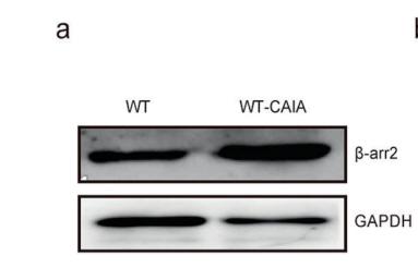
Bands result from membrane strip incubation.")


 Immunohistochemical staining for collagens I, III and α-SMA,scale bars=100 µm. Bar graphs show the percentage of (B) collagens I, (C) III and (D) α-SMA. (E) Western blotting detection of collagens I, III and (F) α-SMA.")
 Immunohistochemical staining for collagens I, III and α-SMA,scale bars=100 µm. Bar graphs show the percentage of (B) collagens I, (C) III and (D) α-SMA. (E) Western blotting detection of collagens I, III and (F) α-SMA.")
 Immunochemistry staining of α-SMA expression in the lungs of wild-type mice, n = 6 in each group. Magnification, ×40 (top panel) and ×200 (bottom panel). Silicotic mice underwent various treatment combinations with DIZE (ACE2 activator), A779 (Mas receptor blocker), and MLN-4760 (ACE2 inhibitor). (B) The proportion of silicotic areas in the lung samples in (A). (C) Western blot showing the protein expression of α-SMA (D), Vim (E), pro-Col I (F), pro-Col III (G), and E-cad (H) in the lungs of mice from the various treatment groups. Values represent the mean ± SD, n = 3 independent experiments, fold change is expressed relative to the control (no treatments), *P < 0.05 vs corresponding group, **P < 0.01 vs corresponding group.")
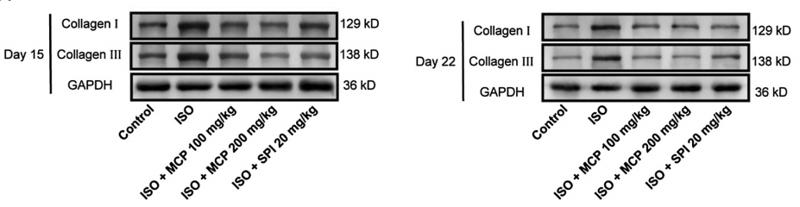
 Western blotting detection of collagen Ⅰ, collagen Ⅲ, and α-smooth muscle actin (α-SMA). Bar graphs show fold changes for the collagen Ⅰ,collagen Ⅲ, and α-SMA expression as analyzed by western blotting. Glyceraldehyde 3-phosphate dehydrogenase (GAPDH) was used as a loading control (n 3). Datawere shown as mean ± SEM. **p < 0.01 vs. control, #p < 0.05 vs. ISO, ##p < 0.01 vs. ISO.")
 The effects of different doses of BEL (15, 30, 60 μM)on α-SMA, collagen Ⅰ, and collagen Ⅲ expression induced by TGF-β1 using immunofluorescence staining.")
. Scale bar,
100 μm. b Representative
immunoblots of Col IA/Col
IIIA/α-SMA and summarized
intensities of blots in the cortex
and medulla (n = 6–8 per
group). c α-SMA/TNF-α/IL-6/
IL-10 mRNA levels in the renal
cortex and medulla (n = 6–7 per
group). DOCA
deoxycorticosterone acetate,
NaBu sodium butyrate, Col IA
collagen type IA, Col IIIA
collagen type IIIA, a-SMA asmooth muscle actin, TNF-a
tumor necrosis factor a; IL-6,
interleukin 6; IL-10, interleukin
10. *P < 0.05; **P < 0.01")

 collagen Ⅰ and (B) collagen III in atrial tissues of heart were determined via immunohistochemistry. At the same time, quantitative analysis was carried out.")

")
 HE staining to detect morphological changes; Masson trichrome staining to detect cardiac collagen deposition (B–D) Western blotting detection of collagen Ⅰ, collagen Ⅲ, and α-smooth muscle actin (α-SMA). Bar graphs show fold changes for the collagen Ⅰ, collagen Ⅲ, and α-SMA expression as analyzed by western blotting. Glyceraldehyde 3-phosphate dehydrogenase (GAPDH) was used as a loading control (n = 3). Data were shown as mean ± SEM. **p < 0.01 vs. control, #p < 0.05 vs. ISO, ##p < 0.01 vs. ISO.")
 Expression of fibrosis-related proteins in serum by western blot analysis.")
 on diabetic-induced cardiac remodeling.(F) The percentage of fibrosis was calculated. Immunohistochemical staining of panel (G) collagen I, (H) collagen III. Scale bar,50 µm. Data are presented as mean ± SD. **P < 0.01 CON vs. SHAM; ##P < 0.01 SG vs. SHAM. n = 3 in each group.")

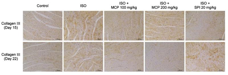
and collagen deposition was detected by masson staining (B). Scale bar = 100 μm. The level of hydroxyproline in the lungs was examined with a hydroxyproline
detection kit (C). The levels of TGF-β1, collagen I, and collagen III in the lungs were detected by western blot (D-E). The results are presented as mean ± SD. N = 6 for
each group.")
 collagen I and (B) collagen III in atrial tissues of heart were determined via immunohistochemistry. At the same time, quantitative analysis was carried out. (C) Protein expression levels of TGF-β1, CTGF, fibronectin and α-SMA in atrial tissues of heart were assessed via western blotting. The results are presented as the mean ± standard deviation (n=6). ###P<0.001 vs. sham group; **P<0.01 and ***P<0.001 vs. AF + LV-NC group. CTGF, connective tissue growth factor; α-SMA, α-smooth muscle actin; AF, atrial fibrillation; LV-NC, lentivirus negative control; miR, microRNA.")
 knockdown reduced myocardial fibrosis 4 weeks after myocardial infarction (MI). (A) Representative images of myocardial tissues with Masson staining (magnification ×200, scale bar=100 µm). Blue staining indicates the collagen. (B) Western blot analysis of collagen I, collagen III, and α-SMA in myocardial tissues. β-actin served as the loading control.")
 Effect of EGCG on obesity-induced myocardial fibrosis in heart tissue by HE and Masson staining; (B) Effect of EGCG on the deposition of collagen I and collagen III in heart tissue by Immunohistochemical staining; (C) The mean OD values of collagen I and collagen III in heart tissue by Immunohistochemical staining; (D) Effect of EGCG on the expression of collagen I and collagen III proteins were detected by western blot with GAPDH as a loading control in the heart. (*p < 0.05, **p < 0.01, ***p < 0.001).")
 Effect of EGCG on obesity-induced myocardial fibrosis in heart tissue by HE and Masson staining; (B) Effect of EGCG on the deposition of collagen I and collagen III in heart tissue by Immunohistochemical staining; (C) The mean OD values of collagen I and collagen III in heart tissue by Immunohistochemical staining; (D) Effect of EGCG on the expression of collagen I and collagen III proteins were detected by western blot with GAPDH as a loading control in the heart. (*p < 0.05, **p < 0.01, ***p < 0.001).")
 Western blot examined the expression of α-SMA, Collagen I, and Collagen III. (b–d) The fold changes of the Western blot results for α-SMA, Collagen I, and Collagen III were analysed; GAPDH was used as a loading control (n = 3). Data were presented as the mean ± standard error of the mean (SEM). ∗p < 0.05 and ∗∗p < 0.01 vs. Control; ##p < 0.01 vs. TGF-β1.")
 Representative (a1) longitudinal and (a2) transverse H&E staining of left ventricle. Scale bar, 50 μm. (b) Staining of Masson trichrome; dark blue indicates collagen fibers. Scale bar, 50 μm. (c) Sirus red staining; note the dense collagen staining. Scale bar, 50 μm. Immunohistochemical staining of (d) collagen I and (e) collagen III. Scale bar, 50 μm. (f) Oil Red O staining quantifies lipid accumulation. Scale bar, 50 μm. (g) The TUNEL assay was performed to identify DNA nicks; red plots indicate TUNEL-positive cells. Scale bar, 50 μm. (h) Representative transmission electron micrographs. Scale bar, 2 μm (h) Quantitative analysis of the cardiomyocyte size. (i) Semiquantification of collagen volume in total left ventricle. Data are presented as mean ± SD. ∗∗P < 0.01 CON vs. SHAM; ##P < 0.01 SG vs. SHAM. n = 6 in each group.")
 Western blotting indicated that the upregulated protein expression levels of collagen I, collagen III, fibronectin, α-SMA and MMP2 in oe-PDCD4-HL-1 cells were attenuated by Pio and CBL0137. (B) Statistical analysis of western blotting. (C) Enzyme-linked immunosorbent assays showed that the upregulated protein concentrations of IL-6, IL-17A, IFN-γ and TNF-α, and downregulated protein concentration of IL-4, in oe-PDCD4-HL-1 cells were reversed by Pio and CBL0137. ***P")
 Representative Masson staining of hearts of mice in the sham (A), 30G TAC (B), and 32G TAC (C) groups. The middle images are high-power fields of the corresponding left images (black square frame), showing interstitial fibrosis. The right images are high-power fields of the corresponding left images, showing perivascular fibrosis (green square frame, up) and endomyocardial fibrosis (blue square frame, down). Scale bar: 100 μm (left images); scale bar: 50 μm (middle and right images). (D) Interstitial fibrosis in the three groups; n = 3 hearts per group. Each heart slide was random selected with five high-power fields for statistical analysis. (E,F) qRT-PCR for the expression of Fn1 and Timp1 in the ventricular tissue; n = 6, sham; n = 6, 30G TAC; n = 6, 32G TAC. (G) Western blot stripes from three independent hearts in the three groups are shown. (H–L) Quantification of protein expression of TGF-β/β-tubulin (H), p-Smad3/Smad3 (I), MMP-9/β-tubulin (J), COL1A1/β-tubulin (K), and COL3/β-tubulin (L) in the hearts of mice in the sham, 30G TAC, and 32G TAC groups 14 days after the surgery. Data are presented as mean ± SEM. *P < 0.05, **P < 0.01, ***P < 0.001, and ****P < 0.0001. Statistical significance was calculated using the unpaired two-tailed t-test.")
, α-SMA (B), Erbb4-IR (C) and miR-29b (D) in different groups was detected by qRT-PCR (n = 6), the results were presented as mean ± SD, *P")
| Product: | Collagen III Antibody |
| Catalog: | AF0136 |
| Description: | Rabbit polyclonal antibody to Collagen III |
| Application: | WB IHC IF/ICC |
| Cited expt.: | WB, IHC, IF/ICC |
| Reactivity: | Human, Mouse, Rat |
| Prediction: | Pig, Bovine, Horse, Sheep, Rabbit, Dog, Chicken, Xenopus |
| Mol.Wt.: | 138kDa(Observed); 139kD(Calculated). |
| Uniprot: | P02461 |
| RRID: | AB_2813770 |
Control Products

Product Info
*The optimal dilutions should be determined by the end user. For optimal experimental results, antibody reuse is not recommended.
*Tips:
WB: For western blot detection of denatured protein samples. IHC: For immunohistochemical detection of paraffin sections (IHC-p) or frozen sections (IHC-f) of tissue samples. IF/ICC: For immunofluorescence detection of cell samples. ELISA(peptide): For ELISA detection of antigenic peptide.
Cite Format: Affinity Biosciences Cat# AF0136, RRID:AB_2813770.
Fold/Unfold
Alpha 1 type III collagen; Alpha1 (III) collagen; CO3A1_HUMAN; COL 3A1; COL3A1; Collagen alpha 1(III) chain; Collagen alpha-1(III) chain; Collagen III alpha 1 chain precursor; Collagen III alpha 1 polypeptide; Collagen type III alpha 1 (Ehlers Danlos syndrome type IV autosomal dominant); Collagen type III alpha 1; Collagen type III alpha 1 chain; Collagen type III alpha; Collagen, fetal; EDS4A; Ehlers Danlos syndrome type IV, autosomal dominant; Fetal collagen; Type III collagen;
Immunogens
A synthesized peptide derived from human Collagen III, corresponding to a region within N-terminal amino acids.
- P02461 CO3A1_HUMAN:
- Protein BLAST With
- NCBI/
- ExPASy/
- Uniprot
MMSFVQKGSWLLLALLHPTIILAQQEAVEGGCSHLGQSYADRDVWKPEPCQICVCDSGSVLCDDIICDDQELDCPNPEIPFGECCAVCPQPPTAPTRPPNGQGPQGPKGDPGPPGIPGRNGDPGIPGQPGSPGSPGPPGICESCPTGPQNYSPQYDSYDVKSGVAVGGLAGYPGPAGPPGPPGPPGTSGHPGSPGSPGYQGPPGEPGQAGPSGPPGPPGAIGPSGPAGKDGESGRPGRPGERGLPGPPGIKGPAGIPGFPGMKGHRGFDGRNGEKGETGAPGLKGENGLPGENGAPGPMGPRGAPGERGRPGLPGAAGARGNDGARGSDGQPGPPGPPGTAGFPGSPGAKGEVGPAGSPGSNGAPGQRGEPGPQGHAGAQGPPGPPGINGSPGGKGEMGPAGIPGAPGLMGARGPPGPAGANGAPGLRGGAGEPGKNGAKGEPGPRGERGEAGIPGVPGAKGEDGKDGSPGEPGANGLPGAAGERGAPGFRGPAGPNGIPGEKGPAGERGAPGPAGPRGAAGEPGRDGVPGGPGMRGMPGSPGGPGSDGKPGPPGSQGESGRPGPPGPSGPRGQPGVMGFPGPKGNDGAPGKNGERGGPGGPGPQGPPGKNGETGPQGPPGPTGPGGDKGDTGPPGPQGLQGLPGTGGPPGENGKPGEPGPKGDAGAPGAPGGKGDAGAPGERGPPGLAGAPGLRGGAGPPGPEGGKGAAGPPGPPGAAGTPGLQGMPGERGGLGSPGPKGDKGEPGGPGADGVPGKDGPRGPTGPIGPPGPAGQPGDKGEGGAPGLPGIAGPRGSPGERGETGPPGPAGFPGAPGQNGEPGGKGERGAPGEKGEGGPPGVAGPPGGSGPAGPPGPQGVKGERGSPGGPGAAGFPGARGLPGPPGSNGNPGPPGPSGSPGKDGPPGPAGNTGAPGSPGVSGPKGDAGQPGEKGSPGAQGPPGAPGPLGIAGITGARGLAGPPGMPGPRGSPGPQGVKGESGKPGANGLSGERGPPGPQGLPGLAGTAGEPGRDGNPGSDGLPGRDGSPGGKGDRGENGSPGAPGAPGHPGPPGPVGPAGKSGDRGESGPAGPAGAPGPAGSRGAPGPQGPRGDKGETGERGAAGIKGHRGFPGNPGAPGSPGPAGQQGAIGSPGPAGPRGPVGPSGPPGKDGTSGHPGPIGPPGPRGNRGERGSEGSPGHPGQPGPPGPPGAPGPCCGGVGAAAIAGIGGEKAGGFAPYYGDEPMDFKINTDEIMTSLKSVNGQIESLISPDGSRKNPARNCRDLKFCHPELKSGEYWVDPNQGCKLDAIKVFCNMETGETCISANPLNVPRKHWWTDSSAEKKHVWFGESMDGGFQFSYGNPELPEDVLDVHLAFLRLLSSRASQNITYHCKNSIAYMDQASGNVKKALKLMGSNEGEFKAEGNSKFTYTVLEDGCTKHTGEWSKTVFEYRTRKAVRLPIVDIAPYDIGGPDQEFGVDVGPVCFL
Predictions
Score>80(red) has high confidence and is suggested to be used for WB detection. *The prediction model is mainly based on the alignment of immunogen sequences, the results are for reference only, not as the basis of quality assurance.
High(score>80) Medium(80>score>50) Low(score<50) No confidence
Research Backgrounds
Collagen type III occurs in most soft connective tissues along with type I collagen. Involved in regulation of cortical development. Is the major ligand of ADGRG1 in the developing brain and binding to ADGRG1 inhibits neuronal migration and activates the RhoA pathway by coupling ADGRG1 to GNA13 and possibly GNA12.
Proline residues at the third position of the tripeptide repeating unit (G-X-Y) are hydroxylated in some or all of the chains.
O-linked glycan consists of a Glc-Gal disaccharide bound to the oxygen atom of a post-translationally added hydroxyl group.
Secreted>Extracellular space>Extracellular matrix.
The C-terminal propeptide, also known as COLFI domain, have crucial roles in tissue growth and repair by controlling both the intracellular assembly of procollagen molecules and the extracellular assembly of collagen fibrils. It binds a calcium ion which is essential for its function.
Belongs to the fibrillar collagen family.
Research Fields
· Human Diseases > Infectious diseases: Parasitic > Amoebiasis.
· Organismal Systems > Immune system > Platelet activation. (View pathway)
· Organismal Systems > Endocrine system > Relaxin signaling pathway.
· Organismal Systems > Digestive system > Protein digestion and absorption.
References
Application: IF/ICC Species: rat Sample: tendon

Application: IHC Species: rat Sample: tendon

Application: WB Species: Mouse Sample:

Application: WB Species: Rat Sample:

Application: WB Species: mouse Sample:
Application: IHC Species: rat Sample: heart
Application: WB Species: rat Sample: heart
Restrictive clause
Affinity Biosciences tests all products strictly. Citations are provided as a resource for additional applications that have not been validated by Affinity Biosciences. Please choose the appropriate format for each application and consult Materials and Methods sections for additional details about the use of any product in these publications.
For Research Use Only.
Not for use in diagnostic or therapeutic procedures. Not for resale. Not for distribution without written consent. Affinity Biosciences will not be held responsible for patent infringement or other violations that may occur with the use of our products. Affinity Biosciences, Affinity Biosciences Logo and all other trademarks are the property of Affinity Biosciences LTD.