

 and mouse anti-beta tubulin Ab(T0023 1:200) for 1 hour at 37°C. An AlexaFluor594 conjugated goat anti-rabbit IgG(H+L) Ab(Red) and an AlexaFluor488 conjugated goat anti-mouse IgG(H+L) Ab(Green) were used as the secondary antibody.
The nuclear counter stain is DAPI(blue).")
Control Products

Related Downloads
Protocols
Product Info
*The optimal dilutions should be determined by the end user. For optimal experimental results, antibody reuse is not recommended.
*Tips:
WB: For western blot detection of denatured protein samples. IHC: For immunohistochemical detection of paraffin sections (IHC-p) or frozen sections (IHC-f) of tissue samples. IF/ICC: For immunofluorescence detection of cell samples. ELISA(peptide): For ELISA detection of antigenic peptide.
Cite Format: Affinity Biosciences Cat# DF10170, RRID:AB_2840749.
Fold/Unfold
AI853584; Blood group--MN locus; CD_antigen=CD235a; CD235a; GLPA_HUMAN; Glycophorin A (MNS blood group); Glycophorin A; Glycophorin A, included; Glycophorin-A; GlycophorinA; GPA; GPErik; GpMiIII; GPSAT; GYPA; GYPA, included; HGpMiIII; HGpMiV; HGpMiX; HGpMiXI; HGpSta(C); MN; MN sialoglycoprotein; MNS; PAS-2; PAS2; Sialoglycoprotein alpha;
Immunogens
A synthesized peptide derived from human Glycophorin A, corresponding to a region within the internal amino acids.
- P02724 GLPA_HUMAN:
- Protein BLAST With
- NCBI/
- ExPASy/
- Uniprot
MYGKIIFVLLLSEIVSISASSTTGVAMHTSTSSSVTKSYISSQTNDTHKRDTYAATPRAHEVSEISVRTVYPPEEETGERVQLAHHFSEPEITLIIFGVMAGVIGTILLISYGIRRLIKKSPSDVKPLPSPDTDVPLSSVEIENPETSDQ
Research Backgrounds
Glycophorin A is the major intrinsic membrane protein of the erythrocyte. The N-terminal glycosylated segment, which lies outside the erythrocyte membrane, has MN blood group receptors. Appears to be important for the function of SLC4A1 and is required for high activity of SLC4A1. May be involved in translocation of SLC4A1 to the plasma membrane. Is a receptor for influenza virus. Is a receptor for Plasmodium falciparum erythrocyte-binding antigen 175 (EBA-175); binding of EBA-175 is dependent on sialic acid residues of the O-linked glycans. Appears to be a receptor for Hepatitis A virus (HAV).
The major O-linked glycan are NeuAc-alpha-(2-3)-Gal-beta-(1-3)-[NeuAc-alpha-(2-6)]-GalNAcOH (about 78 %) and NeuAc-alpha-(2-3)-Gal-beta-(1-3)-GalNAcOH (17 %). Minor O-glycans (5 %) include NeuAc-alpha-(2-3)-Gal-beta-(1-3)-[NeuAc-alpha-(2-6)]-GalNAcOH NeuAc-alpha-(2-8)-NeuAc-alpha-(2-3)-Gal-beta-(1-3)-GalNAcOH. About 1% of all O-linked glycans carry blood group A, B and H determinants. They derive from a type-2 precursor core structure, Gal-beta-(1,3)-GlcNAc-beta-1-R, and the antigens are synthesized by addition of fucose (H antigen-specific) and then N-acetylgalactosamine (A antigen-specific) or galactose (B antigen-specific). Specifically O-linked-glycans are NeuAc-alpha-(2-3)-Gal-beta-(1-3)-GalNAcOH-(6-1)-GlcNAc-beta-(4-1)-[Fuc-alpha-(1-2)]-Gal-beta-(3-1)-GalNAc-alpha (about 1%, B antigen-specific) and NeuAc-alpha-(2-3)-Gal-beta-(1-3)-GalNAcOH-(6-1)-GlcNAc-beta-(4-1)-[Fuc-alpha-(1-2)]-Gal-beta (1 %, O antigen-, A antigen- and B antigen-specific).
Cell membrane>Single-pass type I membrane protein.
Note: Appears to be colocalized with SLC4A1.
Belongs to the glycophorin A family.
Research Fields
· Human Diseases > Infectious diseases: Parasitic > Malaria.
· Organismal Systems > Immune system > Hematopoietic cell lineage. (View pathway)
References
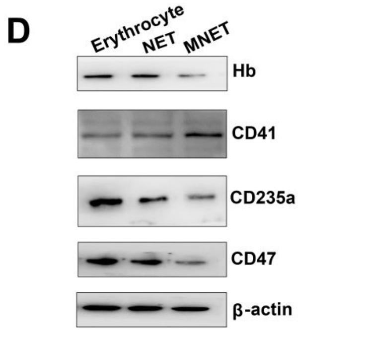
Application: WB Species: mouse Sample: erythrocyte
Restrictive clause
Affinity Biosciences tests all products strictly. Citations are provided as a resource for additional applications that have not been validated by Affinity Biosciences. Please choose the appropriate format for each application and consult Materials and Methods sections for additional details about the use of any product in these publications.
For Research Use Only.
Not for use in diagnostic or therapeutic procedures. Not for resale. Not for distribution without written consent. Affinity Biosciences will not be held responsible for patent infringement or other violations that may occur with the use of our products. Affinity Biosciences, Affinity Biosciences Logo and all other trademarks are the property of Affinity Biosciences LTD.