.
Bands result from membrane strip incubation.")
| Product: | C3AR1 Antibody |
| Catalog: | DF10198 |
| Description: | Rabbit polyclonal antibody to C3AR1 |
| Application: | WB |
| Cited expt.: | WB |
| Reactivity: | Human, Mouse |
| Prediction: | Rabbit |
| Mol.Wt.: | 54 kDa(Observed); 54kD(Calculated). |
| Uniprot: | Q16581 |
| RRID: | AB_2840777 |
Control Products

Related Downloads
Protocols
Product Info
*The optimal dilutions should be determined by the end user. For optimal experimental results, antibody reuse is not recommended.
*Tips:
WB: For western blot detection of denatured protein samples. IHC: For immunohistochemical detection of paraffin sections (IHC-p) or frozen sections (IHC-f) of tissue samples. IF/ICC: For immunofluorescence detection of cell samples. ELISA(peptide): For ELISA detection of antigenic peptide.
Cite Format: Affinity Biosciences Cat# DF10198, RRID:AB_2840777.
Fold/Unfold
AZ 3B; AZ3B; C3a anaphylatoxin chemotactic receptor; C3A R; C3A R1; C3a-R; C3AR; C3AR_HUMAN; C3AR1; C3R1; Complement component 3 receptor 1; Complement component 3a receptor 1; HNFAG09;
Immunogens
A synthesized peptide derived from human C3AR1, corresponding to a region within the internal amino acids.
Widely expressed in several differentiated hematopoietic cell lines, in the lung, spleen, ovary, placenta, small intestine, throughout the brain, heart, and endothelial cells. Mostly expressed in lymphoid tissues.
- Q16581 C3AR_HUMAN:
- Protein BLAST With
- NCBI/
- ExPASy/
- Uniprot
MASFSAETNSTDLLSQPWNEPPVILSMVILSLTFLLGLPGNGLVLWVAGLKMQRTVNTIWFLHLTLADLLCCLSLPFSLAHLALQGQWPYGRFLCKLIPSIIVLNMFASVFLLTAISLDRCLVVFKPIWCQNHRNVGMACSICGCIWVVAFVMCIPVFVYREIFTTDNHNRCGYKFGLSSSLDYPDFYGDPLENRSLENIVQPPGEMNDRLDPSSFQTNDHPWTVPTVFQPQTFQRPSADSLPRGSARLTSQNLYSNVFKPADVVSPKIPSGFPIEDHETSPLDNSDAFLSTHLKLFPSASSNSFYESELPQGFQDYYNLGQFTDDDQVPTPLVAITITRLVVGFLLPSVIMIACYSFIVFRMQRGRFAKSQSKTFRVAVVVVAVFLVCWTPYHIFGVLSLLTDPETPLGKTLMSWDHVCIALASANSCFNPFLYALLGKDFRKKARQSIQGILEAAFSEELTRSTHCPSNNVISERNSTTV
Predictions
Score>80(red) has high confidence and is suggested to be used for WB detection. *The prediction model is mainly based on the alignment of immunogen sequences, the results are for reference only, not as the basis of quality assurance.
High(score>80) Medium(80>score>50) Low(score<50) No confidence
Research Backgrounds
Receptor for the chemotactic and inflammatory peptide anaphylatoxin C3a. This receptor stimulates chemotaxis, granule enzyme release and superoxide anion production.
Among the sulfation sites Tyr-174 is essential for binding of C3a anaphylatoxin.
O-glycosylated.
Cell membrane>Multi-pass membrane protein.
Widely expressed in several differentiated hematopoietic cell lines, in the lung, spleen, ovary, placenta, small intestine, throughout the brain, heart, and endothelial cells. Mostly expressed in lymphoid tissues.
Belongs to the G-protein coupled receptor 1 family.
Research Fields
· Environmental Information Processing > Signaling molecules and interaction > Neuroactive ligand-receptor interaction.
· Human Diseases > Infectious diseases: Bacterial > Staphylococcus aureus infection.
· Organismal Systems > Immune system > Complement and coagulation cascades. (View pathway)
References
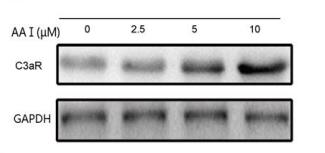
Application: WB Species: mouse Sample: Hepa1-6 cells
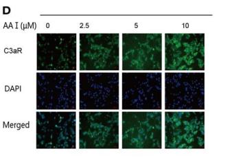
Application: IF/ICC Species: mouse Sample: Hepa1-6 cells
Restrictive clause
Affinity Biosciences tests all products strictly. Citations are provided as a resource for additional applications that have not been validated by Affinity Biosciences. Please choose the appropriate format for each application and consult Materials and Methods sections for additional details about the use of any product in these publications.
For Research Use Only.
Not for use in diagnostic or therapeutic procedures. Not for resale. Not for distribution without written consent. Affinity Biosciences will not be held responsible for patent infringement or other violations that may occur with the use of our products. Affinity Biosciences, Affinity Biosciences Logo and all other trademarks are the property of Affinity Biosciences LTD.