, using OPSB Antibody at 1/1000 dilution.
5ug/NC membrane strip.
Exposure for 2min with Affinity™ ECL Kit(#KF8003).
Bands result from membrane strip incubation.")
| Product: | OPSB Antibody |
| Catalog: | DF10234 |
| Description: | Rabbit polyclonal antibody to OPSB |
| Application: | WB IHC IF/ICC |
| Cited expt.: | WB |
| Reactivity: | Human, Mouse, Rat |
| Prediction: | Pig, Bovine, Horse, Sheep, Rabbit, Dog |
| Mol.Wt.: | 39 kDa(Observed); 39kD(Calculated). |
| Uniprot: | P03999 |
| RRID: | AB_2840812 |
Control Products

Related Downloads
Protocols
Product Info
*The optimal dilutions should be determined by the end user. For optimal experimental results, antibody reuse is not recommended.
*Tips:
WB: For western blot detection of denatured protein samples. IHC: For immunohistochemical detection of paraffin sections (IHC-p) or frozen sections (IHC-f) of tissue samples. IF/ICC: For immunofluorescence detection of cell samples. ELISA(peptide): For ELISA detection of antigenic peptide.
Cite Format: Affinity Biosciences Cat# DF10234, RRID:AB_2840812.
Immunogens
A synthesized peptide derived from human OPSB, corresponding to a region within N-terminal amino acids.
- P03999 OPSB_HUMAN:
- Protein BLAST With
- NCBI/
- ExPASy/
- Uniprot
MRKMSEEEFYLFKNISSVGPWDGPQYHIAPVWAFYLQAAFMGTVFLIGFPLNAMVLVATLRYKKLRQPLNYILVNVSFGGFLLCIFSVFPVFVASCNGYFVFGRHVCALEGFLGTVAGLVTGWSLAFLAFERYIVICKPFGNFRFSSKHALTVVLATWTIGIGVSIPPFFGWSRFIPEGLQCSCGPDWYTVGTKYRSESYTWFLFIFCFIVPLSLICFSYTQLLRALKAVAAQQQESATTQKAEREVSRMVVVMVGSFCVCYVPYAAFAMYMVNNRNHGLDLRLVTIPSFFSKSACIYNPIIYCFMNKQFQACIMKMVCGKAMTDESDTCSSQKTEVSTVSSTQVGPN
Predictions
Score>80(red) has high confidence and is suggested to be used for WB detection. *The prediction model is mainly based on the alignment of immunogen sequences, the results are for reference only, not as the basis of quality assurance.
High(score>80) Medium(80>score>50) Low(score<50) No confidence
Research Backgrounds
Visual pigments are the light-absorbing molecules that mediate vision. They consist of an apoprotein, opsin, covalently linked to cis-retinal (Probable). Required for the maintenance of cone outer segment organization in the ventral retina, but not essential for the maintenance of functioning cone photoreceptors (By similarity). Involved in ensuring correct abundance and localization of retinal membrane proteins (By similarity). May increase spectral sensitivity in dim light (By similarity).
Phosphorylated on some or all of the serine and threonine residues present in the C-terminal region.
Membrane>Multi-pass membrane protein. Photoreceptor inner segment. Cell projection>Cilium>Photoreceptor outer segment.
The three color pigments are found in the cone photoreceptor cells.
Belongs to the G-protein coupled receptor 1 family. Opsin subfamily.
References
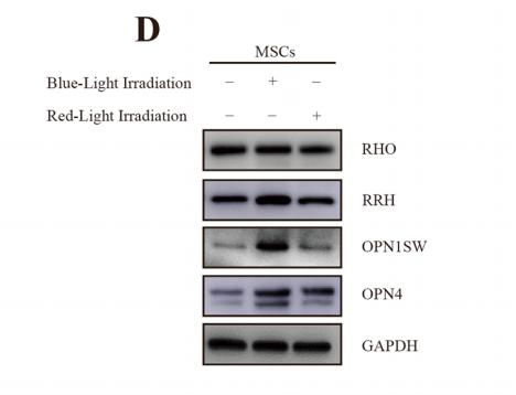
Application: WB Species: Human Sample: mesenchymal stem cells
Restrictive clause
Affinity Biosciences tests all products strictly. Citations are provided as a resource for additional applications that have not been validated by Affinity Biosciences. Please choose the appropriate format for each application and consult Materials and Methods sections for additional details about the use of any product in these publications.
For Research Use Only.
Not for use in diagnostic or therapeutic procedures. Not for resale. Not for distribution without written consent. Affinity Biosciences will not be held responsible for patent infringement or other violations that may occur with the use of our products. Affinity Biosciences, Affinity Biosciences Logo and all other trademarks are the property of Affinity Biosciences LTD.