| Product: | HE4 Antibody |
| Catalog: | DF7703 |
| Description: | Rabbit polyclonal antibody to HE4 |
| Application: | WB |
| Cited expt.: | WB |
| Reactivity: | Human |
| Mol.Wt.: | 23 kDa(Observed); 13kD(Calculated). |
| Uniprot: | Q14508 |
| RRID: | AB_2841174 |
Control Products

Related Downloads
Protocols
Product Info
*The optimal dilutions should be determined by the end user. For optimal experimental results, antibody reuse is not recommended.
*Tips:
WB: For western blot detection of denatured protein samples. IHC: For immunohistochemical detection of paraffin sections (IHC-p) or frozen sections (IHC-f) of tissue samples. IF/ICC: For immunofluorescence detection of cell samples. ELISA(peptide): For ELISA detection of antigenic peptide.
Cite Format: Affinity Biosciences Cat# DF7703, RRID:AB_2841174.
Fold/Unfold
dJ461P17.6; EDDM4; epididymal protein 4; Epididymal secretory protein E4; Epididymis specific whey acidic protein type four disulfide core; HE 4; Major epididymis specific protein E4; Major epididymis-specific protein E4; MGC57529; Putative protease inhibitor WAP5; WAP 5; WAP domain containing protein HE4; WAP domain containing protein HE4 V4; WAP four disulfide core domain 2; WAP four disulfide core domain protein 2; WAP four-disulfide core domain protein 2; WAP5; WFDC 2; WFDC2; WFDC2_HUMAN;
Immunogens
A synthesized peptide derived from human HE4, corresponding to a region within the internal amino acids.
Expressed in a number of normal tissues, including male reproductive system, regions of the respiratory tract and nasopharynx. Highly expressed in a number of tumors cells lines, such ovarian, colon, breast, lung and renal cells lines. Initially described as being exclusively transcribed in the epididymis.
- Q14508 WFDC2_HUMAN:
- Protein BLAST With
- NCBI/
- ExPASy/
- Uniprot
MPACRLGPLAAALLLSLLLFGFTLVSGTGAEKTGVCPELQADQNCTQECVSDSECADNLKCCSAGCATFCSLPNDKEGSCPQVNINFPQLGLCRDQCQVDSQCPGQMKCCRNGCGKVSCVTPNF
Research Backgrounds
Broad range protease inhibitor.
Secreted.
Expressed in a number of normal tissues, including male reproductive system, regions of the respiratory tract and nasopharynx. Highly expressed in a number of tumors cells lines, such ovarian, colon, breast, lung and renal cells lines. Initially described as being exclusively transcribed in the epididymis.
References

Application: WB Species: human Sample: ovarian cancer cells

Application: IF/ICC Species: human Sample: CAOV3 and OVCAR3 cells

Application: WB Species: human Sample: CAOV3 and OVCAR3 cells
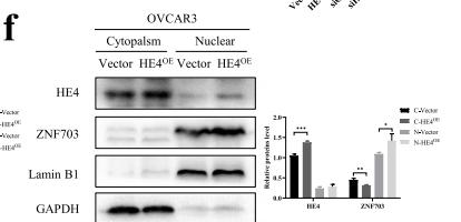
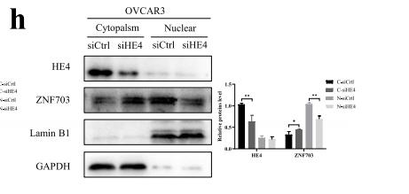
Application: WB Species: human Sample: CAOV3 and OVCAR3 cells

Application: WB Species: human Sample: CAOV3 and OVCAR3 cells
Application: WB Species: human Sample: CAOV3 and OVCAR3 cells
Restrictive clause
Affinity Biosciences tests all products strictly. Citations are provided as a resource for additional applications that have not been validated by Affinity Biosciences. Please choose the appropriate format for each application and consult Materials and Methods sections for additional details about the use of any product in these publications.
For Research Use Only.
Not for use in diagnostic or therapeutic procedures. Not for resale. Not for distribution without written consent. Affinity Biosciences will not be held responsible for patent infringement or other violations that may occur with the use of our products. Affinity Biosciences, Affinity Biosciences Logo and all other trademarks are the property of Affinity Biosciences LTD.