, using LHX3 Antibody at 1/1000 dilution.
5ug/NC membrane strip.
Exposure for 10s with Affinity™ ECL Kit(#KF8003).
Bands result from membrane strip incubation.")
| Product: | LHX3 Antibody |
| Catalog: | DF8362 |
| Description: | Rabbit polyclonal antibody to LHX3 |
| Application: | WB |
| Cited expt.: | |
| Reactivity: | Human, Mouse, Rat |
| Prediction: | Pig, Zebrafish, Bovine, Horse, Sheep, Dog, Chicken, Xenopus |
| Mol.Wt.: | 43 kDa(Observed); 43kD(Calculated). |
| Uniprot: | Q9UBR4 |
| RRID: | AB_2841627 |
Control Products

Related Downloads
Protocols
Product Info
*The optimal dilutions should be determined by the end user. For optimal experimental results, antibody reuse is not recommended.
*Tips:
WB: For western blot detection of denatured protein samples. IHC: For immunohistochemical detection of paraffin sections (IHC-p) or frozen sections (IHC-f) of tissue samples. IF/ICC: For immunofluorescence detection of cell samples. ELISA(peptide): For ELISA detection of antigenic peptide.
Cite Format: Affinity Biosciences Cat# DF8362, RRID:AB_2841627.
Fold/Unfold
CPHD 3; CPHD3; DKFZp762A2013; LHX 3; LHX3; LHX3_HUMAN; LIM 3; LIM homeobox 3; LIM homeobox gene 3; LIM homeobox protein 3; LIM/homeobox protein Lhx3; LIM/homeodomain protein LHX3; Lim3; M2 LHX3; mLim-3; mLIM3; P LIM;
Immunogens
A synthesized peptide derived from human LHX3, corresponding to a region within C-terminal amino acids.
- Q9UBR4 LHX3_HUMAN:
- Protein BLAST With
- NCBI/
- ExPASy/
- Uniprot
MLLETGLERDRARPGAAAVCTLGGTREIPLCAGCDQHILDRFILKALDRHWHSKCLKCSDCHTPLAERCFSRGESVYCKDDFFKRFGTKCAACQLGIPPTQVVRRAQDFVYHLHCFACVVCKRQLATGDEFYLMEDSRLVCKADYETAKQREAEATAKRPRTTITAKQLETLKSAYNTSPKPARHVREQLSSETGLDMRVVQVWFQNRRAKEKRLKKDAGRQRWGQYFRNMKRSRGGSKSDKDSVQEGQDSDAEVSFPDEPSLAEMGPANGLYGSLGEPTQALGRPSGALGNFSLEHGGLAGPEQYRELRPGSPYGVPPSPAAPQSLPGPQPLLSSLVYPDTSLGLVPSGAPGGPPPMRVLAGNGPSSDLSTGSSGGYPDFPASPASWLDEVDHAQF
Predictions
Score>80(red) has high confidence and is suggested to be used for WB detection. *The prediction model is mainly based on the alignment of immunogen sequences, the results are for reference only, not as the basis of quality assurance.
High(score>80) Medium(80>score>50) Low(score<50) No confidence
Research Backgrounds
Acts as a transcriptional activator. Binds to and activates the promoter of the alpha-glycoprotein gene, and synergistically enhances transcription from the prolactin promoter in cooperation with POU1F1/Pit-1 (By similarity). Required for the establishment of the specialized cells of the pituitary gland and the nervous system. Involved in the development of interneurons and motor neurons in cooperation with LDB1 and ISL1.
Nucleus.
The LIM domain specifically interacts with the Pit-1 POU domain and is required for synergistic interactions with Pit-1, but not for basal transcriptional activation events.
References
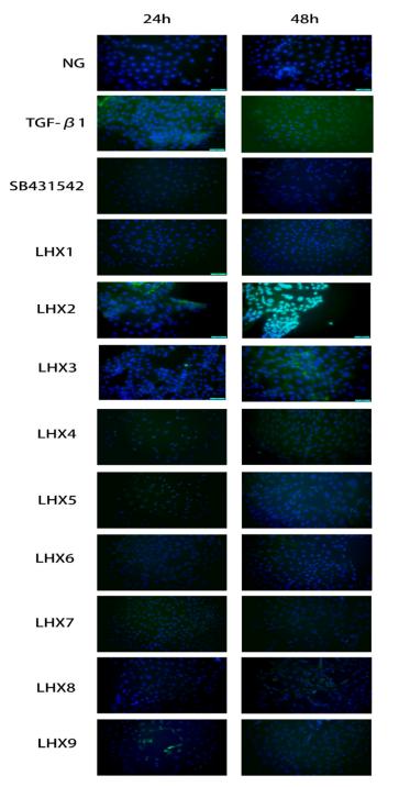
Application: IF/ICC Species: Mouse Sample: MTEC cells
Restrictive clause
Affinity Biosciences tests all products strictly. Citations are provided as a resource for additional applications that have not been validated by Affinity Biosciences. Please choose the appropriate format for each application and consult Materials and Methods sections for additional details about the use of any product in these publications.
For Research Use Only.
Not for use in diagnostic or therapeutic procedures. Not for resale. Not for distribution without written consent. Affinity Biosciences will not be held responsible for patent infringement or other violations that may occur with the use of our products. Affinity Biosciences, Affinity Biosciences Logo and all other trademarks are the property of Affinity Biosciences LTD.