Product: UCP2 Antibody
Catalog: DF8626
Description: Rabbit polyclonal antibody to UCP2
Application: WB IHC IF/ICC
Reactivity: Human, Mouse, Rat
Prediction: Pig, Zebrafish, Bovine, Horse, Sheep, Rabbit, Dog, Xenopus
Mol.Wt.: 33 kDa; 33kD(Calculated).
Uniprot: P55851
RRID: AB_2841830

View similar products>>

   Size Price Inventory
 100ul $280 In stock
 200ul $350 In stock

Lead Time: Same day delivery

For pricing and ordering contact:
Local distributors

Product Info

Source:
Rabbit
Application:
WB 1:1000-3000, IHC 1:50-1:200, IF/ICC 1:100-1:500
*The optimal dilutions should be determined by the end user.
*Tips:

WB: For western blot detection of denatured protein samples. IHC: For immunohistochemical detection of paraffin sections (IHC-p) or frozen sections (IHC-f) of tissue samples. IF/ICC: For immunofluorescence detection of cell samples. ELISA(peptide): For ELISA detection of antigenic peptide.

Reactivity:
Human,Mouse,Rat
Prediction:
Pig(100%), Zebrafish(100%), Bovine(100%), Horse(100%), Sheep(100%), Rabbit(100%), Dog(100%), Xenopus(91%)
Clonality:
Polyclonal
Specificity:
UCP2 Antibody detects endogenous levels of total UCP2.
RRID:
AB_2841830
Cite Format: Affinity Biosciences Cat# DF8626, RRID:AB_2841830.
Conjugate:
Unconjugated.
Purification:
The antiserum was purified by peptide affinity chromatography using SulfoLink™ Coupling Resin (Thermo Fisher Scientific).
Storage:
Rabbit IgG in phosphate buffered saline , pH 7.4, 150mM NaCl, 0.02% sodium azide and 50% glycerol. Store at -20 °C. Stable for 12 months from date of receipt.
Alias:

Fold/Unfold

BMIQ4; Mitochondrial uncoupling protein 2; SLC25A8; Solute carrier family 25 member 8; UCP 2; ucp2; UCP2_HUMAN; UCPH; Uncoupling protein 2; Uncoupling protein 2 mitochondrial proton carrier;

Immunogens

Immunogen:
Uniprot:
Gene(ID):
Expression:
P55851 UCP2_HUMAN:

Widely expressed in adult human tissues, including tissues rich in macrophages. Most expressed in white adipose tissue and skeletal muscle.

Sequence:
MVGFKATDVPPTATVKFLGAGTAACIADLITFPLDTAKVRLQIQGESQGPVRATASAQYRGVMGTILTMVRTEGPRSLYNGLVAGLQRQMSFASVRIGLYDSVKQFYTKGSEHASIGSRLLAGSTTGALAVAVAQPTDVVKVRFQAQARAGGGRRYQSTVNAYKTIAREEGFRGLWKGTSPNVARNAIVNCAELVTYDLIKDALLKANLMTDDLPCHFTSAFGAGFCTTVIASPVDVVKTRYMNSALGQYSSAGHCALTMLQKEGPRAFYKGFMPSFLRLGSWNVVMFVTYEQLKRALMAACTSREAPF

Predictions

Predictions:

Score>80(red) has high confidence and is suggested to be used for WB detection. *The prediction model is mainly based on the alignment of immunogen sequences, the results are for reference only, not as the basis of quality assurance.

Species
Results
Score
Pig
100
Horse
100
Bovine
100
Sheep
100
Dog
100
Zebrafish
100
Rabbit
100
Xenopus
91
Chicken
0
Model Confidence:
High(score>80) Medium(80>score>50) Low(score<50) No confidence

PTMs - P55851 As Substrate

Site PTM Type Enzyme
T14 Phosphorylation
T65 Phosphorylation
T72 Phosphorylation
S77 Phosphorylation
Y79 Phosphorylation
S94 Phosphorylation
S158 Phosphorylation
Y163 Phosphorylation
T165 Phosphorylation
T179 Phosphorylation
S180 Phosphorylation
T229 Phosphorylation
S233 Phosphorylation

Research Backgrounds

Function:

UCP are mitochondrial transporter proteins that create proton leaks across the inner mitochondrial membrane, thus uncoupling oxidative phosphorylation from ATP synthesis. As a result, energy is dissipated in the form of heat.

Subcellular Location:

Mitochondrion inner membrane>Multi-pass membrane protein.

Extracellular region or secreted Cytosol Plasma membrane Cytoskeleton Lysosome Endosome Peroxisome ER Golgi apparatus Nucleus Mitochondrion Manual annotation Automatic computational assertionSubcellular location
Tissue Specificity:

Widely expressed in adult human tissues, including tissues rich in macrophages. Most expressed in white adipose tissue and skeletal muscle.

Subunit Structure:

Acts as a dimer forming a proton channel.

Family&Domains:

Belongs to the mitochondrial carrier (TC 2.A.29) family.

References

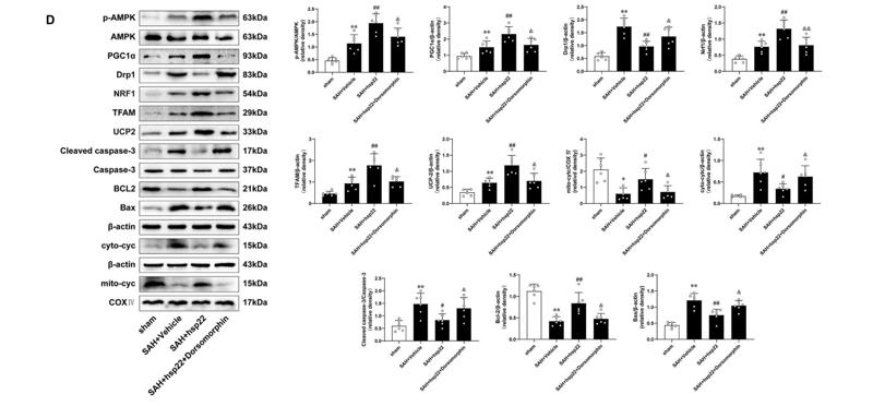
1). Heat shock protein 22 modulates NRF1/TFAM-dependent mitochondrial biogenesis and DRP1-sparked mitochondrial apoptosis through AMPK-PGC1α signaling pathway to alleviate the early brain injury of subarachnoid hemorrhage in rats. Redox Biology, 2021 (PubMed: 33472123) [IF=10.7]

Application: WB    Species: rat    Sample: brain

Fig. 6. Hsp22 regulates PGC1α via AMPK signaling pathway in rats after SAH Beam balance scores, Modified Garcia scores and Brainwater content in various groups. n = 6 per group. (B) Representative photomicrographs of TUNEL staining and quantitative analyses in the indicated groups. n = 4 per group. Scale bar = 100 μm. (C) Typical photomicrographs showing double immunofluorescence staining of PGC1α (green) and NeuN (red) in diverse experimental groups. n = 4 per group. Scale bar = 50 μm. (D) Western blot images and quantitative analyses of p-AMPK/AMPK, PGC1α, Drp1, Nrf1, TFAM, UCP2, Cleaved caspase-3/Caspase-3, Bcl2, Bax, Cytosolic and mitochondrial cytochrome c. n = 6 per group. Bars represent mean ± SD. **P < 0.01, *P < 0.05 vs. Sham group. ##P < 0.01, #P < 0.05 vs. SAH + Vehicle group. &&P < 0.01, &P < 0.05 vs. SAH + hsp22+scramble siRNA. (For interpretation of the references to colour in this figure legend, the reader is referred to the Web version of this article.)

2). Targeted inhibition of CX3CL1 limits podocytes ferroptosis to ameliorate cisplatin-induced acute kidney injury. Molecular medicine (Cambridge, Mass.), 2023 (PubMed: 37875838) [IF=6.0]

Application: WB    Species: Mouse    Sample: kidney tissues

Fig. 4 The mitochondrial function was improved by CX3CL1 deficiency in mice with cisplatin-induced AKI. A TEM images of renal tissues were captured. Scale bar = 500 nm. B The relative expression profiles of the mitochondrial proteins UCP2, Mfn2, and PGC1α in kidney tissues were determined by Western blotting. C DHE staining was conducted to determine the ROS level in renal tissues. Scale bar = 20 μm. D TEM was employed to capture images of podocytes. Scale bar = 500 nm. E Western blot analysis illustrating the relative expression patterns of the mitochondrial proteins UCP2, Mfn2, and PGC1α in podocytes. F The ROS level was detected through DCFH-DA labeling. Scale bar = 10 μm. G JC-1 staining was conducted to detect MMP in podocytes. Scale bar = 10 μm. (AKI, acute kidney injury; TEM, transmission electron microscopy; UCP2, uncoupling protein 2; Mfn2, Mitofusin 2; PGC1α, peroxisome proliferators-activated receptor γ coactivator l-alpha; DHE, dihydroethidium; ROS, reactive oxygen species; DCFH-DA, 2,7-dichlorodihydrofluorescein diacetate; MMP, matrix metalloproteinase; P value was calculated by one-way analysis of variance and Tukey’s test. *p 

3). Gut Flora Mediates the Rapid Tolerance of Electroacupuncture on Ischemic Stroke by Activating Melatonin Receptor through Regulating Indole-3-Propionic Acid. The American journal of Chinese medicine, 2022 (PubMed: 35475976) [IF=4.8]

4). Caffeine Citrate Protects Against Sepsis-Associated Encephalopathy and Inhibits the UCP2/NLRP3 Axis in Astrocytes. Journal of interferon & cytokine research : the official journal of the International Society for Interferon and Cytokine Research, 2022 (PubMed: 35420462) [IF=1.9]

5). Melatonin Alleviates Acute Kidney Injury by Inhibiting NRF2/Slc7a11 Axis-Mediated Ferroptosis. Oxidative Medicine and Cellular Longevity, 2022 (PubMed: 35979396)

Application: WB    Species: Mice    Sample: kidney

Figure 4 Melatonin improves mitochondrial function and inhibits ferroptosis in the IR-induced acute kidney injury. (a) Renal tissues were imaged using transmission electron microscopy (TEM). Red arrows indicate mitochondrial cristae disappearance and outer membrane rupture. Scale bar = 200 nm. (b) Western blot analysis of mitochondrial proteins and their relative protein levels of UCP2 (c) and MFN2 (d). (e) Renal relative PGC-1a and TFAM levels were evaluated by RT-PCR. (f) Quantification of mRNA levels of Acsl4, Cox-2, and GPX4 kidney tissue by real-time PCR. (g) Western blots of NRF2, Slc7a11, and GPX4 proteins and their relative protein levels of NRF2 (h), Slc7a11 (i), and GPX4 (j). Data represent the mean ± SEM of six mice in each group. ∗p < 0.05,  ∗∗p < 0.01, and∗∗∗p < 0.001 compared to the Sham and Sham+MT (20 mg/kg) groups. #p < 0.05,  ##p < 0.01, and###p < 0.001 compared to the IR-treated group.

Restrictive clause


Affinity Biosciences tests all products strictly. Citations are provided as a resource for additional applications that have not been validated by Affinity Biosciences. Please choose the appropriate format for each application and consult Materials and Methods sections for additional details about the use of any product in these publications.

For Research Use Only.
Not for use in diagnostic or therapeutic procedures. Not for resale. Not for distribution without written consent. Affinity Biosciences will not be held responsible for patent infringement or other violations that may occur with the use of our products. Affinity Biosciences, Affinity Biosciences Logo and all other trademarks are the property of Affinity Biosciences LTD.