Product: Cytokeratin 7 Antibody
Catalog: AF0195
Description: Rabbit polyclonal antibody to Cytokeratin 7
Application: WB IHC IF/ICC
Cited expt.: WB, IHC, IF/ICC
Reactivity: Human
Mol.Wt.: 51kDa; 51kD(Calculated).
Uniprot: P08729
RRID: AB_2833388

View similar products>>

   Size Price Inventory
 100ul $280 In stock
 200ul $350 In stock

Lead Time: Same day delivery

For pricing and ordering contact:
Local distributors

Product Info

Source:
Rabbit
Application:
WB 1:500-1:3000, IHC 1:50-1:200, IF/ICC: 1:100-1:500
*The optimal dilutions should be determined by the end user.
*Tips:

WB: For western blot detection of denatured protein samples. IHC: For immunohistochemical detection of paraffin sections (IHC-p) or frozen sections (IHC-f) of tissue samples. IF/ICC: For immunofluorescence detection of cell samples. ELISA(peptide): For ELISA detection of antigenic peptide.

Reactivity:
Human
Clonality:
Polyclonal
Specificity:
Cytokeratin 7 Antibody detects endogenous levels of total Cytokeratin 7.
RRID:
AB_2833388
Cite Format: Affinity Biosciences Cat# AF0195, RRID:AB_2833388.
Conjugate:
Unconjugated.
Purification:
The antiserum was purified by peptide affinity chromatography using SulfoLink™ Coupling Resin (Thermo Fisher Scientific).
Storage:
Rabbit IgG in phosphate buffered saline , pH 7.4, 150mM NaCl, 0.02% sodium azide and 50% glycerol. Store at -20 °C. Stable for 12 months from date of receipt.
Alias:

Fold/Unfold

CK 7; CK-7; CK7; Cytokeratin 7; Cytokeratin-7; D15Wsu77e; K2C7; K2C7_HUMAN; K7; Keratin 7; Keratin 7, type II; Keratin type II cytoskeletal 7; Keratin, 55K type II cytoskeletal; Keratin, simple epithelial; Keratin, simple epithelial type I, K7; Keratin, type II cytoskeletal 7; Keratin-7; Krt2-7; KRT7; MGC11625; MGC129731; MGC3625; Sarcolectin; SCL; Type II mesothelial keratin K7; Type-II keratin Kb7;

Immunogens

Immunogen:

A synthesized peptide derived from human Cytokeratin 7, corresponding to a region within C-terminal amino acids.

Uniprot:
Gene(ID):
Expression:
P08729 K2C7_HUMAN:

Expressed in cultured epidermal, bronchial and mesothelial cells but absent in colon, ectocervix and liver. Observed throughout the glandular cells in the junction between stomach and esophagus but is absent in the esophagus.

Description:
K7 a type II cytoskeletal keratin. The keratins are intermediate filament proteins responsible for the structural integrity of epithelial cells and are subdivided into cytokeratins and hair keratins. Phosphorylation of keratins at specific sites affects their organization, assembly dynamics, and their interaction with signaling molecules. Specifically expressed in the simple epithelia lining the cavities of the internal organs and in the gland ducts and blood vessels
Sequence:
MSIHFSSPVFTSRSAAFSGRGAQVRLSSARPGGLGSSSLYGLGASRPRVAVRSAYGGPVGAGIREVTINQSLLAPLRLDADPSLQRVRQEESEQIKTLNNKFASFIDKVRFLEQQNKLLETKWTLLQEQKSAKSSRLPDIFEAQIAGLRGQLEALQVDGGRLEAELRSMQDVVEDFKNKYEDEINHRTAAENEFVVLKKDVDAAYMSKVELEAKVDALNDEINFLRTLNETELTELQSQISDTSVVLSMDNSRSLDLDGIIAEVKAQYEEMAKCSRAEAEAWYQTKFETLQAQAGKHGDDLRNTRNEISEMNRAIQRLQAEIDNIKNQRAKLEAAIAEAEERGELALKDARAKQEELEAALQRGKQDMARQLREYQELMSVKLALDIEIATYRKLLEGEESRLAGDGVGAVNISVMNSTGGSSSGGGIGLTLGGTMGSNALSFSSSAGPGLLKAYSIRTASASRRSARD

Research Backgrounds

Function:

Blocks interferon-dependent interphase and stimulates DNA synthesis in cells. Involved in the translational regulation of the human papillomavirus type 16 E7 mRNA (HPV16 E7).

PTMs:

Arg-20 is dimethylated, probably to asymmetric dimethylarginine.

Subcellular Location:

Cytoplasm.

Extracellular region or secreted Cytosol Plasma membrane Cytoskeleton Lysosome Endosome Peroxisome ER Golgi apparatus Nucleus Mitochondrion Manual annotation Automatic computational assertionSubcellular location
Tissue Specificity:

Expressed in cultured epidermal, bronchial and mesothelial cells but absent in colon, ectocervix and liver. Observed throughout the glandular cells in the junction between stomach and esophagus but is absent in the esophagus.

Family&Domains:

Belongs to the intermediate filament family.

References

1). KRT7 promotes epithelial‑mesenchymal transition in ovarian cancer via the TGF‑β/Smad2/3 signaling pathway. ONCOLOGY REPORTS, 2021 (PubMed: 33416175) [IF=3.8]

Application: WB    Species: Human    Sample: ovarian cell

Figure 2. - Effect of KRT7 on HEY and OVCAR433 cell proliferation and colony formation. (A) Protein expression levels of KRT7 in ovarian cell lines were assessed using western blotting. (B) Overexpression of KRT7 in HEY cells was confirmed by western blotting. (C) Overexpression of KRT7 significantly increased the proliferative capacity of HEY cells. (D) Overexpression of KRT7 in HEY cells significantly increased colony formation. (E) Knockdown of KRT7 in OVCAR433 cells was confirmed by western blotting. Knockdown of KRT7 in OVCAR433 cells significantly reduced (F) cell proliferation and (G) colony formation. All experiments were performed at least three times. Results are presented as the mean ± standard deviation. **P<0.01. KRT7, keratin 7; sh, short hairpin RNA; NC, negative control; OD, optical density.

Application: IHC    Species: Human    Sample: ovarian tumor tissues

Figure 1. - KRT7 expression is indicative of a poor prognosis in patients with ovarian cancer. (A) Representative images of KRT7 staining in normal and ovarian tumor tissues, and immunohistochemical score of KRT7 in normal and ovarian tumor tissues. (B) mRNA expression levels of KRT7 in normal and ovarian tumor tissues. Associations between the (C) pathological grades and (D) clinical stages of ovarian cancer with KRT7 expression. (E) Effect of mRNA expression levels of KRT7 on the median survival time of patients with ovarian cancer. (F) Gene Ontology analysis of the differentially expressed genes between the high- and low-expression KRT7 groups in patients with ovarian cancer. Data were obtained from the Human Protein Atlas and The Cancer Genome Atlas database. *P<0.05, **P<0.01. KRT7, keratin 7; ECM, extracellular matrix.

2). microRNA-210/Long non-coding RNA MEG3 axis inhibits trophoblast cell migration and invasion by suppressing EMT process. PLACENTA, 2021 (PubMed: 33990028) [IF=3.0]

3). Isolation, culture, and identification of ceruminous gland cells. Journal of Molecular Histology, 2022 (PubMed: 35113280) [IF=2.9]

4). Upregulation of ITGB6 in primary palmar hyperhidrosis. Advances in clinical and experimental medicine : official organ Wroclaw Medical University, 2023 (PubMed: 37212774) [IF=2.1]

Application: IF/ICC    Species: Human    Sample:

Fig. 2. Identification of human sweat gland cells and verification of integrin β6 (ITGB6) overexpression transfection efficiency. A. Immunofluorescence staining of CEA (green) and CK7 (red) in sweat gland cells extracted from patients with primary palmar hyperhidrosis. The cell nuclei were stained with 4’,6-diamidino-2-phenylindole (DAPI) (blue); B. Quantitative polymerase chain reaction (ITGB6 compared to control, Tukey’s honest significant difference (HSD), p < 0.001; ITGB6 compared to negative control (NC), Tukey’s HSD, p < 0.001); C,D. Western blot detection of the transfection efficiency of ITGB6 overexpression analysis results (ITGB6 compared to control, Tukey’s HSD, p < 0.001; ITGB6 compared to NC, Tukey’s HSD, p < 0.001). The data are presented as mean ± 95% confidence interval (95% CI)

5). Comparative study of the external auditory canal in humans and large mammals. The Anatomical Record, 2022 (PubMed: 34050614) [IF=1.8]

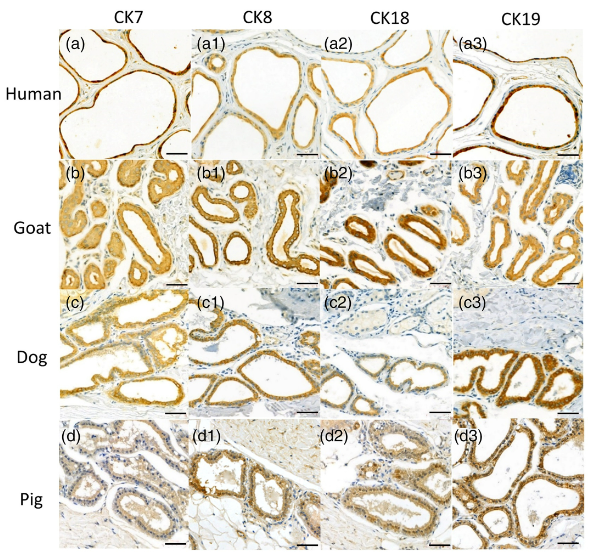
Application: IHC    Species: Human,Goat,Dog,Pig    Sample: EAC glandular cells

FIGURE 4 Immunostaining of cytoskeletal proteins of the ceruminous gland of a (a) human, (b) goat, (c) dog, and (d) pig. CK7, CK8, CK18, and CK19 are expressed in the EAC glandular cells of humans, goats, dogs, and pig. The positive expressions appeared to be brown or dark brown. Scale bar = 50 μm

Restrictive clause


Affinity Biosciences tests all products strictly. Citations are provided as a resource for additional applications that have not been validated by Affinity Biosciences. Please choose the appropriate format for each application and consult Materials and Methods sections for additional details about the use of any product in these publications.

For Research Use Only.
Not for use in diagnostic or therapeutic procedures. Not for resale. Not for distribution without written consent. Affinity Biosciences will not be held responsible for patent infringement or other violations that may occur with the use of our products. Affinity Biosciences, Affinity Biosciences Logo and all other trademarks are the property of Affinity Biosciences LTD.