, using G-CSF Antibody at 1/1000 dilution.
5ug/NC membrane strip.
Exposure for 1min with Affinity™ ECL Kit(#KF8003).
Bands result from membrane strip incubation.")
| Product: | G-CSF Antibody |
| Catalog: | DF9542 |
| Description: | Rabbit polyclonal antibody to G-CSF |
| Application: | WB IHC |
| Cited expt.: | |
| Reactivity: | Human, Rat |
| Prediction: | Bovine, Sheep, Rabbit |
| Mol.Wt.: | 22 kDa(Observed); 22kD(Calculated). |
| Uniprot: | P09919 |
| RRID: | AB_2842738 |
Control Products

Product Info
*The optimal dilutions should be determined by the end user. For optimal experimental results, antibody reuse is not recommended.
*Tips:
WB: For western blot detection of denatured protein samples. IHC: For immunohistochemical detection of paraffin sections (IHC-p) or frozen sections (IHC-f) of tissue samples. IF/ICC: For immunofluorescence detection of cell samples. ELISA(peptide): For ELISA detection of antigenic peptide.
Cite Format: Affinity Biosciences Cat# DF9542, RRID:AB_2842738.
Fold/Unfold
C17orf33; Colony stimulating factor 3 (granulocyte); Colony stimulating factor 3; CSF 3; CSF beta; CSF3; CSF3_HUMAN; CSF3OS; Csfg; Filgrastim; G-CSF; GCSA; GCSF; Granulocyte colony stimulating factor; Granulocyte colony-stimulating factor; Lenograstim; Macrophage granulocyte inducer 2; MGC45931; MGI 2; Pluripoietin;
Immunogens
A synthesized peptide derived from human G-CSF, corresponding to a region within the internal amino acids.
- P09919 CSF3_HUMAN:
- Protein BLAST With
- NCBI/
- ExPASy/
- Uniprot
MAGPATQSPMKLMALQLLLWHSALWTVQEATPLGPASSLPQSFLLKCLEQVRKIQGDGAALQEKLVSECATYKLCHPEELVLLGHSLGIPWAPLSSCPSQALQLAGCLSQLHSGLFLYQGLLQALEGISPELGPTLDTLQLDVADFATTIWQQMEELGMAPALQPTQGAMPAFASAFQRRAGGVLVASHLQSFLEVSYRVLRHLAQP
Predictions
Score>80(red) has high confidence and is suggested to be used for WB detection. *The prediction model is mainly based on the alignment of immunogen sequences, the results are for reference only, not as the basis of quality assurance.
High(score>80) Medium(80>score>50) Low(score<50) No confidence
Research Backgrounds
Granulocyte/macrophage colony-stimulating factors are cytokines that act in hematopoiesis by controlling the production, differentiation, and function of 2 related white cell populations of the blood, the granulocytes and the monocytes-macrophages. This CSF induces granulocytes.
O-glycan consists of Gal-GalNAc disaccharide which can be modified with up to two sialic acid residues (done in recombinantly expressed G-CSF from CHO cells).
Secreted.
Belongs to the IL-6 superfamily.
Research Fields
· Environmental Information Processing > Signaling molecules and interaction > Cytokine-cytokine receptor interaction. (View pathway)
· Environmental Information Processing > Signal transduction > PI3K-Akt signaling pathway. (View pathway)
· Environmental Information Processing > Signal transduction > Jak-STAT signaling pathway. (View pathway)
· Human Diseases > Infectious diseases: Parasitic > Malaria.
· Organismal Systems > Immune system > Hematopoietic cell lineage. (View pathway)
· Organismal Systems > Immune system > IL-17 signaling pathway. (View pathway)
References
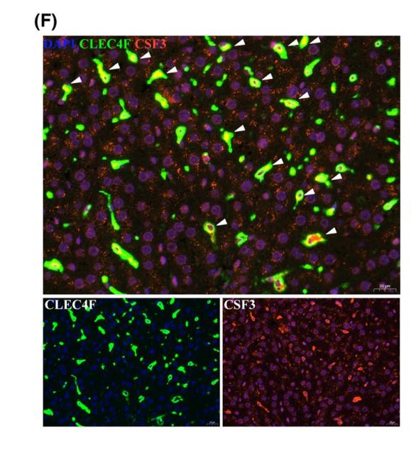
Application: IF/ICC Species: Rat Sample: Kupffer cells
Restrictive clause
Affinity Biosciences tests all products strictly. Citations are provided as a resource for additional applications that have not been validated by Affinity Biosciences. Please choose the appropriate format for each application and consult Materials and Methods sections for additional details about the use of any product in these publications.
For Research Use Only.
Not for use in diagnostic or therapeutic procedures. Not for resale. Not for distribution without written consent. Affinity Biosciences will not be held responsible for patent infringement or other violations that may occur with the use of our products. Affinity Biosciences, Affinity Biosciences Logo and all other trademarks are the property of Affinity Biosciences LTD.