

| Product: | Phospho-MOB1A (Thr35) Antibody |
| Catalog: | AF4481 |
| Description: | Rabbit polyclonal antibody to Phospho-MOB1A (Thr35) |
| Application: | WB IF/ICC |
| Cited expt.: | WB |
| Reactivity: | Human, Mouse, Rat |
| Prediction: | Bovine, Horse, Sheep, Rabbit, Dog, Xenopus |
| Mol.Wt.: | 25kDa(Observed); 25kD(Calculated). |
| Uniprot: | Q7L9L4 |
| RRID: | AB_2844532 |
Control Products

Related Downloads
Protocols
Product Info
*The optimal dilutions should be determined by the end user. For optimal experimental results, antibody reuse is not recommended.
*Tips:
WB: For western blot detection of denatured protein samples. IHC: For immunohistochemical detection of paraffin sections (IHC-p) or frozen sections (IHC-f) of tissue samples. IF/ICC: For immunofluorescence detection of cell samples. ELISA(peptide): For ELISA detection of antigenic peptide.
Cite Format: Affinity Biosciences Cat# AF4481, RRID:AB_2844532.
Fold/Unfold
MATS 2; MATS2; MGC33910; Mob 1A; Mob 1B; MOB 4A; MOB kinase activator 1B; Mob1 homolog 1A; MOB1 Mps One Binder homolog B; MOB1 Mps one binder kinase activator like 1A; Mob1A; Mob1B; MOBKL 1A; MOBKL1A; MOL1A_HUMAN; Mps one binder kinase activator like 1A; Mps one binder kinase activator-like 1A; Protein Mob4A;
Immunogens
A synthesized peptide derived from human MOB1A around the phosphorylation site of Thr35.
Adrenal gland, bone marrow, brain, lung, placenta, prostate, salivary gland, skeletal muscle, testis, thymus, thyroid gland, uterus, colon with mucosa, fetal brain and fetal liver.
- Q7L9L4 MOB1B_HUMAN:
- Protein BLAST With
- NCBI/
- ExPASy/
- Uniprot
MSFLFGSRSSKTFKPKKNIPEGSHQYELLKHAEATLGSGNLRMAVMLPEGEDLNEWVAVNTVDFFNQINMLYGTITDFCTEESCPVMSAGPKYEYHWADGTNIKKPIKCSAPKYIDYLMTWVQDQLDDETLFPSKIGVPFPKNFMSVAKTILKRLFRVYAHIYHQHFDPVIQLQEEAHLNTSFKHFIFFVQEFNLIDRRELAPLQELIEKLTSKDR
Predictions
Score>80(red) has high confidence and is suggested to be used for WB detection. *The prediction model is mainly based on the alignment of immunogen sequences, the results are for reference only, not as the basis of quality assurance.
High(score>80) Medium(80>score>50) Low(score<50) No confidence
Research Backgrounds
Activator of LATS1/2 in the Hippo signaling pathway which plays a pivotal role in organ size control and tumor suppression by restricting proliferation and promoting apoptosis. The core of this pathway is composed of a kinase cascade wherein STK3/MST2 and STK4/MST1, in complex with its regulatory protein SAV1, phosphorylates and activates LATS1/2 in complex with its regulatory protein MOB1, which in turn phosphorylates and inactivates YAP1 oncoprotein and WWTR1/TAZ. Phosphorylation of YAP1 by LATS1/2 inhibits its translocation into the nucleus to regulate cellular genes important for cell proliferation, cell death, and cell migration. Stimulates the kinase activity of STK38L.
Phosphorylated by STK3/MST2 and STK4/MST1 and this phosphorylation enhances its binding to LATS1.
Cytoplasm. Nucleus.
Adrenal gland, bone marrow, brain, lung, placenta, prostate, salivary gland, skeletal muscle, testis, thymus, thyroid gland, uterus, colon with mucosa, fetal brain and fetal liver.
Belongs to the MOB1/phocein family.
Research Fields
· Environmental Information Processing > Signal transduction > Hippo signaling pathway. (View pathway)
· Environmental Information Processing > Signal transduction > Hippo signaling pathway - multiple species. (View pathway)
References
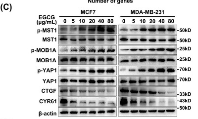
Application: WB Species: Human Sample: MCF7 and MDA‐MB‐231 cells
Restrictive clause
Affinity Biosciences tests all products strictly. Citations are provided as a resource for additional applications that have not been validated by Affinity Biosciences. Please choose the appropriate format for each application and consult Materials and Methods sections for additional details about the use of any product in these publications.
For Research Use Only.
Not for use in diagnostic or therapeutic procedures. Not for resale. Not for distribution without written consent. Affinity Biosciences will not be held responsible for patent infringement or other violations that may occur with the use of our products. Affinity Biosciences, Affinity Biosciences Logo and all other trademarks are the property of Affinity Biosciences LTD.