
.
Bands result from membrane strip incubation.")


 and mouse anti-beta tubulin Ab(#T0023) for 1 hour at 37°C. An AlexaFluor594 conjugated goat anti-rabbit IgG Ab(Red) and an AlexaFluor488 conjugated goat anti-mouse IgG Ab(Green) were used as the secondary antibody.
The nuclear counter stain is DAPI (blue).")
| Product: | SMCR7L/MID51 Antibody |
| Catalog: | DF12019 |
| Description: | Rabbit polyclonal antibody to SMCR7L/MID51 |
| Application: | WB IHC IF/ICC |
| Cited expt.: | WB |
| Reactivity: | Human, Mouse, Rat |
| Prediction: | Pig, Zebrafish, Bovine, Horse, Sheep, Rabbit, Dog, Chicken, Xenopus |
| Mol.Wt.: | 48-51 kDa(Observed); 51kD(Calculated). |
| Uniprot: | Q9NQG6 |
| RRID: | AB_2844824 |
Control Products

Related Downloads
Protocols
Product Info
*The optimal dilutions should be determined by the end user. For optimal experimental results, antibody reuse is not recommended.
*Tips:
WB: For western blot detection of denatured protein samples. IHC: For immunohistochemical detection of paraffin sections (IHC-p) or frozen sections (IHC-f) of tissue samples. IF/ICC: For immunofluorescence detection of cell samples. ELISA(peptide): For ELISA detection of antigenic peptide.
Cite Format: Affinity Biosciences Cat# DF12019, RRID:AB_2844824.
Fold/Unfold
dJ1104E15.3; HSU79252; Hypothetical protein LOC54471; MID51; MIEF1; mitochondrial dynamic protein MID51; mitochondrial dynamic protein of 51 kDa; mitochondrial dynamics protein MID51; mitochondrial dynamics protein of 51 kDa; mitochondrial elongation factor 1; SMC7L_HUMAN; SMCR7-like protein; Smcr7l; Smith Magenis syndrome chromosome region candidate gene 7 protein like; Smith-Magenis syndrome chromosomal region candidate gene 7 protein-like; Smith-Magenis syndrome chromosome region, candidate 7-like;
Immunogens
A synthesized peptide derived from human SMCR7L/MID51, corresponding to a region within the internal amino acids.
- Q9NQG6 MID51_HUMAN:
- Protein BLAST With
- NCBI/
- ExPASy/
- Uniprot
MAGAGERKGKKDDNGIGTAIDFVLSNARLVLGVGGAAMLGIATLAVKRMYDRAISAPTSPTRLSHSGKRSWEEPNWMGSPRLLNRDMKTGLSRSLQTLPTDSSTFDTDTFCPPRPKPVARKGQVDLKKSRLRMSLQEKLLTYYRNRAAIPAGEQARAKQAAVDICAELRSFLRAKLPDMPLRDMYLSGSLYDDLQVVTADHIQLIVPLVLEQNLWSCIPGEDTIMNVPGFFLVRRENPEYFPRGSSYWDRCVVGGYLSPKTVADTFEKVVAGSINWPAIGSLLDYVIRPAPPPEALTLEVQYERDKHLFIDFLPSVTLGDTVLVAKPHRLAQYDNLWRLSLRPAETARLRALDQADSGCRSLCLKILKAICKSTPALGHLTASQLTNVILHLAQEEADWSPDMLADRFLQALRGLISYLEAGVLPSALNPKVNLFAELTPEEIDELGYTLYCSLSEPEVLLQT
Predictions
Score>80(red) has high confidence and is suggested to be used for WB detection. *The prediction model is mainly based on the alignment of immunogen sequences, the results are for reference only, not as the basis of quality assurance.
High(score>80) Medium(80>score>50) Low(score<50) No confidence
Research Backgrounds
Mitochondrial outer membrane protein which regulates mitochondrial fission. Promotes the recruitment and association of the fission mediator dynamin-related protein 1 (DNM1L) to the mitochondrial surface independently of the mitochondrial fission FIS1 and MFF proteins. Regulates DNM1L GTPase activity and DNM1L oligomerization. Binds ADP and can also bind GDP, although with lower affinity. Does not bind CDP, UDP, ATP, AMP or GTP. Inhibits DNM1L GTPase activity in the absence of bound ADP. Requires ADP to stimulate DNM1L GTPase activity and the assembly of DNM1L into long, oligomeric tubules with a spiral pattern, as opposed to the ring-like DNM1L oligomers observed in the absence of bound ADP. Does not require ADP for its function in recruiting DNM1L.
Mitochondrion outer membrane>Single-pass membrane protein.
Expression is relatively high in heart, skeletal muscle, pancreas and kidney.
Belongs to the SMCR7 family.
References

Application: WB Species: Mouse Sample:

Application: WB Species: mouse Sample: hippocampus
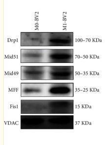
Application: WB Species: Mouse Sample: M0-BV2 and M1-BV2 cells
Restrictive clause
Affinity Biosciences tests all products strictly. Citations are provided as a resource for additional applications that have not been validated by Affinity Biosciences. Please choose the appropriate format for each application and consult Materials and Methods sections for additional details about the use of any product in these publications.
For Research Use Only.
Not for use in diagnostic or therapeutic procedures. Not for resale. Not for distribution without written consent. Affinity Biosciences will not be held responsible for patent infringement or other violations that may occur with the use of our products. Affinity Biosciences, Affinity Biosciences Logo and all other trademarks are the property of Affinity Biosciences LTD.