


 and mouse anti-beta tubulin Ab(T0023 1:200) for 1 hour at 37°C. An AlexaFluor594 conjugated goat anti-rabbit IgG(H+L) Ab(Red) and an AlexaFluor488 conjugated goat anti-mouse IgG(H+L) Ab(Green) were used as the secondary antibody.
The nuclear counter stain is DAPI(blue).")
| Product: | NG2 Antibody |
| Catalog: | DF12589 |
| Description: | Rabbit polyclonal antibody to NG2 |
| Application: | WB IHC IF/ICC |
| Cited expt.: | WB, IF/ICC |
| Reactivity: | Human, Mouse, Rat |
| Prediction: | Pig, Bovine, Horse, Sheep, Rabbit |
| Mol.Wt.: | 251 kDa,320kDa(Observed); 251kD(Calculated). |
| Uniprot: | Q6UVK1 |
| RRID: | AB_2845551 |
Control Products

Related Downloads
Protocols
Product Info
*The optimal dilutions should be determined by the end user. For optimal experimental results, antibody reuse is not recommended.
*Tips:
WB: For western blot detection of denatured protein samples. IHC: For immunohistochemical detection of paraffin sections (IHC-p) or frozen sections (IHC-f) of tissue samples. IF/ICC: For immunofluorescence detection of cell samples. ELISA(peptide): For ELISA detection of antigenic peptide.
Cite Format: Affinity Biosciences Cat# DF12589, RRID:AB_2845551.
Fold/Unfold
4732461B14Rik; AN2; AN2 proteoglycan; Chondroitin sulfate proteoglycan 4 (melanoma-associated); Chondroitin sulfate proteoglycan 4; Chondroitin sulfate proteoglycan NG2; Cspg4; Cspg4 chondroitin sulfate proteoglycan 4; CSPG4_HUMAN; HMW-MAA; HSN tumor-specific antigen; Kiaa4232; MCSP; MCSPG; MEL-CSPG; Melanoma chondroitin sulfate proteoglycan; Melanoma-associated chondroitin sulfate proteoglycan; MELCSPG; MSK16; NG2;
Immunogens
A synthesized peptide derived from human NG2, corresponding to a region within the internal amino acids.
- Q6UVK1 CSPG4_HUMAN:
- Protein BLAST With
- NCBI/
- ExPASy/
- Uniprot
MQSGPRPPLPAPGLALALTLTMLARLASAASFFGENHLEVPVATALTDIDLQLQFSTSQPEALLLLAAGPADHLLLQLYSGRLQVRLVLGQEELRLQTPAETLLSDSIPHTVVLTVVEGWATLSVDGFLNASSAVPGAPLEVPYGLFVGGTGTLGLPYLRGTSRPLRGCLHAATLNGRSLLRPLTPDVHEGCAEEFSASDDVALGFSGPHSLAAFPAWGTQDEGTLEFTLTTQSRQAPLAFQAGGRRGDFIYVDIFEGHLRAVVEKGQGTVLLHNSVPVADGQPHEVSVHINAHRLEISVDQYPTHTSNRGVLSYLEPRGSLLLGGLDAEASRHLQEHRLGLTPEATNASLLGCMEDLSVNGQRRGLREALLTRNMAAGCRLEEEEYEDDAYGHYEAFSTLAPEAWPAMELPEPCVPEPGLPPVFANFTQLLTISPLVVAEGGTAWLEWRHVQPTLDLMEAELRKSQVLFSVTRGARHGELELDIPGAQARKMFTLLDVVNRKARFIHDGSEDTSDQLVLEVSVTARVPMPSCLRRGQTYLLPIQVNPVNDPPHIIFPHGSLMVILEHTQKPLGPEVFQAYDPDSACEGLTFQVLGTSSGLPVERRDQPGEPATEFSCRELEAGSLVYVHRGGPAQDLTFRVSDGLQASPPATLKVVAIRPAIQIHRSTGLRLAQGSAMPILPANLSVETNAVGQDVSVLFRVTGALQFGELQKQGAGGVEGAEWWATQAFHQRDVEQGRVRYLSTDPQHHAYDTVENLALEVQVGQEILSNLSFPVTIQRATVWMLRLEPLHTQNTQQETLTTAHLEATLEEAGPSPPTFHYEVVQAPRKGNLQLQGTRLSDGQGFTQDDIQAGRVTYGATARASEAVEDTFRFRVTAPPYFSPLYTFPIHIGGDPDAPVLTNVLLVVPEGGEGVLSADHLFVKSLNSASYLYEVMERPRHGRLAWRGTQDKTTMVTSFTNEDLLRGRLVYQHDDSETTEDDIPFVATRQGESSGDMAWEEVRGVFRVAIQPVNDHAPVQTISRIFHVARGGRRLLTTDDVAFSDADSGFADAQLVLTRKDLLFGSIVAVDEPTRPIYRFTQEDLRKRRVLFVHSGADRGWIQLQVSDGQHQATALLEVQASEPYLRVANGSSLVVPQGGQGTIDTAVLHLDTNLDIRSGDEVHYHVTAGPRWGQLVRAGQPATAFSQQDLLDGAVLYSHNGSLSPRDTMAFSVEAGPVHTDATLQVTIALEGPLAPLKLVRHKKIYVFQGEAAEIRRDQLEAAQEAVPPADIVFSVKSPPSAGYLVMVSRGALADEPPSLDPVQSFSQEAVDTGRVLYLHSRPEAWSDAFSLDVASGLGAPLEGVLVELEVLPAAIPLEAQNFSVPEGGSLTLAPPLLRVSGPYFPTLLGLSLQVLEPPQHGALQKEDGPQARTLSAFSWRMVEEQLIRYVHDGSETLTDSFVLMANASEMDRQSHPVAFTVTVLPVNDQPPILTTNTGLQMWEGATAPIPAEALRSTDGDSGSEDLVYTIEQPSNGRVVLRGAPGTEVRSFTQAQLDGGLVLFSHRGTLDGGFRFRLSDGEHTSPGHFFRVTAQKQVLLSLKGSQTLTVCPGSVQPLSSQTLRASSSAGTDPQLLLYRVVRGPQLGRLFHAQQDSTGEALVNFTQAEVYAGNILYEHEMPPEPFWEAHDTLELQLSSPPARDVAATLAVAVSFEAACPQRPSHLWKNKGLWVPEGQRARITVAALDASNLLASVPSPQRSEHDVLFQVTQFPSRGQLLVSEEPLHAGQPHFLQSQLAAGQLVYAHGGGGTQQDGFHFRAHLQGPAGASVAGPQTSEAFAITVRDVNERPPQPQASVPLRLTRGSRAPISRAQLSVVDPDSAPGEIEYEVQRAPHNGFLSLVGGGLGPVTRFTQADVDSGRLAFVANGSSVAGIFQLSMSDGASPPLPMSLAVDILPSAIEVQLRAPLEVPQALGRSSLSQQQLRVVSDREEPEAAYRLIQGPQYGHLLVGGRPTSAFSQFQIDQGEVVFAFTNFSSSHDHFRVLALARGVNASAVVNVTVRALLHVWAGGPWPQGATLRLDPTVLDAGELANRTGSVPRFRLLEGPRHGRVVRVPRARTEPGGSQLVEQFTQQDLEDGRLGLEVGRPEGRAPGPAGDSLTLELWAQGVPPAVASLDFATEPYNAARPYSVALLSVPEAARTEAGKPESSTPTGEPGPMASSPEPAVAKGGFLSFLEANMFSVIIPMCLVLLLLALILPLLFYLRKRNKTGKHDVQVLTAKPRNGLAGDTETFRKVEPGQAIPLTAVPGQGPPPGGQPDPELLQFCRTPNPALKNGQYWV
Predictions
Score>80(red) has high confidence and is suggested to be used for WB detection. *The prediction model is mainly based on the alignment of immunogen sequences, the results are for reference only, not as the basis of quality assurance.
High(score>80) Medium(80>score>50) Low(score<50) No confidence
Research Backgrounds
Proteoglycan playing a role in cell proliferation and migration which stimulates endothelial cells motility during microvascular morphogenesis. May also inhibit neurite outgrowth and growth cone collapse during axon regeneration. Cell surface receptor for collagen alpha 2(VI) which may confer cells ability to migrate on that substrate. Binds through its extracellular N-terminus growth factors, extracellular matrix proteases modulating their activity. May regulate MPP16-dependent degradation and invasion of type I collagen participating in melanoma cells invasion properties. May modulate the plasminogen system by enhancing plasminogen activation and inhibiting angiostatin. Functions also as a signal transducing protein by binding through its cytoplasmic C-terminus scaffolding and signaling proteins. May promote retraction fiber formation and cell polarization through Rho GTPase activation. May stimulate alpha-4, beta-1 integrin-mediated adhesion and spreading by recruiting and activating a signaling cascade through CDC42, ACK1 and BCAR1. May activate FAK and ERK1/ERK2 signaling cascades.
O-glycosylated; contains glycosaminoglycan chondroitin sulfate which are required for proper localization and function in stress fiber formation (By similarity). Involved in interaction with MMP16 and ITGA4.
Phosphorylation by PRKCA regulates its subcellular location and function in cell motility.
Cell membrane>Single-pass type I membrane protein>Extracellular side. Apical cell membrane>Single-pass type I membrane protein>Extracellular side. Cell projection>Lamellipodium membrane>Single-pass type I membrane protein>Extracellular side. Cell surface.
Note: Localized at the apical plasma membrane it relocalizes to the lamellipodia of astrocytoma upon phosphorylation by PRKCA. Localizes to the retraction fibers. Localizes to the plasma membrane of oligodendrocytes (By similarity).
Detected only in malignant melanoma cells.
References
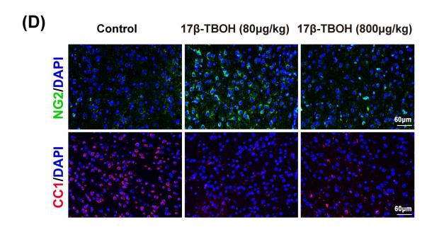
Application: IF/ICC Species: mice Sample: medial prefrontal cortex (mPFC)

Application: WB Species: Mouse Sample:

Application: IF/ICC Species: Mouse Sample:

Application: IF/ICC Species: Mice Sample: retinas
Restrictive clause
Affinity Biosciences tests all products strictly. Citations are provided as a resource for additional applications that have not been validated by Affinity Biosciences. Please choose the appropriate format for each application and consult Materials and Methods sections for additional details about the use of any product in these publications.
For Research Use Only.
Not for use in diagnostic or therapeutic procedures. Not for resale. Not for distribution without written consent. Affinity Biosciences will not be held responsible for patent infringement or other violations that may occur with the use of our products. Affinity Biosciences, Affinity Biosciences Logo and all other trademarks are the property of Affinity Biosciences LTD.