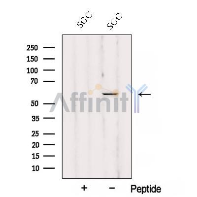
Ajuba Antibody - #DF12817
| Product: | Ajuba Antibody |
| Catalog: | DF12817 |
| Description: | Rabbit polyclonal antibody to Ajuba |
| Application: | WB |
| Reactivity: | Human, Mouse, Rat |
| Prediction: | Pig, Zebrafish, Bovine, Horse, Sheep, Dog |
| Mol.Wt.: | 57 kDa; 57kD(Calculated). |
| Uniprot: | Q96IF1 |
| RRID: | AB_2845778 |
Related Downloads
Protocols
Product Info
*The optimal dilutions should be determined by the end user. For optimal experimental results, antibody reuse is not recommended.
*Tips:
WB: For western blot detection of denatured protein samples. IHC: For immunohistochemical detection of paraffin sections (IHC-p) or frozen sections (IHC-f) of tissue samples. IF/ICC: For immunofluorescence detection of cell samples. ELISA(peptide): For ELISA detection of antigenic peptide.
Cite Format: Affinity Biosciences Cat# DF12817, RRID:AB_2845778.
Fold/Unfold
Ajuba LIM protein; Ajuba, mouse, homolog of; Jub ajuba homolog; Jub; LIM domain containing protein ajuba; MGC15563; Protein ajuba;
Immunogens
A synthesized peptide derived from human Ajuba, corresponding to a region within the internal amino acids.
- Q96IF1 AJUBA_HUMAN:
- Protein BLAST With
- NCBI/
- ExPASy/
- Uniprot
MERLGEKASRLLEKFGRRKGESSRSGSDGTPGPGKGRLSGLGGPRKSGPRGATGGPGDEPLEPAREQGSLDAERNQRGSFEAPRYEGSFPAGPPPTRALPLPQSLPPDFRLEPTAPALSPRSSFASSSASDASKPSSPRGSLLLDGAGAGGAGGSRPCSNRTSGISMGYDQRHGSPLPAGPCLFGPPLAGAPAGYSPGGVPSAYPELHAALDRLYAQRPAGFGCQESRHSYPPALGSPGALAGAGVGAAGPLERRGAQPGRHSVTGYGDCAVGARYQDELTALLRLTVGTGGREAGARGEPSGIEPSGLEEPPGPFVPEAARARMREPEAREDYFGTCIKCNKGIYGQSNACQALDSLYHTQCFVCCSCGRTLRCKAFYSVNGSVYCEEDYLFSGFQEAAEKCCVCGHLILEKILQAMGKSYHPGCFRCIVCNKCLDGIPFTVDFSNQVYCVTDYHKNYAPKCAACGQPILPSEGCEDIVRVISMDRDYHFECYHCEDCRMQLSDEEGCCCFPLDGHLLCHGCHMQRLNARQPPANYI
Predictions
Score>80(red) has high confidence and is suggested to be used for WB detection. *The prediction model is mainly based on the alignment of immunogen sequences, the results are for reference only, not as the basis of quality assurance.
High(score>80) Medium(80>score>50) Low(score<50) No confidence
Research Backgrounds
Adapter or scaffold protein which participates in the assembly of numerous protein complexes and is involved in several cellular processes such as cell fate determination, cytoskeletal organization, repression of gene transcription, mitosis, cell-cell adhesion, cell differentiation, proliferation and migration. Contributes to the linking and/or strengthening of epithelia cell-cell junctions in part by linking adhesive receptors to the actin cytoskeleton. May be involved in signal transduction from cell adhesion sites to the nucleus. Plays an important role in regulation of the kinase activity of AURKA for mitotic commitment. Also a component of the IL-1 signaling pathway modulating IL-1-induced NFKB1 activation by influencing the assembly and activity of the PRKCZ-SQSTM1-TRAF6 multiprotein signaling complex. Functions as an HDAC-dependent corepressor for a subset of GFI1 target genes. Acts as a transcriptional corepressor for SNAI1 and SNAI2/SLUG-dependent repression of E-cadherin transcription. Acts as a hypoxic regulator by bridging an association between the prolyl hydroxylases and VHL enabling efficient degradation of HIF1A. Positively regulates microRNA (miRNA)-mediated gene silencing. Negatively regulates the Hippo signaling pathway and antagonizes phosphorylation of YAP1.
Phosphorylated by LATS2 during mitosis. Phosphorylated by AURKA.
Cytoplasm>Cytoskeleton. Cell membrane. Cell junction. Nucleus. Cytoplasm>Cytoskeleton>Microtubule organizing center>Centrosome. Cytoplasm>P-body.
Note: Shuttles between the cytoplasm and the nucleus. Localizes on centrosomes during G2-M phase. Preferentially co- localizes with cadherin-adhesive complexes at sites of cell-cell contacts. Colocalizes with GFI1 in the nucleus.
LIM region interacts with CTNNA1. The preLIM region binds directly actin filaments.
LIM-2 and LIM-3 domains mediate the interaction with the N-terminal region of AURKA. The association between LATS2 and AJUBA required the second LIM domain of AJUBA.
Belongs to the zyxin/ajuba family.
Research Fields
· Environmental Information Processing > Signal transduction > Hippo signaling pathway. (View pathway)
· Environmental Information Processing > Signal transduction > Hippo signaling pathway - multiple species. (View pathway)
Restrictive clause
Affinity Biosciences tests all products strictly. Citations are provided as a resource for additional applications that have not been validated by Affinity Biosciences. Please choose the appropriate format for each application and consult Materials and Methods sections for additional details about the use of any product in these publications.
For Research Use Only.
Not for use in diagnostic or therapeutic procedures. Not for resale. Not for distribution without written consent. Affinity Biosciences will not be held responsible for patent infringement or other violations that may occur with the use of our products. Affinity Biosciences, Affinity Biosciences Logo and all other trademarks are the property of Affinity Biosciences LTD.
