
, using Survivin Antibody at 1/1000 dilution.
5ug/NC membrane strip.
Exposure for 5s with Affinity™ ECL Kit(#KF8003).
Bands result from membrane strip incubation.")

 and mouse anti-beta tubulin Ab(T0023) for 1 hour at 37°C. An AlexaFluor594 conjugated goat anti-rabbit IgG(H+L) Ab(Red) and an AlexaFluor488 conjugated goat anti-mouse IgG(H+L) Ab(Green) were used as the secondary antibody.
The nuclear counter stain is DAPI(blue).")
| Product: | Survivin Antibody |
| Catalog: | AF6017 |
| Description: | Rabbit polyclonal antibody to Survivin |
| Application: | WB IHC IF/ICC |
| Cited expt.: | WB, IHC |
| Reactivity: | Human, Mouse, Rat |
| Prediction: | Pig, Bovine, Horse, Sheep, Dog |
| Mol.Wt.: | 20kDa(Observed); 16kD(Calculated). |
| Uniprot: | O15392 |
| RRID: | AB_2834951 |
Control Products

Related Downloads
Protocols
Product Info
*The optimal dilutions should be determined by the end user. For optimal experimental results, antibody reuse is not recommended.
*Tips:
WB: For western blot detection of denatured protein samples. IHC: For immunohistochemical detection of paraffin sections (IHC-p) or frozen sections (IHC-f) of tissue samples. IF/ICC: For immunofluorescence detection of cell samples. ELISA(peptide): For ELISA detection of antigenic peptide.
Cite Format: Affinity Biosciences Cat# AF6017, RRID:AB_2834951.
Fold/Unfold
API4; Apoptosis inhibitor 4; Apoptosis inhibitor survivin; Apoptosis inhibitor4; Baculoviral IAP repeat containing 5; Baculoviral IAP repeat containing protein 5; Baculoviral IAP repeat-containing protein 5; BIRC 5; BIRC5; BIRC5_HUMAN; EPR 1; IAP4; Survivin variant 3 alpha; SVV; TIAP;
Immunogens
A synthesized peptide derived from human Survivin, corresponding to a region within the internal amino acids.
Expressed only in fetal kidney and liver, and to lesser extent, lung and brain (PubMed:10626797). Abundantly expressed in adenocarcinoma (lung, pancreas, colon, breast, and prostate) and in high-grade lymphomas (PubMed:14741722, PubMed:16329164). Also expressed in various renal cell carcinoma cell lines (PubMed:10626797). Expressed in cochlea including the organ of Corti, the lateral wall, the interdental cells of the Limbus as well as in Schwann cells and cells of the cochlear nerve and the spiral ganglions (at protein level). Not expressed in cells of the inner and outer sulcus or the Reissner's membrane (at protein level) (PubMed:21364656, PubMed:20627126).
- O15392 BIRC5_HUMAN:
- Protein BLAST With
- NCBI/
- ExPASy/
- Uniprot
MGAPTLPPAWQPFLKDHRISTFKNWPFLEGCACTPERMAEAGFIHCPTENEPDLAQCFFCFKELEGWEPDDDPIEEHKKHSSGCAFLSVKKQFEELTLGEFLKLDRERAKNKIAKETNNKKKEFEETAKKVRRAIEQLAAMD
Predictions
Score>80(red) has high confidence and is suggested to be used for WB detection. *The prediction model is mainly based on the alignment of immunogen sequences, the results are for reference only, not as the basis of quality assurance.
High(score>80) Medium(80>score>50) Low(score<50) No confidence
Research Backgrounds
Multitasking protein that has dual roles in promoting cell proliferation and preventing apoptosis. Component of a chromosome passage protein complex (CPC) which is essential for chromosome alignment and segregation during mitosis and cytokinesis. Acts as an important regulator of the localization of this complex; directs CPC movement to different locations from the inner centromere during prometaphase to midbody during cytokinesis and participates in the organization of the center spindle by associating with polymerized microtubules. Involved in the recruitment of CPC to centromeres during early mitosis via association with histone H3 phosphorylated at 'Thr-3' (H3pT3) during mitosis. The complex with RAN plays a role in mitotic spindle formation by serving as a physical scaffold to help deliver the RAN effector molecule TPX2 to microtubules. May counteract a default induction of apoptosis in G2/M phase. The acetylated form represses STAT3 transactivation of target gene promoters. May play a role in neoplasia. Inhibitor of CASP3 and CASP7. Essential for the maintenance of mitochondrial integrity and function. Isoform 2 and isoform 3 do not appear to play vital roles in mitosis. Isoform 3 shows a marked reduction in its anti-apoptotic effects when compared with the displayed wild-type isoform.
Ubiquitinated by the Cul9-RING ubiquitin-protein ligase complex, leading to its degradation. Ubiquitination is required for centrosomal targeting.
In vitro phosphorylation at Thr-117 by AURKB prevents interaction with INCENP and localization to mitotic chromosomes. Phosphorylation at Thr-48 by CK2 is critical for its mitotic and anti-apoptotic activities. Phosphorylation at Thr-34 by CDK15 is critical for its anti-apoptotic activity. Phosphorylation at Ser-20 by AURKC is critical for regulation of proper chromosome alignment and segregation, and possibly cytokinesis.
Acetylation at Lys-129 by CBP results in its homodimerization, while deacetylation promotes the formation of monomers which heterodimerize with XPO1/CRM1 which facilitates its nuclear export. The acetylated form represses STAT3 transactivation. The dynamic equilibrium between its acetylation and deacetylation at Lys-129 determines its interaction with XPO1/CRM1, its subsequent subcellular localization, and its ability to inhibit STAT3 transactivation.
Cytoplasm. Nucleus. Chromosome. Chromosome>Centromere. Cytoplasm>Cytoskeleton>Spindle. Chromosome>Centromere>Kinetochore. Midbody.
Note: Localizes at the centromeres from prophase to metaphase, at the spindle midzone during anaphase and a the midbody during telophase and cytokinesis. Accumulates in the nucleus upon treatment with leptomycin B (LMB), a XPO1/CRM1 nuclear export inhibitor (By similarity). Localizes on chromosome arms and inner centromeres from prophase through metaphase. Localizes to kinetochores in metaphase, distributes to the midzone microtubules in anaphase and at telophase, localizes exclusively to the midbody (PubMed:11084331). Colocalizes with AURKB at mitotic chromosomes (PubMed:14610074). Acetylation at Lys-129 directs its localization to the nucleus by enhancing homodimerization and thereby inhibiting XPO1/CRM1-mediated nuclear export (PubMed:20826784).
Expressed only in fetal kidney and liver, and to lesser extent, lung and brain. Abundantly expressed in adenocarcinoma (lung, pancreas, colon, breast, and prostate) and in high-grade lymphomas. Also expressed in various renal cell carcinoma cell lines. Expressed in cochlea including the organ of Corti, the lateral wall, the interdental cells of the Limbus as well as in Schwann cells and cells of the cochlear nerve and the spiral ganglions (at protein level). Not expressed in cells of the inner and outer sulcus or the Reissner's membrane (at protein level).
The BIR repeat is necessary and sufficient for LAMTOR5 binding.
Belongs to the IAP family.
Research Fields
· Cellular Processes > Cell growth and death > Apoptosis. (View pathway)
· Cellular Processes > Cell growth and death > Apoptosis - multiple species. (View pathway)
· Environmental Information Processing > Signal transduction > Hippo signaling pathway. (View pathway)
· Human Diseases > Drug resistance: Antineoplastic > Platinum drug resistance.
· Human Diseases > Infectious diseases: Viral > Hepatitis B.
· Human Diseases > Cancers: Overview > Pathways in cancer. (View pathway)
· Human Diseases > Cancers: Specific types > Colorectal cancer. (View pathway)
References
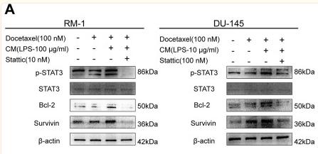
Application: WB Species: Mouse Sample: tumor tissue
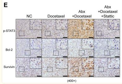
Application: IHC Species: Mouse Sample: tumor tissue
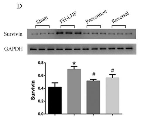
Application: WB Species: rat Sample: lung
Application: WB Species: Human Sample: bladder cancer cells
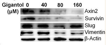
Application: WB Species: Human Sample: A549 and H460 cell
Restrictive clause
Affinity Biosciences tests all products strictly. Citations are provided as a resource for additional applications that have not been validated by Affinity Biosciences. Please choose the appropriate format for each application and consult Materials and Methods sections for additional details about the use of any product in these publications.
For Research Use Only.
Not for use in diagnostic or therapeutic procedures. Not for resale. Not for distribution without written consent. Affinity Biosciences will not be held responsible for patent infringement or other violations that may occur with the use of our products. Affinity Biosciences, Affinity Biosciences Logo and all other trademarks are the property of Affinity Biosciences LTD.