
, using SOX9 Antibody at 1/1000 dilution.
5ug/NC membrane strip.
Exposure for 3s with Affinity™ ECL Kit(#KF8003).
Bands result from membrane strip incubation.")


 Relative mRNA levels of Sox9 and C/EBP in three groups. (b and c) Detection and quantification of Sox9 by Western blot and image J. (e and f) Detection and quantification of C/EBP by Western blot and image J. All data were expressed as means ± SD. *p < 0.05, **p < 0.01, ***p < 0.0001, compared to scrambled group. t")
 for 24 h.
(A) Representative Oil Red O-stained images of macrophages and quantification of the staining. (B) Immunoblots of SRA1, CD36, SRB1, ABCA1, and ABCG1 in
macrophages. (C) Quantification of relative mRNA expression of SRA1, CD36, SRB1, ABCA1, and ABCG1. **p < 0.01 denote significant differences between the
Control and the ox-LDL groups, &&p < 0.01 denote significant differences between the ox-LDL shRNA NC and ox-LDL + shFABP3 groups.")
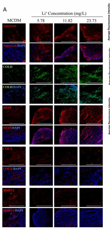
 Protein expression of Aggrecan, COL II, Sox-9, and HIF-1α as determined using western blotting; β-actin was used as a loading control. The expression of Aggrecan (B), COL II (C), Sox-9 (D) and HIF1-α (E) genes was determined using real-time PCR. GAPDH was used as a housekeeping gene. Data are presented as the mean ± standard deviation (n=3), *P")
 for 24 h and protein expression of FGF23 and SOX9 in chondrocytes was determined by WB (a – e). Data are expressed with mean ± SD; (b – e) *p")
 in the study groups at postoperative (A) days 1, 3, and 7 and (B) weeks 3, 6, 12, and 24. Scale bars indicate 100 µm. (C) Percentage of TGF-β–positive cells at the tendon-to-bone interface; error bars represent SDs. Part A: A-1 day study group; B-3 day study group; C-7 day study group; D-1day control group; E-3 day control group; F-7 day control group. Part B: A-3 week study group; B-6 week study group; C-12 week study group; D-24 week study group; E-3 week control group; F-6 week control group; G-12 week control group; H-24 week control group. *Significant difference between groups (P < .05). ACLR, anterior cruciate ligament reconstruction; FHT, free hamstring tendon; IP-HT, insertion-preserved hamstring tendon.")
 Relative mRNA, (B) representative western blot bands, and (C) statistical results of knee cartilage 7-day male mice offspring in the control group versus obese group. O-WT-P7: offspring 7-day male mice from the control mothers. O-OB-P7: offspring 7-day male mice from the obese mothers. (A) n = 6 represents six mice in each group from at least three litters of different mothers. (B) n = 6, each band represents the expression of cartilage tissue proteins in two mice from at least three litters of different mothers. Results are expressed as mean ± SD. ns not significant, * p < 0.05, ** p < 0.01, and *** p < 0.001.")
 IL-1β and (B) IL-18 levels in rat hearts. (C, D) NLRP3 protein localization in cardiac tissue sections by immunohistochemistry (scale bar = 50 μm). (E) Representative immunoblots of SOX9 and NLRP3 inflammasome components in rat hearts. (F) Densitometric quantification of protein expression. N = 10 animals per group. Data are presented as the mean ± standard deviation.")
| Product: | SOX9 Antibody |
| Catalog: | AF6330 |
| Description: | Rabbit polyclonal antibody to SOX9 |
| Application: | WB IHC IF/ICC |
| Cited expt.: | WB, IHC, IF/ICC |
| Reactivity: | Human, Mouse, Rat, Monkey |
| Prediction: | Pig, Horse, Sheep, Rabbit, Dog, Chicken, Xenopus |
| Mol.Wt.: | 50,70kDa(Observed); 56kD(Calculated). |
| Uniprot: | P48436 |
| RRID: | AB_2835186 |
Control Products

Related Downloads
Protocols
Product Info
*The optimal dilutions should be determined by the end user. For optimal experimental results, antibody reuse is not recommended.
*Tips:
WB: For western blot detection of denatured protein samples. IHC: For immunohistochemical detection of paraffin sections (IHC-p) or frozen sections (IHC-f) of tissue samples. IF/ICC: For immunofluorescence detection of cell samples. ELISA(peptide): For ELISA detection of antigenic peptide.
Cite Format: Affinity Biosciences Cat# AF6330, RRID:AB_2835186.
Fold/Unfold
campomelic dysplasia autosomal sex reversal; CMD 1; CMD1; CMPD 1; CMPD1; SOX 9; Sox9; SOX9_HUMAN; SRA 1; SRA1; SRXX2; SRXY10; SRY (sex determining region Y) box 9 (campomelic dysplasia autosomal; SRY (sex determining region Y) box 9; SRY (sex determining region Y)-box 9; SRY (sex-determining region Y)-box 9 protein; SRY related HMG box gene 9; Transcription factor SOX 9; Transcription factor SOX-9; transcription factor SOX9;
Immunogens
A synthesized peptide derived from human SOX9, corresponding to a region within the internal amino acids.
- P48436 SOX9_HUMAN:
- Protein BLAST With
- NCBI/
- ExPASy/
- Uniprot
MNLLDPFMKMTDEQEKGLSGAPSPTMSEDSAGSPCPSGSGSDTENTRPQENTFPKGEPDLKKESEEDKFPVCIREAVSQVLKGYDWTLVPMPVRVNGSSKNKPHVKRPMNAFMVWAQAARRKLADQYPHLHNAELSKTLGKLWRLLNESEKRPFVEEAERLRVQHKKDHPDYKYQPRRRKSVKNGQAEAEEATEQTHISPNAIFKALQADSPHSSSGMSEVHSPGEHSGQSQGPPTPPTTPKTDVQPGKADLKREGRPLPEGGRQPPIDFRDVDIGELSSDVISNIETFDVNEFDQYLPPNGHPGVPATHGQVTYTGSYGISSTAATPASAGHVWMSKQQAPPPPPQQPPQAPPAPQAPPQPQAAPPQQPAAPPQQPQAHTLTTLSSEPGQSQRTHIKTEQLSPSHYSEQQQHSPQQIAYSPFNLPHYSPSYPPITRSQYDYTDHQNSSSYYSHAAGQGTGLYSTFTYMNPAQRPMYTPIADTSGVPSIPQTHSPQHWEQPVYTQLTRP
Predictions
Score>80(red) has high confidence and is suggested to be used for WB detection. *The prediction model is mainly based on the alignment of immunogen sequences, the results are for reference only, not as the basis of quality assurance.
High(score>80) Medium(80>score>50) Low(score<50) No confidence
Research Backgrounds
Transcriptional regulator that plays a role in chondrocytes differentiation and skeletal development. Binds to the COL2A1 promoter and activates COL2A1 expression, as part of a complex with ZNF219 (By similarity).
Ubiquitinated. Ubiquitination leads to proteasomal degradation and is negatively regulated by DDRGK1.
Nucleus.
Research Fields
· Environmental Information Processing > Signal transduction > cAMP signaling pathway. (View pathway)
References

Application: IF/ICC Species: Mouse Sample:

Application: IF/ICC Species: Rat Sample:
Application: IF/ICC Species: Human Sample: iPSCs

Application: WB Species: Rat Sample:
Restrictive clause
Affinity Biosciences tests all products strictly. Citations are provided as a resource for additional applications that have not been validated by Affinity Biosciences. Please choose the appropriate format for each application and consult Materials and Methods sections for additional details about the use of any product in these publications.
For Research Use Only.
Not for use in diagnostic or therapeutic procedures. Not for resale. Not for distribution without written consent. Affinity Biosciences will not be held responsible for patent infringement or other violations that may occur with the use of our products. Affinity Biosciences, Affinity Biosciences Logo and all other trademarks are the property of Affinity Biosciences LTD.