
.")

, diluted 1/600 was used as secondary antibody.")
 and mouse anti-beta tubulin Ab(T0023 1:200) for 1 hour at 37°C. An AlexaFluor594 conjugated goat anti-rabbit IgG(H+L) Ab(Red) and an AlexaFluor488 conjugated goat anti-mouse IgG(H+L) Ab(Green) were used as the secondary antibody.
The nuclear counter stain is DAPI(blue).")
 microscopy at x200 magnification was used to assess cell morphology. The A549 cells (parental cells) had an epithelioid, rounded cobblestone appearance and there was limited formation of pseudopodia. A549/PTX and A549/DDP cells exhibited a spindle-shaped morphology and an increased formation of pseudopodia, indicating a loss of cell polarity. (B) E-cadherin, β-catenin, vimentin, MMP-2 and MMP-9 which are EMT-related proteins, were assessed in terms of expression levels. EMT-related transcription factors (Snail, Slug, Twist and ZEB1) were measured in A549/PTX and A549/DDP cells using western blot analysis. (C) The expression changes were confirmed at the mRNA level by qRT-PCR. Expression was standardized to the expression of GAPDH and normalized to 1.0 in the parental cells (compared with the parental A549 cells, means ± SEM, n=3, * P<0.05)")


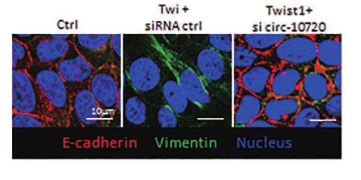
 inhibited JAM-A-transfected cell invasion (B) and EMT (C).")
 Representative photographs of treated and untreated cells. PTL treatment reduced p-ERK 2, NF-κB, and Snail staining compared with sections obtained from control mice. (B and D) Representative photographs of treated and untreated cells. PTL treatment increased E-cadherin and Occludin staining and reduced Vimentin and N-cadherin staining compared with sections obtained from control mice. Each experiment was performed in triplicate. Results show the means of the three experiments, and the error bars represent standard deviation (*P < 0.05 and **P < 0.01).")


 Phase contrast photomicrographs of confluent cultures of cells were captured after treatment for 48 h. Scale bar: 200 μm. (B) Western blot analysis levels of of ZO-1, E-cadherin, Vimentin, α-SMA and the housekeeping protein GAPDH in the lysates of ARPE-19 cells after treatment for 48 h. *P< 0.05, **P< 0.01, ***P< 0.001. The data are presented as the mean ± S.D. (n = 3/group).")


 Immunofluorescence assay conducted in the HepG2 cancer cell line demonstrated that Downloaded from mct.aacrjournals.org on October 10, 2018. © 2018 American Association for Cancer Research. Author manuscripts have been peer reviewed and accepted for publication but have not yet been edited. Author Manuscript Published OnlineFirst on October 8, 2018; DOI: 10.1158/1535-7163.MCT-18-0448 22 OA can enhance fluorescence intensity of E-cadherin and attenuate that of vimentin, whereas treatment of NO scavenger (C-PTIO) or transfection of siRNA iNOS exhibits the opposite effect. This outcome is consistent with the finding of Western blot assay. The results were obtained from three independent experiments, each performed in triplicate. Data are represented as mean ± standard error of the mean (*P < 0.05, **P < 0.01).")
 Results of IHC assays. The expression levels of E-cadherin were significantly upregulated, whereas those of vimentin, MMP2, and MMP9 were downregulated by OA or regorafenib treatment, and OA enhanced the effects of regorafenib. The expression levels of iNOS and NT were upregulated by OA but not by regorafenib. (G) Staining indexes of IHC assays. Data are represented as mean ± standard error of the mean (*P < 0.05, **P < 0.01).")
 Effect of Pyr or MTX on the migration of NCI-H460 at 24 and 48 h. (B) Effects of Pyr and MTX on the migration of A549 cells at 24 and 48 h. (C) Transwell chambers were utilized for the invasion assay, and images were obtained under 200× magnification. NCI-H460 and A549 cells were treated with Pyr or MTX. (D) Changes of E-cadherin and vimentin expression in NCI-H460 cells treated with Pyr or MTX (Western blot assay). β-actin was used as the loading control. (E) Changes of E-cadherin and vimentin expression in NCI-H460 cells treated with Pyr or MTX (immunofluorescence assay). Each experiment was performed in triplicate. Results are shown as means ± SD (*P < 0.05, **P < 0.01).")
 Effect of Pyr or MTX on the migration of NCI-H460 at 24 and 48 h. (B) Effects of Pyr and MTX on the migration of A549 cells at 24 and 48 h. (C) Transwell chambers were utilized for the invasion assay, and images were obtained under 200× magnification. NCI-H460 and A549 cells were treated with Pyr or MTX. (D) Changes of E-cadherin and vimentin expression in NCI-H460 cells treated with Pyr or MTX (Western blot assay). β-actin was used as the loading control. (E) Changes of E-cadherin and vimentin expression in NCI-H460 cells treated with Pyr or MTX (immunofluorescence assay). Each experiment was performed in triplicate. Results are shown as means ± SD (*P < 0.05, **P < 0.01).")
 xenografts, whereas MTX can only inhibit the tumor growth.(G) Effect of Pyr and MTX on the expression of EMT markers in LLC xenograft tumor tissues, as observed through immunohistochemistry analysis (40×). Brown or yellow staining was considered as positive expression. Each experiment was performed in triplicate. The results are shown as means ± SD (*P < 0.05, **P < 0.01).")


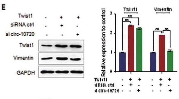
 Expression of E-cadherin, β-catenin, vimentin, MMP-2, MMP-9, Snail, Slug, Twist and ZEB1 in A549/DDP cells was measured with western blotting.")
 Representative photographs of treated and untreated cells. PTL treatment reduced p-ERK 2, NF-κB, and Snail staining compared with sections obtained from control mice. (B and D) Representative photographs of treated and untreated cells. PTL treatment increased E-cadherin and Occludin staining and reduced Vimentin and N-cadherin staining compared with sections obtained from control mice. Each experiment was performed in triplicate. Results show the means of the three experiments, and the error bars represent standard deviation (*P < 0.05 and **P < 0.01).")
 and Vimentin (red) was performed. The nucleus was staining with DAPI.")

 mRNA levels of EMA, CK and CD49f in EnSCs. (B) mRNA levels of THY‑1, Col I, 5B5 and vimentin in EnSCs. (C) Protein expression of EMA, CK and CD49f in EnSCs. (D) Protein expression of THY‑1, Col I, 5B5 and vimentin in EnSCs. *P<0.05 vs. control. EMA, epithelial membrane antigen; CK, cytokeratin; Col I, collagen type 1; CD49F, integrin α‑6; THY‑1, Thy‑1 membrane glycoprotein; EnSCs, endometrial stem cells.")

 Expression levels of EMT-associated markers (Zeb1, Zeb2, E-cadherin and Vimentin) with E2 treatment for different doses (0, 10−12, 10−10, 10−8, 10−6 mol/L) and time (0, 24, 48 or 72 h) in EECs detected by Western blot.(M: mol/L); (C and D) Expression levels of MALAT1 and miR200s under E2 for various doses (0, 10−12, 10−10, 10−8, 10−6 mol/L) for 24 h in EECs measured by qRT-PCR. Data were evaluated by one-way ANOVA analysis (*P < 0.05, **P < 0.01, ***P < 0.001 compared with untreated group).")
 Overexpression of KRT7 increased Snail, vimentin and N-cadherin expression, and decreased E-cadherin expression in HEY cells as detected by western blotting. (C and D) Knockdown of KRT7 resulted in increased E-cadherin expression and reduced vimentin and Snail expression in OVCAR433 cells as detected by western blotting. All experiments were performed at least three times. Results are presented as the mean ± standard deviation. **P<0.01. KRT7, keratin 7; sh, short hairpin RNA; NC, negative control.")
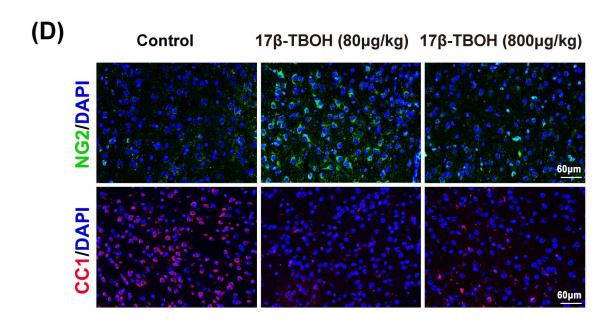
 Neuronal cell death and
reactive astrocytes in the cortex tissue, as determined by Fluoro-Jade B (FJB) staining and glial fibrillary acidic protein (GFAP) immunofluorescence staining,
respectively, at 20×. (B) Number of FJB-positive neurons per area, calculated using ImageJ. (C) Mean fluorescence intensity (relative to the normal control (NC)) of
GFAP, as determined using ImageJ. (D) The expression of GFAP, glyceraldehyde-3-phosphate dehydrogenase (GAPDH), vimentin, and β-actin, as measured using
western blotting. (E–F) Relative protein levels of GFAP and vimentin, as determined by ImageJ. *P < 0.05 vs. NC, **P < 0.01 vs. NC, #P < 0.05 vs. diabetes mellitus
(DM), ##P < 0.05 vs. DM.")
 Representative bands of caspase‑3, cleaved‑caspase‑3, vimentin, α‑SMA and E‑cadherin protein expression in the individual groups.")
.")
 Immunohistochemical staining to identify EMT biomarkers and doxycycline inhibited proteins in treated and untreated cells. Doxycycline-treated cells display stronger E-cadherin and α-SMA staining but reduced vimentin,MMP-2 and MMP-9 staining;")
 Crystal violet staining of the shNS and control group cells that crossed the polycarbonate membrane of the Transwell chamber to detect cell migration. (B) The number of cells that crossed the Transwell migration chamber in different groups. (C) Crystal violet staining of the shNS and control group cells that crossed the Matrigel-coated polycarbonate membrane of the Transwell chamber to detect cell invasion. (D) The number of cells that crossed the Transwell invasion chamber in different groups. (E) Representative Western blotting results indicate the EMT marker expressions in the different groups. The results are presented as the means ± SD, as based on three independent experiments. Statistical significance was determined using Student's t-test. *P < 0.05. Scale: 100 mm.")
 The shNS and control group cells were inoculated s.c. into the right flanks of BALB/c nude mice. Tumor volumes were monitored and recorded weekly (n ¼ 3). The results are presented as the means ± SD, as based on three independent experiments. ****P < 0.0001. (B) Examples of the tumors excised from the shNS and control mice on the 19th week following injection. Scale: 2 cm. (C) Immunohistochemistry staining of the tumor sections obtained from mice in the shNS and control groups. Antibodies: NS, Ki-67, cyclin D1, and Vimentin.")

 The protein expression of c-Myc, vimentin, E-cadherin, HIF-1α, CXCR4, and SDF-1 in PANC-1 and SW1990 pancreatic cancer cells under different treatments was detected via western blot. (B) Quantification results of protein expressions of c-Myc, vimentin, E-cadherin, HIF-1α, CXCR4, and SDF-1 in PANC-1 pancreatic cancer cells. (C) Quantification results of protein expression of c-Myc, vimentin, E-cadherin, HIF-1α, CXCR4, and SDF-1 in SW1990 pancreatic cancer cells (* p < 0.05, ** p < 0.01 vs control, ▲ p < 0.05, ▲▲ p < 0.01 vs bufalin treatment group, n = 3).")
 The downregulation of NEAT1 in si-NEAT1 cell lines (A549 and H460) detected by qRT-PCR. (B, C) The
cell proliferation of A549 and H460 cells with si-NEAT1 measured by CCK8 assay. (D) The regulation of si-NEAT1 on
cell apoptosis tested by flow cytometry. (E, F) Transwell experiments on cell migration, invasion of si-NEAT1 cell lines
(A549 and H460). (G) The expression of EMT marker protein by Western blot normalized to GAPDH. *p < 0.05. CCK8,
Cell Counting Kit-8; EMT, epithelial–mesenchymal transition; qRT-PCR, quantitative real-time polymerase chain
reaction.")
 Relative LINP1 expression in EC9706 sh1, sh2, sh3, and sh4 was verified by qRT-PCR. The LINP1 sh2 cell line had the most significant knockdown effect compared with the control cell line (61.75%, P<0.001). (B) The Alamar Blue proliferation assay indicated that the proliferation of shRNA-LINP1 cells was significantly inhibited compared with that of the corresponding control cells and shRNA-scr cells (P<0.001). (C,D) Colony formation assays showing that the number of colonies formed was significantly lower in shRNA-LINP1 cells than in NC and shRNA-scr cells in macroscopic view (71.7 vs. 73.3 vs. 24.7, P<0.001). (E,F) Wound healing assays showed that the migration rate was slower in shRNA-LINP1 cells than in NC and shRNA-scr cells after 48 h (19.1% vs. 46.4% vs. 46.6%; P<0.001) (magnification 100×). (G,H) Transwell migration assays demonstrated that the number of migratory cells was lower in shRNA-LINP1 than in the two control cells (60.3 vs. 236.3 vs. 238.7; P<0.001) (magnification 100×). (I,J,K) qRT-PCR and western blot analyses of the expression of EMT markers. E-cadherin was significantly upregulated, whereas N-cadherin, vimentin, snail and slug were significantly downregulated in shRNA-LINP1 cells compared with NC and shRNA-scr cells (all P<0.05). **, P<0.01; ***, P<0.001.")
 qRT-PCR showing the knock down of lncRNA AL161431.1, and the corresponding changes in E-cadherin, N-cadherin, and vimentin in SW1990 and BxPC-3 cells; (B) Western blots showing the changes in protein level in E-cadherin, N-cadherin, and vimentin in SW1990 and BxPC-3 cells after transfection of scrambled siRNA or siRNA1; (C) Immunofluorescence analysis showing the change of expression of E-cadherin, N-cadherin, and vimentin in SW1990 and BxPC-3 cells after transfection of scrambled siRNA or siRNA1 (200x). *p < 0.05, **p < 0.01, ***p < 0.001.")
 qRT-PCR showing the knock down of lncRNA AL161431.1, and the corresponding changes in E-cadherin, N-cadherin, and vimentin in SW1990 and BxPC-3 cells; (B) Western blots showing the changes in protein level in E-cadherin, N-cadherin, and vimentin in SW1990 and BxPC-3 cells after transfection of scrambled siRNA or siRNA1; (C) Immunofluorescence analysis showing the change of expression of E-cadherin, N-cadherin, and vimentin in SW1990 and BxPC-3 cells after transfection of scrambled siRNA or siRNA1 (200x). *p < 0.05, **p < 0.01, ***p < 0.001.")
, negative control (NC), overexpression (OE), and knockdown (KD) groups. B-C: The colony-forming assay showed the colony-forming ability of HTR8 cells in the four groups. D-E: The scratch wound healing assay showed that the closed wound area of HTR8 cells in the four groups. F and I: Transwell assay that the migration and invasion number of HTR8 cells in the four groups. J: Western blot assay determined the expression of epithelial-mesenchymal transition (EMT) markers in the four groups. K-L: Flow cytometry showed that the percentage of apoptosis cells in the four groups. M: Western blot assay determined the expression of apoptosis-related proteins in the four groups. All experiments were conducted with three biological replicates. Data were expressed as mean ± SD. * P < 0.05 compared to NC group.")

 Cell invasion was measured by transwell assay in bladder cancer cells treated with increasing concentrations of gigantol (magnification ×100). (B) qRT-PCR analysis of the Wnt target genes and EMT markers in bladder cancer cells treated with gigantol. (C) Western blot analysis of Wnt/EMT markers in bladder cancer cells treated with indicated concentrations of gigantol. Data in (B) were shown as means ± SD (n = 3), the statistically significant differences were considered at *p<0.05, **p<0.01, ***p<0.001.")
 in esophageal squamous cell carcinoma (ESCC) cells. (E) The protein levels of EMT markers (E-cadherin and Vimentin) were detected by Western blot in stable Tet-on inducible RECQL4 knockdown cell lines (KYSE30 and TE-1 cells) (+Dox) and controls (–Dox).")
immunofluorescence analysis showing the change of expression of E-cadherin, N-cadherin, and vimentin in SW1990 and BxPC-3 cells after transfection of scrambled siRNA or siRNA1 (200×). *P<0.05, **P<0.01.")
 western blots showing the changes in protein level in E-cadherin, N-cadherin, and vimentin in SW1990 and BxPC-3 cells after transfection of scrambled siRNA or siRNA1")
 NUDCD1 knockdown decreased the expression of Ncadherin and vimentin and upregulated the expression of E-cadherin in PANC-1 cells.")

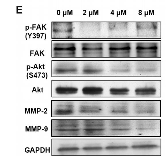
Expression of γH2AX, 53BP1, and p21CIP in A549 cells exposed to SiO2 for the
indicated times (0–48 h) (C) Flow cytometry quantitation of cells undergoing DNA
synthesis (BrdU). A549 cells were exposed to SiO2 for the indicated times with
respect to controls with no SiO2. (D-F) IHC for γH2AX and α-SMA in (D) A549 cells,
(E) MLE12 cells, and (F) MRC-5 cells after exposure to SiO2 for 48 h. Scale bar = 50
μm. (G). Expression of Vimentin, α-SMA and Col I in A549 cells exposed to SiO2 for
the indicated times (0–48 h). Data are presented as the mean ± SD. n = 3 per group.
*p < 0.05.")
 cells. Expression analyses of RECQL4 mRNA and protein in 5 ESCC cell lines by (A) qRT-PCR and (B) Western blot. Each bar represents the mean ± SD of 3 replicates. (C) Tet-on inducible shRNA lentiviral vector [LV3 (H1/GFP&Puro)] and tet-on inducible RECQL4 shRNA lentiviral vectors were transfected into 2 higher RECQL4 expression cell lines (KYSE30 and TE-1 cells). Stably transfected cells were obtained by selection with puromycin. Cells were treated with doxycycline (Dox) (500 ng/mL) for 24 h to repress the expression of RECQL4. The cells without Dox treatment were used as controls. The RECQL4 knockout efficiency was examined by Western blot. (D) The clone formation assay in stable RECQL4 knockdown cell lines (KYSE30 and TE-1 cells) with Dox treatment (+Dox) and controls (–Dox). (E) The EdU incorporation assay in stable RECQL4 knockdown cell lines (KYSE30 and TE-1 cells) with Dox treatment (+Dox) and controls (–Dox). EdU incorporation was measured by immunofluorescence staining of EdU (red) and 4′,6-diamidino-2-phenylindole (blue) under the same microscopic magnification (×200). Scale bar, 50 μm. Left, representative EdU incorporation; right, quantitation of EdU incorporation. The number of EdU-positive cells per 200 nucleated cells was determined. *P < 0.05 vs. the control. (F–H) RECQL4 inhibited the proliferation of ESCC in vivo. (F) A plot of tumor volume over time. Tumor xenograft volumes in Tet-on inducible shRNA-RECQL4-treated nude mice were smaller than those in the scramble group. Tumor graft mass was measured every 3 days, and the volume was calculated using the following formula: volume = length × width2/2. (G and H) The dissected xenografts were photographed and weighed at the endpoint. *P < 0.05 vs. the control.")
 Immunofluorescence staining against α-SMA was used to identify
VSMCs. B, C) VSMCs were starved for 24 h and subsequently incubated with PDGF-BB (10, 20 and 30 ng/ml) for 24 h. The mRNA and protein levels of HOXA5 were
determined by qRT-PCR and Western blot. D, E) HOXA5 mRNA and protein levels were examined in VSMCs infected with NC or HOXA5 overexpression lentivirus
using qRT-PCR and Western blot. VSMCs were infected with NC or HOXA5 overexpression lentivirus for 48 h and then cultured with 10 ng/ml PDGF-BB for 24 h
(F–K). F, G) Relative expression of HOXA5 mRNA and protein was determined by qRT-PCR and Western blot. H) Western blot analysis of the protein levels of
calponin, α-SMA, SM22α, vimentin, PCNA and thrombospondin. I, J) Immunofluorescence staining was utilized to assess levels of calponin and vimentin in VSMCs.
The nucleus was stained with DAPI. K) VSMC proliferation was detected using the MTT assay. Scale bar, 50 μm &, p < 0.05.")
. D, F, G. The expression levels of MMP2 (D, F) and MMP9 (D, G) in lung cancer tissues (n=6). D, H. The expression level of TGF-β1 in lung cancer tissues (n=6). D, I. The expression level of collagen type I in lung cancer tissues(n=6). Note: &, &&&& respectively represent a significant difference compared with the Saline group (P<0.05), (P<0.0001).")
 qRT-PCR showing the knock down of lncRNA ELFN1-AS1, and the corresponding changes in E-cadherin, N-cadherin, and vimentin in SW1990 and BxPC-3 cells; (B) western blots showing the changes in protein level in E-cadherin, N-cadherin, and vimentin in SW1990 and BxPC-3 cells after transfection of scrambled siRNA or siRNA1; (C) immunofluorescence analysis showing the change of expression of E-cadherin, N-cadherin, and vimentin in SW1990 and BxPC-3 cells after transfection of scrambled siRNA or siRNA1 (200×). *P<0.05, **P<0.01.")
 qRT-PCR showing the knock down of lncRNA ELFN1-AS1, and the corresponding changes in E-cadherin, N-cadherin, and vimentin in SW1990 and BxPC-3 cells; (B) western blots showing the changes in protein level in E-cadherin, N-cadherin, and vimentin in SW1990 and BxPC-3 cells after transfection of scrambled siRNA or siRNA1; (C) immunofluorescence analysis showing the change of expression of E-cadherin, N-cadherin, and vimentin in SW1990 and BxPC-3 cells after transfection of scrambled siRNA or siRNA1 (200×). *P<0.05, **P<0.01.")
 in the supernatants were detected by ELISA. (B) STRING database used to analyze the interactions between PTPN2 and FSP-1. qRT-PCR (C) and Western blot (D) to detect the JAK-STAT signaling pathway, Mean ± SD, **P < 0.001, were compared with HCT116 shNC+TAFs control group; ##P < 0.001, were compared with LoVo shNC+TAFs control group.")
 NUDCD1 knockdown decreased the expression of N-cadherin and vimentin and upregulated the expression of E-cadherin in PANC-1 cells. (E–H) NUDCD1 knockdown decreased the expression of N-cadherin and vimentin, and upregulated the expression of E-cadherin in Patu8988 cells. ***p<0.001, ****p<0.0001.")
 in vitro. B Western blot analysis of E-cadherin, Snail1, ZEB2, and Vimentin expression in TC cells compared with PC-knockdown TC cells; the full blot is provided in the supplementary file")
. To fit into the manuscript properly, the gel was reasonably trimmed. aa, P < 0.01 vs the control group; b, P < 0.05, bb, P < 0.01 vs the Wnt-1 group. ##, P < 0.01 vs the normal group; *, P < 0.05 and **, P < 0.01 vs the model group")
 cell proliferation, migration, invasion, and epithelial-mesenchymal transition (EMT) in vitro. a CCK-8
assays were conducted after transfection with miR-107 mimics or inhibitors in HNE1 and 5–8F cell lines. b Colony-forming capacity was determined after transfection
with miR-107 mimics or inhibitors in HNE1 and 5–8F cell lines. c Migratory potential was examined in cells transfected with miR-107 mimics or inhibitors by wound
healing assay. d Invasive potential of cells was examined in cells transfected with miR-107 mimics or inhibitors using Transwell chambers with Matrigel. e Apoptosis
was evaluated using cells transfected with miR-107 mimics or inhibitors. f Protein markers of EMT (E-cadherin, N-cadherin, and vimentin) were detected by Western
blot analysis after transfection with miR-107 mimics or inhibitors in HNE1 and 5–8F cell lines. The values are expressed as the mean ± SD of three independent
experiments. *P < 0.05; **P < 0.01; ***P < 0.001.")
 markers in the four groups.")
 and endothelium‐dependent vessels. The yellow arrow pointed to the VM+ channels (PAS‐positive and CD34‐negative channels). Scale bars: 50 μm. B and C, Immunohistochemical staining was used to distinguishing EMT‐related markers (E‐cadherin and vimentin) between the ND and HFD groups. Scale bars: 100 μm. **P < .01, ***P < .001. n = 30 for each group")
 induced the epithelial‐mesenchymal transition in Apc min/+ mice. A and B, Real‐time PCR analysis revealed DCA downregulated epithelial markers (claudin‐4 and E‐cadherin) and upregulated mesenchymal markers (vimentin and fibronectin) expression, respectively. C and D, vimentin expression was higher in the DCA group than in the control group, whereas the expression of E‐cadherin was decreased. n = 4. E, Immunohistochemical analysis; n = 10 for each group. Scale bars: 50 μm. *P < .05, **P < .01, ***P < .001")
 Growth curves of tumor volume in sh‐NC and sh‐hsa_circ_0017620 groups. (B) The tumor weight was measured in sh‐NC and sh‐hsa_circ_0017620 groups. (C and D) The expression of hsa_circ_0017620 and miR‐520a‐5p was measured in sh‐NC and sh‐hsa_circ_0017620 groups with qRT‐PCR. (E–G) The mRNA and protein expression of KRT5 were measured in sh‐NC and sh‐hsa_circ_0017620 groups with western blot. (H) The positive expression rates of Ki67 and Vimentin in the forming tumors from sh‐NC and sh‐hsa_circ_0017620 groups were detected by IHC assay. *p < 0.05")
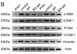
, vimentin (B), α-SMA (C) and Collagen I (F)
were determined by RT-qPCR. (D) The protein expression level of E-cadherin analyzed by Western blot. (E) The protein expression of E-cadherin relative to GADPH
protein expression. (G) The protein expression levels of vimentin and α-SMA analyzed by Western blot. The protein expression of vimentin (H) and α-SMA (I) relative
to GADPH protein expression. Data are expressed as the mean ± S.E.M. (n = 3–7), *P < 0.05 vs Sham; **P < 0.01 vs Sham; ***P < 0.001 vs Sham; ##P < 0.01 vs 5/6
Nx; ###P < 0.001 vs 5/6 Nx; ns, no significance.")
 Western blot assessment of EMT-linked proteins in Patu-8988, as well as PANC-1 cells inoculated with 0, 50 and 100μM fisetin for 24 h. (B) Histogram illustrating E-cadherin protein contents. (C) Histogram illustrating N-cadherin protein contents. (D) Histogram illustrating Vimentin protein contents. (E) Histogram illustrating alpha-SMA protein contents. (F) Histogram illustrating Collagen I protein contents. (G) Histogram illustrating Collagen III protein contents. All assays were replicated thrice, and data are given as means±SD.*p<.05, **p<0.01, ***p<.001, ****p<.0001, in contrast with the controls.")
 The extracellular acidification rate of the LAD cells transfected by si-ALDOA and si-AC122108.1. (E,F) GSK-3β interacts with ALDOA proteins by co-immunoprecipitation with anti-FLAG M2 beads. (G-J) The differential expression of β-catenin and p-β-catenin in LAD cells transfected by si-ALDOA and si-AC122108.1. (K-N) The differential expression of EMT (pithelial-mesenchymaltransition)-related proteins and downstream-target proteins of the WNT/β-Catenin signaling pathway in LAD cells transfected by si-ALDOA and si-AC122108.1. (O,P) The differential expression of β-catenin and p-β-catenin in LAD cells transfected by oe-ALDOA and si-AC122108.1. (Q,R) The differential expression of EMT-related proteins and downstream target proteins of the WNT/β-Catenin signaling pathway in LAD cells transfected by oe-ALDOA and si-AC122108.1 (*P<0.05, **P<0.01, ***P<0.001, ****P<0.0001). ALDOA, aldolase A; LAD, lung adenocarcinoma; EMT, epithelial-mesenchymal transition.")
 Representative images of HDPCs at passage 3. HDPCs are (B) positive for vimentin and (C) negative for cytokeratin. (D) Representative image of mineralized nodules in HDPCs after osteogenic induction for 28 days following Alizarin Red staining. (E) Representative image of chondroblasts in HDPCs after chondrogenic induction for 21 days following Alcian Blue staining. (F) Representative image of lipid droplets in HDPCs after adipogenic induction for 28 days following Oil Red O staining. Scale bars, 50 µm. (G) Representative flow cytometry histograms showing the expression profile of markers in the p3 HDPCs. HDPCs, human dental pulp cells; VIM, vimentin; CK, cytokeratin.")
 Cell morphology of A2780 and SKOV3 after CA and EGF treatment. (B) Expression of E-cadherin, N-cadherin, vimentin, and Snail were detected by Western blotting in A2780 and SKOV3 cells after CA and EGF treatment. Representative fluorescence images of E-cadherin (C) and N-cadherin (D) in A2780 and SKOV3 cells. At least three independent experiments were performed.")
 Overexpression of LAMC2 promoted the expression of N-cadherin and vimentin proteins in TU177 cells and inhibited the expression of E-cadherin protein. Western blot was performed to detect the effect of LAMC2 overexpression on the EMT in LSCC cells. (b) LAMC2 knockdown inhibited N-cadherin and vimentin protein expression and promoted E-cadherin protein expression in AMC-HN-8 cells. Western blot was performed to detect the effect of LAMC2 knockdown on the EMT in LSCC cells. Data were expressed as mean ± SD, n = 3. Compared to the vector group, ##P < 0.01. Compared to the shNC group, ∗∗P < 0.01.")
 and (b). The BHLHE41, hypoxia-inducible factor-1alpha (HIF-1α), and epithelial-mesenchymal transition- (EMT-) related factor levels in hypoxia-induced CC cells were tested by Western blot. All experiments have been performed in triplicate, and data were expressed as mean ± SD. ∗∗P < 0.01 vs. hypoxia;")

 and immunochemical staining (B) showed that cytokeratin 19 and E-cadherin expression was significantly decreased, and vimentin increased, as compared to the control group (P < 0.05). Scale bar: 200 μm.")
 and body weight (B) were measured at the indicated weeks (n = 6). 24-h urine volume (C), 24-h urinary albumin (D), ACR (E), and urinary NAG levels (F) were detected at 0 and 10 weeks (n = 6). Histopathological changes were determined by H&E staining, PAS staining, and Masson staining (n = 6) (G). The mesangial scores and the degree of fibrosis were quantified (n = 6) (H). The qRT-PCR results of E-cadherin, Vimentin, and α-SMA in each group (n = 3) (I). The WB analysis of the indicated molecules among groups (n = 3) (J - K). The immunohistochemistry analysis of the EMT-related molecules in the renal tissues of the indicated groups (n = 3) (L). Data are expressed as the mean ± SEM.")
 and body weight (B) were measured at the indicated weeks (n = 6). 24-h urine volume (C), 24-h urinary albumin (D), ACR (E), and urinary NAG levels (F) were detected at 0 and 10 weeks (n = 6). Histopathological changes were determined by H&E staining, PAS staining, and Masson staining (n = 6) (G). The mesangial scores and the degree of fibrosis were quantified (n = 6) (H). The qRT-PCR results of E-cadherin, Vimentin, and α-SMA in each group (n = 3) (I). The WB analysis of the indicated molecules among groups (n = 3) (J - K). The immunohistochemistry analysis of the EMT-related molecules in the renal tissues of the indicated groups (n = 3) (L). Data are expressed as the mean ± SEM.")
, collagen I (b), vimentin (c), N-cadherin (d), and a-SMA (e) protein and their protein band (f) in the lung tissue of mice in each group. NC, normal control group; BLM, bleomycin-induced systemic sclerosis model group; PESV-L, low-dose PESV intervention group; PESV-M, medium-dose PESV intervention group; PESV-H, high-dose PESV intervention group; DXM, dexamethasone intervention group.")
 miR-22-5p inhibitor could inhibit the content of miR-22-5p in NCI-H1299 cells, and miR-22-5p mimics could increase the content of miR-22-5p in A549 cells. (b) miR-22-5p downregulation promoted the proliferation of NCI-H1299 cells, and miR-22-5p upregulation inhibited the proliferation of A549 cells. (c) miR-22-5p inhibitor promoted the wound healing speed of NCI-H1299 cells, and miR-22-5p upregulation decreased the migration ability of A549 cells. (d) miR-22-5p inhibitor promoted the invasion of NCI-H1299 cells, and miR-22-5p upregulation decreased the invasion ability of A549 cells. (e) miR-22-5p inhibitor promoted vimentin expression and inhibited E-cadherin expression in NCI-H1299 cells, whereas miR-22-5p upregulation inhibited vimentin expression and increased E-cadherin expression in A549 cells")
 Flow cytometry assay for the analysis of cell apoptosis. (B) Cell migration via Transwell assay (scale bar, 50 µm). (C) CCK-8 cell viability assay. (D) Expression level of vimentin and E-cadherin via western blotting. *P")
. Cells stained by vimentin antibody. (B). Nucleus labeled with DAPI. (C). Merged images.")
 Sirius red staining of lung tissues and the expression levels of KIF3A measured by IHC staining in rats exposed to silica, with or without Ac-SDKP intervention (scale bar = 100 µm). (B) Primary cilia in lung tissue observed by IF staining. The primary cilia were marked by ARL13B (green), and the silicitic nodules were marked by vimentin (red) (scale bar = 50 mm). (C) Protein expression levels of COL I, α-SMA, and E-cadherin in rats exposed to silica with or without Ac-SDKP. Quantification of the Western blots normalized to the loading control, α-Tub; data are presented as mean ± SD.")
 Immunofluorescence staining showed that BV-LPS could abolish EC-LPS-induced EMT in A549 cells (magnification, ×600). (C) Reverse transcription-quantitative PCR assay showed that BV-LPS could abolish EC-LPS-induced inflammation in A549 cells. (D and E) Western blot analysis showed that BV-LPS could abolish EC-LPS-induced EMT in A549 cells. **P")
 The expression levels of the synthetic proteins (vimentin and collagen I) were detected by western blotting. (B) The expression levels of the contractile proteins (α-SMA and SM22α) were detected by western blotting. (C) The expression levels of vimentin were assessed by immunofluorescence analysis. The nuclei were stained with DAPI. Scale bar, 100 µm. ***P")
 H&E and Masson staining results of the skin tissue (obtained on day 9 after wounding) in the ImP-treated normal and T2DM groups. ImP-L: 700 nM, ImP-H: 1.5 μΜ, dermal topical administration. (C and D) H&E and Masson staining results of the skin tissues (obtained on day 9 after wounding) in the PGF-T2DM group (ImP 2 mg/mL, intragastric administration). (E) The effect of ImP on the expression of Ki67, vimentin, and CD31 in skin tissues detected by immunohistochemistry. ImP-L: 700 nM, ImP-H: 1.5 μΜ, dermal topical administration. (F) The statistic results of Ki67, vimentin, and CD31 immunohistochemical scores. Data were expressed as mean ± SD (∗p < 0.05, ∗∗p < 0.01). The number of sample replicates for all experiments was 6 (n = 6).")
 mRNA levels in NC and SYNPO2-overexpressing 5637 cells was detected by reverse transcription-quantitative PCR. MYC, P=0.0019; TP53, P=0.0023; CDK4, P=0.0221; CDK6, P=0.0022 and SYNPO2, P")
 The effect of SEC on the migration of BC cells (Wound healing assay, 100 × ). (B) The effect of SEC on the migration of BC cells (Transwell assay, 100 × ). (C) The effect of SEC on the invasion of BC cells (Matrigel-coated transwell assay, 100 × ). (D) The effect of SEC on the protein levels of EMT markers and MMPs of BC cells (Western blot). Data are shown as mean ± SD from three independent experiments.")
 and Transwell assays (scale = 50 μm), respectively. 2 F: Changes in the levels of proteins related to the proliferation, apoptosis, and migration of HCT116 and SW480 cells after lncRNA RP11-197K6.1 knockdown, as analyzed by western blotting assay (**P")
 The impact of the DEM treatment was examined by using the transwell assay in NSCLCs. H460, P = 0.0397 (4 µM), P = 0.0006 (8 µM), P < 0.0001 (16 µM); H1975, P < 0.0001 (8,16 µM); PC-9, P = 0.0117 (4 µM), P = 0.0001 (8 µM), P < 0.0001 (16 µM) (B) The impact of DEM treatment was assessed by a wound healing assay in H460 cells. H460, P = 0.0143 (8 µM), P = 0.0045 (16 µM); H1975, P = 0.0162 (8 µM), P = 0.0057 (16 µM); PC-9, P < 0.0001 (4,8,16 µM) (C) The DEM-treated NSCLCs were evaluated for the E-cadherin, N-cadherin, and vimentin expressions. *P < 0.05, **P < 0.01, ***P < 0.001, ****P < 0.0001.")
 Quantification of RVSP and mPAP in rats; (C) The ratio of right ventricle weight to left ventricle plus ventricular septum weight (RV/LV + S) was measured; (D) Morphological analysis of the pulmonary artery was performed using HE and Masson staining; (E) The expression of endothelial markers (CD31 or vWf) and mesothelial markers (α-SMA or vimentin) was detected using double-labelling immunofluorescence ; (F) The expression of H19 in pulmonary artery of rats was detected by qRT-PCR; (G) The expression of H19 in lung tissue of rats in each group was detected by in situ hybridization (brown)")
 Protein levels of Smad2/3, p-Smad2/3, Smad4, Vimentin, α-SMA, and E-cadherin in type II AECs co-cultured with Tregs supernatant were detected by Western blot. *P<0.05, **P<0.01, ***P<0.001, n = 3.")
.")

 The expression of CD44 and CD133 in WT-Huh7 and LenR-Huh7. (B) Tumor sphere-forming capacity in both LenR cell lines and the WT (Scale bar = 50 μm). (C,D) A wound-healing assay showing the migration of corn oil, DSF, CuO NPs, lenvatinib, and DSF@CuO-treated LenR Huh7 cells in 0, 24, and 48 h. (E,F) A transwell assay was used to evaluate the impact of different treatment groups on the invasion ability of LenR Huh7. (G,H) The impact of different treatment groups on tumor spheroid formation in LenR-Huh7 (scale bar = 50 μm). (I,K) Western blot analysis of EpCAM, SOX9, and CD24 protein levels in LenR Huh7 cells after incubation with different groups. (J,L) Western blot analysis of E-cadherin, N-cadherin, and Vimentin protein levels in LenR Huh7 cells after incubation with different groups. (G1: corn oil, G2: DSF, G3: CuO NPs, G4: Len@CuO, and G5: DSF@CuO). Data are presented as mean ± SD and are representative of three independent experiments. *** p < 0.001.")

 Representative images of wound healing experiments and quantitative data of migration of HCC cells with upregulated or downregulated HOXB4 expression. Scale bar, 200 μm; (e-f) Invasion ability of HCC cells with upregulated or downregulated HOXB4 expression was determined by Transwell assays. Scale bar, 100 μm; (g) Immunoblots analysis of active MMP-2, active MMP-9, E-cadherin, N-cadherin, and Vimentin in Li-7 cells with upregulated or downregulated HOXB4 expression; (h) Representative immunofluorescence images of E-cadherin expression in Li-7 cells with upregulated or downregulated HOXB4 expression. Scale bar, 50 μm. Data are expressed as mean ± SD, N = 3. **p")
 Down-regulation of GLI2 is accompanied by down-regulation of PD-L1, TGF-beta, IL6 and snail1. (E–H) Up-regulation of GLI2 is accompanied with increased expression of PD-L1, TGF-beta, and IL6. (I) Co-culture process. (J–L) Down-regulation of tumor-derived GLI2 decreases tumor-derived TGF-beta, PDL1 and IL6, likewise the NK-derived TIM-3 and PD1, while NK-derived IFN-gamma is increased. TGF-beta active protein powder restores the expression of tumor-derived PDL1 and IL6, and NK-derived TIM-3 and PD1. (M) Down-regulation of tumor-derived GIL2 leads increased secretion of NK-derived IFN-gamma, detected by Elisa.")
 GSEA enrichment score curve. (B) We used a microscope to observe the morphology of Huh/SR cells. (C) Western blot detection of EMT-related protein expression in Huh7 and Huh7/SR cells. Using Image J to analyze the band. Data are mean ± SD and analyzed by unpaired t test. n = 3. (D) Transwell assay of Huh7 and Huh7/SR cells. bar= 20 µm. n = 3. (E) IB analysis of WCL and anti-XPO1 IPs derived from Huh7 cells. IgG served as a negative control. (F) Molecular docking of XPO1 and Vimentin. (G) The subcellular localization of endogenous XPO1 and Vimentin was analyzed by fluorescence microscopy in Huh7/SR cells. The arrows highlight the nuclear colocalization of XPO1 and Vimentin by immunofluorescence staining. bar= 5 µm. n = 3. (H) R analysis of the correlation between XPO1 and VIM in TCGA HCC dataset. (I) Western blot detection of EMT-related protein expression. n = 3. (J) R analysis of the correlation between XPO1 and EMT markers in the TCGA HCC dataset.")

 and the corresponding statistical graphs of sphere numbers and diameters (bottom), comparing the sphere-forming abilities of the three cell lines, Bar = 100 μm. D, E Western blot analysis showing differences in EMT-related protein expression among the cell lines. F Transwell invasion assay (top, Bar = 50 μm) and wound healing assay (bottom, Bar = 100 μm) results demonstrate the invasion and migration abilities of the different cell lines. The number of invasive cells and the percentage of unhealed areas are compared among the three treatments; ① indicates Tu177/CDDP cells, ② indicates Tu177/CDDPTNFAIP2-/- cells, and ③ indicates Tu177/CDDPTNFAIP2-/-+oe-NRF2 cells. * represents p")
 Representative images of Masson-stained lung sections (scale bar: 100 μm) with quantitative analysis of collagen deposition. Green dashed lines outline areas of collagen accumulation, and green arrows indicate airway wall thickening caused by extracellular matrix remodeling. Collagen deposition is a percentage of the total tissue area (mean ± SD). (C–F) Western blot analysis of EMT-associated proteins (Vimentin, E-Cadherin, α-SMA) in lung tissue samples. Protein expression levels were normalized to GAPDH and are shown as fold changes relative to sham controls (mean ± SD). (G) Immunofluorescence staining of E-cadherin and α-SMA in lung tissues (scale bar: 100 μm). (H–I) Quantification of E-cadherin intensity and α-SMA intensity (RFU). N = 8 mice group. ###p")
 Nrf2−/− mice genotype identification. (B) Protein bands and analysis of Nrf2 in mice mammary tissue. (C) Expression of COL1, α-SMA, Vim mRNA in mammary tissue. (D-G) Protein bands and analysis of E-cad, α-SMA, Vim and β-actin in mice mammary tissue. (H-I) Results of immunofluorescence staining and analysis of mammary tissue with α-SMA, 200 μm. The data were presented as Mean ± SD. There were 6 mice in each group (n = 6). Three independent repeatable experiments were performed. ** represents P")
 Results from proteomic analysis indicated that MDK affected the Wnt/β-catenin signalling pathway and protein ubiquitination. The raw data are shown in Table S8. (A) Volcano plot analysis results showed differentially expressed proteins (Table S9). The red dots represent upregulated expression in the siMDK group compared with the control group, and the yellow dots represent downregulated expression in the siMDK group compared with the control group. (B) Cluster analysis results showed similarities and differences among the groups (control vs. siMDK) and the stability of the mass spectrometry results of the three repeated experiments (Table S10). (C) KEGG pathway enrichment analysis revealed the signalling pathway affected by MDK knockdown (Table S11). (D) After treatment with 10 mM MG132 for 4 h, siMDK-U118MG and siMDK-SF126 cell lysates were immunoprecipitated with c-Myc and immunoblotted with ubiquitination antibodies. (E) After treatment with 10 mM MG132 for 4 h, OE-MDK-SHG44 and OE-MDK-U87 cell lysates were immunoprecipitated with c-Myc and immunoblotted with ubiquitination antibodies. (F–G) 50 mg/mL cycloheximide was added (at different time points: 0, 2, 4, 6, and 8 h) to glioma cells transfected with siRNA to block protein synthesis. The expression of c-Myc was then detected by Western blot. (H) MDK knockdown was performed on SF126, U118MG, and U251, and then a Western blot was used to detect the Wnt/β-catenin signalling pathway and EMT pathway markers. (I) MDK was overexpressed in U87, BT325, and SHG44, and Western blot was used to detect the Wnt/β-catenin signalling pathway and EMT pathway markers.")
; scale bar: 50 μm. B Immunohistochemical analysis showing SREBP-1 expression in kidneys and 3β-HSD expression in testicular tissues across different groups (400×); scale bar: 50 μm. C Western blot bands representing protein expression levels of AQP1, SIRT1, SREBP-1, E-cadherin, and Vimentin in kidneys in different experimental setups.")
 assays. b Quantitation of the number of invaded cells. *** indicates P")
 The knockdown effectiveness of circNRIP1 was determined by qRT–PCR. (B and C) Cell migration was determined by wound healing assay in U87 and LN229 cells. The scale bars are 600 μm. (D and E) Cell invasion was determined by transwell assay in U87 and LN229 cells. The scale bars are 400 μm. (F) Cell proliferation was detected by CCK-8 assay in U87 and LN229 cells. (G and H) Protein levels of E-cadherin, N-cadherin, Fibronectin, and Vimentin were detected by western blotting. Data are presented as the mean ± SD of three independent experiments.")
 Image of subcutaneous xenograft tumors (n = 6 for each group). (B) Tumor volume was measured every 2 days. (C) Tumor weight was examined 28 days after injection. (D) Ki67, Fibronectin, N-cadherin, Vimentin, GPR133 IHC staining of xenograft tumors. The scale bars are 100 μm.")
 Effects of doxazosin (Dox; 5 or 10 mg/kg), finasteride (Fin; 10 mg/kg), and the combination (doxazosin + finasteride) on the expression of N-cadherin, E-cadherin, vimentin, α-SMA and fibronectin in the testosterone propionate (TP; 7.5 mg/kg)-induced prostate growth in mice at the 28th day (A-C). Effects of doxazosin (Dox; 1-50 μM) in the absence or presence of testosterone (T; 10 nM) on the expression of N-cadherin, E-cadherin, vimentin in WPMY-1 cells for 24 h (D). Western blot analysis of protein expressions (E-H). (*p < 0.05 compared with the control, #p < 0.05 compared with TP group, †p < 0.05 compared with the T group).")

| Product: | Vimentin Antibody |
| Catalog: | AF7013 |
| Description: | Rabbit polyclonal antibody to Vimentin |
| Application: | WB IHC IF/ICC |
| Cited expt.: | WB, IHC, IF/ICC |
| Reactivity: | Human, Mouse, Rat |
| Prediction: | Pig, Bovine, Horse, Rabbit, Dog, Chicken, Xenopus |
| Mol.Wt.: | 53kDa(Observed); 54kD(Calculated). |
| Uniprot: | P08670 |
| RRID: | AB_2835318 |
Control Products

Related Downloads
Protocols
Product Info
*The optimal dilutions should be determined by the end user. For optimal experimental results, antibody reuse is not recommended.
*Tips:
WB: For western blot detection of denatured protein samples. IHC: For immunohistochemical detection of paraffin sections (IHC-p) or frozen sections (IHC-f) of tissue samples. IF/ICC: For immunofluorescence detection of cell samples. ELISA(peptide): For ELISA detection of antigenic peptide.
Cite Format: Affinity Biosciences Cat# AF7013, RRID:AB_2835318.
Fold/Unfold
CTRCT30; Epididymis luminal protein 113; FLJ36605; HEL113; VIM; VIME_HUMAN; Vimentin;
Immunogens
A synthesized peptide derived from human Vimentin
Highly expressed in fibroblasts, some expression in T- and B-lymphocytes, and little or no expression in Burkitt's lymphoma cell lines. Expressed in many hormone-independent mammary carcinoma cell lines.
- P08670 VIME_HUMAN:
- Protein BLAST With
- NCBI/
- ExPASy/
- Uniprot
MSTRSVSSSSYRRMFGGPGTASRPSSSRSYVTTSTRTYSLGSALRPSTSRSLYASSPGGVYATRSSAVRLRSSVPGVRLLQDSVDFSLADAINTEFKNTRTNEKVELQELNDRFANYIDKVRFLEQQNKILLAELEQLKGQGKSRLGDLYEEEMRELRRQVDQLTNDKARVEVERDNLAEDIMRLREKLQEEMLQREEAENTLQSFRQDVDNASLARLDLERKVESLQEEIAFLKKLHEEEIQELQAQIQEQHVQIDVDVSKPDLTAALRDVRQQYESVAAKNLQEAEEWYKSKFADLSEAANRNNDALRQAKQESTEYRRQVQSLTCEVDALKGTNESLERQMREMEENFAVEAANYQDTIGRLQDEIQNMKEEMARHLREYQDLLNVKMALDIEIATYRKLLEGEESRISLPLPNFSSLNLRETNLDSLPLVDTHSKRTLLIKTVETRDGQVINETSQHHDDLE
Predictions
Score>80(red) has high confidence and is suggested to be used for WB detection. *The prediction model is mainly based on the alignment of immunogen sequences, the results are for reference only, not as the basis of quality assurance.
High(score>80) Medium(80>score>50) Low(score<50) No confidence
Research Backgrounds
Vimentins are class-III intermediate filaments found in various non-epithelial cells, especially mesenchymal cells. Vimentin is attached to the nucleus, endoplasmic reticulum, and mitochondria, either laterally or terminally.
Involved with LARP6 in the stabilization of type I collagen mRNAs for CO1A1 and CO1A2.
Filament disassembly during mitosis is promoted by phosphorylation at Ser-55 as well as by nestin (By similarity). One of the most prominent phosphoproteins in various cells of mesenchymal origin. Phosphorylation is enhanced during cell division, at which time vimentin filaments are significantly reorganized. Phosphorylation by PKN1 inhibits the formation of filaments. Phosphorylated at Ser-56 by CDK5 during neutrophil secretion in the cytoplasm. Phosphorylated by STK33. Phosphorylated on tyrosine residues by SRMS.
O-glycosylated during cytokinesis at sites identical or close to phosphorylation sites, this interferes with the phosphorylation status.
S-nitrosylation is induced by interferon-gamma and oxidatively-modified low-densitity lipoprotein (LDL(ox)) possibly implicating the iNOS-S100A8/9 transnitrosylase complex.
Cytoplasm. Cytoplasm>Cytoskeleton. Nucleus matrix.
Highly expressed in fibroblasts, some expression in T- and B-lymphocytes, and little or no expression in Burkitt's lymphoma cell lines. Expressed in many hormone-independent mammary carcinoma cell lines.
The central alpha-helical coiled-coil IF rod domain mediates elementary homodimerization.
The [IL]-x-C-x-x-[DE] motif is a proposed target motif for cysteine S-nitrosylation mediated by the iNOS-S100A8/A9 transnitrosylase complex.
Belongs to the intermediate filament family.
Research Fields
· Human Diseases > Infectious diseases: Viral > Epstein-Barr virus infection.
· Human Diseases > Cancers: Overview > MicroRNAs in cancer.
References

Application: IF/ICC Species: human Sample: NK-92 cells

Application: WB Species: Human Sample: HepG2 and Hep3B cells
Application: WB Species: Rat Sample:
Application: WB Species: human Sample: PLC-PRF-5 cells
Application: IF/ICC Species: human Sample: PLC-PRF-5 cells

Application: IHC Species: mouse Sample: tumor tissues
Application: IF/ICC Species: human Sample: PLC cells
Application: WB Species: human Sample: PLC cells
Application: IHC Species: human Sample: tumor

Application: IHC Species: Mouse Sample:
Application: WB Species: mouse Sample: B16F10
Application: WB Species: human Sample: CRC

Application: WB Species: Mouse Sample:
Restrictive clause
Affinity Biosciences tests all products strictly. Citations are provided as a resource for additional applications that have not been validated by Affinity Biosciences. Please choose the appropriate format for each application and consult Materials and Methods sections for additional details about the use of any product in these publications.
For Research Use Only.
Not for use in diagnostic or therapeutic procedures. Not for resale. Not for distribution without written consent. Affinity Biosciences will not be held responsible for patent infringement or other violations that may occur with the use of our products. Affinity Biosciences, Affinity Biosciences Logo and all other trademarks are the property of Affinity Biosciences LTD.