
 Antibody.
Lane 1: Rat heart, blocked with antigen-specific peptides,
Lane 2: Rat heart,
Lane 3: Mouse brain,
Lane 4: A549 cells(serum starvation treatment),
Lane 5: Raji cells(serum starvation treatment).")
, using Cleaved-PARP (Asp214) Antibody at 1/1000 dilution.
5ug/NC membrane strip.
Exposure for 10s with Affinity™ ECL Kit(#KF8001).
Bands result from membrane strip incubation.")
 Antibody at 1/1000 dilution.
5ug/NC membrane strip.
Exposure for 10s with Affinity™ ECL Kit(#KF8001).
Bands result from membrane strip incubation.")
 Antibody at 1/1000 dilution.
5ug/NC membrane strip.
Exposure for 10s with Affinity™ ECL Kit(#KF8001).
Bands result from membrane strip incubation.")
, using Cleaved-PARP (Asp214) Antibody at 1/1000 dilution.
5ug/NC membrane strip.
Exposure for 10s with Affinity™ ECL Kit(#KF8001).
Bands result from membrane strip incubation.")
 were used as negative control. (C) Immunoblot analysis to assess the expression of cleaved-PARP, cleaved-caspase3 and Bax. Semiquantitative analyses of protein expression in following histogram (mean ± S.E.M., three independent replicates per groups). * p < 0.05; ** p < 0.01.")
. B, AKR1C2 expression level significantly affected the IC50 of cisplatin in AKR1C2 knockdown cell (left) and overexpression cell (right). C, MTS assays were performed to detected cells viability after cisplatin, LY294002 single or combinational use in indicated cells for 72 h. D, Indicated cells were treated with cisplatin and LY294002 at different combination scheme for 72 h, the pAKT, cleaved‐caspase3, cleaved‐PARP protein levels were analysed by Western blotting. GAPDH was used as the loading control. E, After drug exposure for 72 h, KYSE30L and KYSE180H were double stained with propidium iodide (PI) and Annexin V‐FITC and analysed by flow cytometry. F, Tumour volumes of KYSE180H xenografts treated with different combination scheme by intraperitoneal injection every 5 d and for three cycles. Vehicle, PBS. G, Representative images of pAKT and c‐aspase3 IHC staining of xenograft tumours. 200× original magnification. Scale bar, 50 μm. Cisp, cisplatin; C‐casp, cleaved‐caspase3. Superscript “L” and “H” represent low expression and high expression, respectively. *P < .05, **P < .01, ***P < .001, NS, not significant. Student t test")
 The ratio of red-to green-stained cells after different treatment is shown (n = 5–6). In the flow diagram, the abscissa represents the green fluorescence of JC-1, and the ordinate represents the red fluorescence of JC-1, and P2 (xx%) represents xx% of the cells under green fluorescence. (C, D) The levels of apoptosis signaling proteins (cPARP, c-caspase3, Bcl2, Bax and cytc) were measured by western blotting (n = 6). *p < 0.05, **p < 0.01 and ***p < 0.001 vs. control group.")
 Cell viability was examined using a Cell Counting Kit-8 assay. Absorbance was measured at 450 nm. (B) Capacity of cell proliferation was assessed using an EdU assay. (C) Total cells were stained by Hoechst 33342 (blue). EdU-positive cells were labeled in red fluorescence. The percentage of EdU-positive cells was calculated in the merged image. Scale bar, 50 µm. (D and E) Cell apoptosis was examined by flow cytometry via Annexin V-FITC/PI staining. (F) Western blotting was used to determine the relative expression levels of (G) cleaved caspase-3, (H) cleaved PARP and (I) Bcl-2. GAPDH was used as the internal control. Data are presented as the mean ± SD. *P<0.05. #P<0.05. EdU, 5-ethynyl-2′-deoxyuridine; Hyp, hyperoside; PARP, poly(ADP)-ribose polymerase; NC, negative control; OD, optical density.")
 promotes apoptosis induction of lenalidomide (Len) in MM cells. (A and B) Annexin V-FITC/PI double staining analysis of ARP1 and RPMI-8226 cells treated with 1 μM chidamide and/ or 4 μM lenalidomide for 24 hours. Percentages of MM cell apoptosis based on three independent experiments. (C and D) MM cells derived from 3 MM patients were treated with 1 μM chidamide and/ or 4 μM lenalidomide in vitro for 24 hours, using flow cytometry for analysis. (E and F) expression of apoptosis biomarkers cleaved caspase-3, cleaved PARP were determined using Western blotting, β-actin as reference control. *, p < 0.05; **, p < 0.01 vs combination.")
 ARP-1 cells were pretreated with or without NAC (15.0 mmol/L) for 2 hours at 37°C and then incubated with CHI (1.0 µmol/L) and/or BTZ (5.0 nmol/L) for 24 hours, then ROS generation was detected. (B) ARP-1 cells were pretreated with or without 15 mmol/L NAC and then treated with Chidamide or Bortezomib alone or in combination, and cell viabilities were evaluated using CCK-8 assays. (C) The expression of γ-H2AX in ARP-1 cells treated with CHI (1.0 µmol/L) and/or BTZ (5.0 nmol/L) were determined by Western blot. (D) Representative images of γ-H2AX (Red) and nuclei (Blue) in ARP-1 cells treated with single agent or combination for 24 hours by immunofluorescence assay. Scale bars represent 20 µm. (E, F) Western blot analysis of the expressions of cleaved caspase3, cleaved caspase8, cleaved PARP-1 and HDAC1 in XG1 (E) and ARP-1 (F) cells after 48 hours treatment with single agent or in combination. Error bars indicate mean ± SD. **p < 0.01.")
 Efficacy of single-agent and combination treatment of CHI with BTZ in a human myeloma model in B-NDG mice. Tumor volumes, tumor weight and mice weight in ARP-1 cells xenografted mice model following treatment with single agent CHI and combinations with BTZ were shown (n=3). (C) Immunohistochemical analyses with anti-cleaved caspase-3, anti-cleaved PARP and anti-Ki-67 induced by the treatment with single agent and in combinations in vivo. (D) Western blot analysis of the expression of HDAC1 in the xenografts treated with single agent and in combinations. Error bars indicate mean ± SD. **p < 0.01; NS, not significant.")
 MTT assays of NIH-3T3 cells. Cells were exposed to the indicated doses of Remdesivir (0–250 μM) for 24 h, IC50 = 60.32 μM (n = 4 per group). (B–C) NIH-3T3 cells were co-treated with TGF-β1 (5 ng ml−1) and Remdesivir (12.5, 25, 50 μM) for 24 h to detect the Ki67 expression levels by immunofluorescence. The analyses of mean gray value were shown below. Scale bar = 60 μm (n = 3 per group). (D) NIH-3T3 cells were co-treated with TGF-β1 (5 ng ml−1) and Remdesivir (12.5, 25, 50 μM) for 24 h. Cleaved PARP were assessed using western blot. GAPDH was used as the internal control, n = 3. Data was presented as the means ± SD. *p < 0.05, **p < 0.01, ***p < 0.001, **** p < 0.0001.")
 (i) Immunofluorescent staining of CD4 (red), KI67 (green), and 4′,6-diamidino-2-phenylindole (DAPI, blue) in grafts. (ii) Immunofluorescent staining of CD8 (red), KI67 (green), and DAPI in grafts (Scale bar = 200 μm; original magnification: ×200). (B) Proportion and absolute number of graft-infiltrating (i) CD4+ T cells and their expression of (ii) KI67, and proportion and absolute number of graft-infiltrating (iii) CD8+ T cells and their expression of (iv) KI67 (n = 3 mice/group). (C) Proportion of (i) IFN-γ and (ii) cleaved-caspase-3 in graft-infiltrating CD3+ T cells (n = 3 mice/group). (D) (i) Cleaved-caspase-3 and cleaved-PARP protein expression in grafts. Myocardial cell apoptosis co-immunofluorescence staining and expression of (ii) cleaved-caspase-3 and (iii) cleaved-PARP (n = 3 mice/group). (E) Relative mRNA expression of IFN-γ, IL-6, IL-10, Foxp3, and FasL in grafts measured by qPCR (n = 3 mice/group). SPCs, spleen cells; LNCs, lymph node cells; POD, post-operative day. *p < 0.05, **p < 0.01, ***p < 0.001 compared to the normal saline-treated group.")

 The apoptosis of DB and SU-DHL-4 cells was measured using flow cytometry with Annexin V and PI staining. Annexin V+PI− cells were considered as early apoptotic cells, and Annexin V+PI+ cells as late apoptotic cells. (C) The levels of several apoptosis markers, cleaved caspase-3, cleaved caspase-9 and cleaved PARP-1, were examined in CNN3-silenced cells. **P")


 The cell cycle was measured by flow cytometry. (C–D) The protein expression of p21 and p27 was detected by Western blot. Apoptosis detection by (E) flow cytometry and (F) quantitative analysis of the apoptosis rate. (G) The protein expression of cleaved PARP and cleaved caspase-3 was detected by Western blot. Values were mean ± SD. **p < 0.01. Cell experiments were performed at least three biological repetitions per group.")
 Expression level of GLI2 after siRNA treatment in colorectal cancer cell (lovo) and gastric cancer by (a) PCR assay and (B) WB. (C) CCK-8 assay. Alive and dead cell staining in (D) colorectal cancer and (E) gastric cancer.")
 qRT-PCR measurement of apoptosis related protein mRNA expression in 143B and U2Os cells treated with indicated concentration of GA (*P")
 NCI-H1395 and (B) HCC-827 cells to cisplatin at the indicated time points. Quantitative analysis demonstrates the effect of RNF125 (C) knockdown or (D) overexpression on caspase-3 activity in NCI-H1395 and HCC-827 cells in the presence of cisplatin. The concentration of pNA indicates the activity of caspase-3. Western blotting determines the levels of cleaved PARP in (E) RNF125-silenced or (F) RNF125-overexpressing NCI-H1395 and HCC-827 cells in the presence of cisplatin. Data are presented as mean ± SD. *P")
 FPHPE increased apoptosis rates in HepG2 and SMMC7721 cells. Cells were treated with FPHPE at the indicated concentrations for 48 h, and apoptotic rates were assessed by Annexin V-FITC/PI flow cytometry (A) with quantitative analysis shown in (B). (C, D) FPHPE enhanced the expression of apoptosis-associated proteins. Western blot analysis of cleaved PARP and cleaved caspase-3 in HCC cell lines (C) and tumor tissues (E), with densitometric quantification of three independent replicates shown in (D). β-Actin served as the loading control. Results are expressed as mean ± SEM (*p")
: MDM2 and p53 were detected in the nucleus and cytoplasm of lung cancer and cancer-adjacent tissues. (b): Quantitative IHC analysis revealed markedly higher p53 expression in cancer tissues than in cancer-adjacent tissues. (c): Western blot confirmed MDM2 expression in cancerous (Ca) and noncancerous lung tissues derived from pulmonary bullae bullectomy (N). (d1-d4): In the presence of 5-FU, increasing nutlin-3 concentrations (0–10 μM) reduced the number of pyroptotic cells with plasma membrane vacuoles in A549 cells. (e1-e2): nutlin-3 treatment decreased A549 cell survival (P = 0.001, 0 μM vs. 5 μM; P = 7.52 × 10⁻⁵, 0 μM vs. 10 μM; P = 0.038, 2 μM vs. 5 μM; P = 0.016, 5 μM vs. 10 μM) and reduced the percentage of pyroptotic cells among dead cells (P = 0.019, 0 μM vs. 2 μM; P = 0.003, 0 μM vs. 5 μM; P = 5.02 × 10⁻⁴, 0 μM vs. 10 μM; P = 0.038, 2 μM vs. 5 μM; P = 4.76 × 10⁻⁴, 2 μM vs. 10 μM, P = 0.029, 5 μM vs. 10 μM) in a dose-dependent manner (0–10 μM). (e3-e4): In GSDME-overexpressing HBE cells, nutlin-3 similarly decreased cell survival (P = 0.031, 0 μM vs. 2 μM; P = 2.67 × 10⁻³, 2 μM vs. 5 μM; P = 4.50 × 10⁻³, 5 μM vs. 10 μM) and the proportion of pyroptotic cells (P = 6.68 × 10⁻⁴, 0 μM vs. 2 μM; P = 2.77 × 10⁻³, 2 μM vs. 5 μM; P = 7.44 × 10⁻⁴, 5 μM vs. 10 μM). (f1), (g1-g2): Nutlin-3 treatment (2–10 μM) decreased GSDME-NT levels (P = 0.019, 0 μM vs. 2 μM; P = 0.014, 2 μM vs. 5 μM; P = 0.017, 5 μM vs. 10 μM) and increased cleaved PARP levels (P = 0.005, 0 μM vs. 2 μM; P = 0.004, 2 μM vs. 5 μM; P = 0.03, 5 μM vs. 10 μM), indicating a shift from pyroptosis to apoptosis. (f2-f3): Similar results were observed in A549 cells treated with cisplatin (5 mg/mL) or pemetrexed (10 mmol/L), where nutlin-3 administration (2–10 μM) reduced GSDME-NT levels and increased cleaved PARP levels. (f4), (g3-g4): GSDME-overexpressing HBE cells also exhibited decreased GSDME NT levels (P = 0.001, 0 μM vs. 2 μM; P = 0.001, 2 μM vs. 5 μM; P = 0.003, 5 μM vs. 10 μM) and increased cleaved PARP levels (P = 0.028, 0 μM vs. 2 μM; P = 0.025, 2 μM vs. 5 μM; P = 0.027, 5 μM vs. 10 μM) following nutlin-3 treatment (2–10 μM). (OE: overexpression, #P < 0.05, ##P < 0.01, n = 4–6, scale bar = 50 μm)")
| Product: | Cleaved-PARP (Asp214) Antibody |
| Catalog: | AF7023 |
| Description: | Rabbit polyclonal antibody to Cleaved-PARP (Asp214) |
| Application: | WB |
| Cited expt.: | WB |
| Reactivity: | Human, Mouse, Rat |
| Prediction: | Pig, Zebrafish, Bovine, Horse, Sheep, Rabbit, Dog, Chicken, Xenopus |
| Mol.Wt.: | 85kDa(cleaved), 115kDa(precursor)(Observed); 113kD(Calculated). |
| Uniprot: | P09874 |
| RRID: | AB_2835327 |
Control Products

Related Downloads
Protocols
Product Info
*The optimal dilutions should be determined by the end user. For optimal experimental results, antibody reuse is not recommended.
*Tips:
WB: For western blot detection of denatured protein samples. IHC: For immunohistochemical detection of paraffin sections (IHC-p) or frozen sections (IHC-f) of tissue samples. IF/ICC: For immunofluorescence detection of cell samples. ELISA(peptide): For ELISA detection of antigenic peptide.
Cite Format: Affinity Biosciences Cat# AF7023, RRID:AB_2835327.
Fold/Unfold
ADP-ribosyltransferase diphtheria toxin-like 1; ADPRT 1; ADPRT; ADPRT1; APOPAIN; ARTD1; NAD(+) ADP-ribosyltransferase 1; PARP; PARP-1; PARP1; PARP1_HUMAN; Poly [ADP-ribose] polymerase 1; Poly ADP ribose polymerase 1; Poly[ADP-ribose] synthase 1; PPOL; SCA1;
Immunogens
The antiserum was produced against synthesized peptide derived from human PARP.
- P09874 PARP1_HUMAN:
- Protein BLAST With
- NCBI/
- ExPASy/
- Uniprot
MAESSDKLYRVEYAKSGRASCKKCSESIPKDSLRMAIMVQSPMFDGKVPHWYHFSCFWKVGHSIRHPDVEVDGFSELRWDDQQKVKKTAEAGGVTGKGQDGIGSKAEKTLGDFAAEYAKSNRSTCKGCMEKIEKGQVRLSKKMVDPEKPQLGMIDRWYHPGCFVKNREELGFRPEYSASQLKGFSLLATEDKEALKKQLPGVKSEGKRKGDEVDGVDEVAKKKSKKEKDKDSKLEKALKAQNDLIWNIKDELKKVCSTNDLKELLIFNKQQVPSGESAILDRVADGMVFGALLPCEECSGQLVFKSDAYYCTGDVTAWTKCMVKTQTPNRKEWVTPKEFREISYLKKLKVKKQDRIFPPETSASVAATPPPSTASAPAAVNSSASADKPLSNMKILTLGKLSRNKDEVKAMIEKLGGKLTGTANKASLCISTKKEVEKMNKKMEEVKEANIRVVSEDFLQDVSASTKSLQELFLAHILSPWGAEVKAEPVEVVAPRGKSGAALSKKSKGQVKEEGINKSEKRMKLTLKGGAAVDPDSGLEHSAHVLEKGGKVFSATLGLVDIVKGTNSYYKLQLLEDDKENRYWIFRSWGRVGTVIGSNKLEQMPSKEDAIEHFMKLYEEKTGNAWHSKNFTKYPKKFYPLEIDYGQDEEAVKKLTVNPGTKSKLPKPVQDLIKMIFDVESMKKAMVEYEIDLQKMPLGKLSKRQIQAAYSILSEVQQAVSQGSSDSQILDLSNRFYTLIPHDFGMKKPPLLNNADSVQAKVEMLDNLLDIEVAYSLLRGGSDDSSKDPIDVNYEKLKTDIKVVDRDSEEAEIIRKYVKNTHATTHNAYDLEVIDIFKIEREGECQRYKPFKQLHNRRLLWHGSRTTNFAGILSQGLRIAPPEAPVTGYMFGKGIYFADMVSKSANYCHTSQGDPIGLILLGEVALGNMYELKHASHISKLPKGKHSVKGLGKTTPDPSANISLDGVDVPLGTGISSGVNDTSLLYNEYIVYDIAQVNLKYLLKLKFNFKTSLW
Predictions
Score>80(red) has high confidence and is suggested to be used for WB detection. *The prediction model is mainly based on the alignment of immunogen sequences, the results are for reference only, not as the basis of quality assurance.
High(score>80) Medium(80>score>50) Low(score<50) No confidence
Research Backgrounds
Poly-ADP-ribosyltransferase that mediates poly-ADP-ribosylation of proteins and plays a key role in DNA repair. Mainly mediates glutamate and aspartate ADP-ribosylation of target proteins: the ADP-D-ribosyl group of NAD(+) is transferred to the acceptor carboxyl group of glutamate and aspartate residues and further ADP-ribosyl groups are transferred to the 2'-position of the terminal adenosine moiety, building up a polymer with an average chain length of 20-30 units. Mediates the poly(ADP-ribosyl)ation of a number of proteins, including itself, APLF and CHFR. Also mediates serine ADP-ribosylation of target proteins following interaction with HPF1; HPF1 conferring serine specificity. Probably also catalyzes tyrosine ADP-ribosylation of target proteins following interaction with HPF1. Catalyzes the poly-ADP-ribosylation of histones in a HPF1-dependent manner. Involved in the base excision repair (BER) pathway by catalyzing the poly-ADP-ribosylation of a limited number of acceptor proteins involved in chromatin architecture and in DNA metabolism. ADP-ribosylation follows DNA damage and appears as an obligatory step in a detection/signaling pathway leading to the reparation of DNA strand breaks. In addition to base excision repair (BER) pathway, also involved in double-strand breaks (DSBs) repair: together with TIMELESS, accumulates at DNA damage sites and promotes homologous recombination repair by mediating poly-ADP-ribosylation. In addition to proteins, also able to ADP-ribosylate DNA: catalyzes ADP-ribosylation of DNA strand break termini containing terminal phosphates and a 2'-OH group in single- and double-stranded DNA, respectively. Required for PARP9 and DTX3L recruitment to DNA damage sites. PARP1-dependent PARP9-DTX3L-mediated ubiquitination promotes the rapid and specific recruitment of 53BP1/TP53BP1, UIMC1/RAP80, and BRCA1 to DNA damage sites. Acts as a regulator of transcription: positively regulates the transcription of MTUS1 and negatively regulates the transcription of MTUS2/TIP150. With EEF1A1 and TXK, forms a complex that acts as a T-helper 1 (Th1) cell-specific transcription factor and binds the promoter of IFN-gamma to directly regulate its transcription, and is thus involved importantly in Th1 cytokine production. Involved in the synthesis of ATP in the nucleus, together with NMNAT1, PARG and NUDT5. Nuclear ATP generation is required for extensive chromatin remodeling events that are energy-consuming.
Phosphorylated by PRKDC and TXK.
Poly-ADP-ribosylated on glutamate and aspartate residues by autocatalysis. Poly-ADP-ribosylated by PARP2; poly-ADP-ribosylation mediates the recruitment of CHD1L to DNA damage sites. ADP-ribosylated on serine by autocatalysis; serine ADP-ribosylation takes place following interaction with HPF1.
S-nitrosylated, leading to inhibit transcription regulation activity.
Nucleus. Nucleus>Nucleolus. Chromosome.
Note: Localizes to sites of DNA damage.
Research Fields
· Cellular Processes > Cell growth and death > Apoptosis. (View pathway)
· Cellular Processes > Cell growth and death > Necroptosis. (View pathway)
· Environmental Information Processing > Signal transduction > NF-kappa B signaling pathway. (View pathway)
· Genetic Information Processing > Replication and repair > Base excision repair.
References
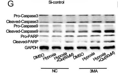
Application: WB Species: rat Sample:
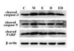
Application: WB Species: Rat Sample: lung tissues
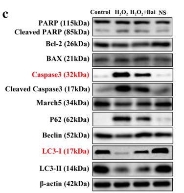
Application: WB Species: human Sample: L02 cells
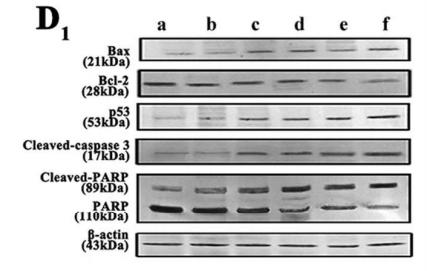
Application: WB Species: Mouse Sample: C6 cells
Restrictive clause
Affinity Biosciences tests all products strictly. Citations are provided as a resource for additional applications that have not been validated by Affinity Biosciences. Please choose the appropriate format for each application and consult Materials and Methods sections for additional details about the use of any product in these publications.
For Research Use Only.
Not for use in diagnostic or therapeutic procedures. Not for resale. Not for distribution without written consent. Affinity Biosciences will not be held responsible for patent infringement or other violations that may occur with the use of our products. Affinity Biosciences, Affinity Biosciences Logo and all other trademarks are the property of Affinity Biosciences LTD.